Veterinary Science
http://dx.doi.org/10.4142/jvs.2012.13.4.377
Received: 19 Oct. 2011, Revised: 27 Dec. 2011, Accepted: 4 Apr. 2012
Original Article
*Corresponding author: Tel: +82-31-463-4578; Fax: 82-31-463-4516; E-mail: [email protected]
ⓒ 2012 The Korean Society of Veterinary Science.
This is an Open Access article distributed under the terms of the Creative Commons Attribution Non-Commercial License (http://creativecommons.org/licenses/by-nc/3.0) which permits
unrestricted non-commercial use, distribution, and reproduction in any medium, provided the original work is properly cited.
A survey of porcine reproductive and respiratory syndrome among wild boar populations in Korea
Eun-Jin Choi, Chang-Hee Lee, Bang-Hun Hyun, Jae-Jo Kim, Seong-In Lim, Jae-Young Song, Yeun-Kyung Shin*
Animal, Plant and Fisheries Quarantine and Inspection Agency, Anyang 430-855, Korea
No information is currently available on porcine reproductive and respiratory syndrome virus (PRRSV) infection in wild boars (Sus scrofa) in Korea. In this study, the status of PRRS in wild boars was investigated. Blood samples were collected from 267 wild boars from eight provinces in Korea. Four of the samples tested (1.5%) were positive for PRRSV antibodies and eight (3.0%) were positive for antigens. Of the virus-positive samples, three and five samples were typed as containing European (EU, type 1) or North American (NA, type 2) viruses, respectively.
Two amplicons (one from type 1 and one from type 2) were used to analyze the PRRSV open reading frame 7 (ORF7) sequence. The nucleotide sequences of type 1 PRRSV ORF7 had identities between 96.1% and 98.4% with PRRSVs from domestic pigs in Korea. The sequences of type 2 PRRSV ORF7 had identities of 100% with the PRRSV strain VR-2332, which was prototypic North American strain.
These results show that PRRSVs are present in wild boars in Korea, and effective PRRSV surveillance of the wild boar population might therefore be useful for disease control.
Keywords: ELISA, Korea, porcine reproductive and respiratory syndrome, RT-PCR, wild boar (Sus scrofa)
Introduction
Porcine reproductive and respiratory syndrome (PRRS) is an infectious disease characterized by reproductive disorders in sows along with respiratory signs in piglets and fatteners resulting in significant economic losses in the pig industry worldwide [21,35]. The disease is caused by the PRRS virus (PRRSV), which is classified as a member of the order Nidovirales, family Arteriviridae, and genus Arterivirus [6]. The genome of PRRSV is approximately 15 kb in length and consists of at least nine open reading
frames (ORFs) [2,10]. ORF1a and 1b encode the enzymes responsible for replication; ORF2a and ORFs 3, 4, and 5 encode the membrane-associated glycoproteins; ORF2b and 6 encode the non-glycosylated membrane proteins, and ORF7 encodes the nucleoprotein (N) protein [34].
PRRSVs are divided into two genotypes: European (type 1) and North American (type 2) strains. The two genotypes share an approximately 67% similarity at the nucleotide level over the full genome [20,22]. The virus is primarily transmitted by contact with infected pigs but also through feces, urine, semen, and fomites. Additionally, it can be spread indirectly, presumably via aerosol routes and possibly by mechanical vectors [37].
The habitat of wild boar (Sus scrofa) has been destructed with community development and, consequently, at some area, the density and distribution of the wild boar have increased from 2010 to 2011. Without predators or competing animals, the numbers of wild boars have increased regionally [13]. Direct contact between wild boars and domestic pigs may occur rarely because all domestic pigs are reared within farming facilities in Korea.
The potential role of wild boars as a reservoir for PRRSV has been reported in France, Germany, and the USA with serological evidence of infection [3,24]. Since the emergence of PRRSV in 1993, PRRS has been widespread in domestic pigs throughout Korea [6,15,17,18,35].
Furthermore, wild boars and domestic pigs have been reported to have the same susceptibility to PRRSV [1].
Monitoring PRRS in wild boars might therefore be an
important factor for disease control in domestic pigs. The
present study was performed to assess the prevalence of
PRRSV in wild boars in Korea and provide information for
developing effective PRRS surveillance programs.
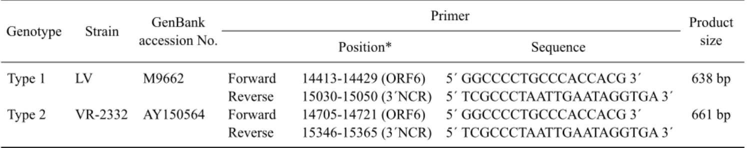
Table 1. Primers for the detection and differentiation of porcine reproductive and respiratory syndrome virus (PRRSV) Genotype Strain GenBank
accession No.
Primer Product
Position* Sequence size
Type 1 Type 2
LV VR-2332
M9662 AY150564
Forward Reverse Forward Reverse
14653-14671 (ORF7) 15030-15050 (3´NCR) 14933-14951 (ORF7) 15346-15365 (3´NCR)
5´ ATGGCCAGCCAGTCAATCA 3´;
5´ TCGCCCTAATTGAATAGGTGA 3´
5´ ATGGCCAGCCAGTCAATCA 3´;
5´ TCGCCCTAATTGAATAGGTGA 3´
398 bp 433 bp
*Primer position: forward primers are from ORF7 and reverse primers are from the 3´NCR region. ORF: open reading frame, NCR:
non-coding region.
Table 2. Primers for propagating the complete ORF7 region of PRRSV Genotype Strain GenBank
accession No.
Primer Product
Position* Sequence size
Type 1 Type 2
LV VR-2332
M9662 AY150564
Forward Reverse Forward Reverse
14413-14429 (ORF6) 15030-15050 (3´NCR) 14705-14721 (ORF6) 15346-15365 (3´NCR)
5´ GGCCCCTGCCCACCACG 3´
5´ TCGCCCTAATTGAATAGGTGA 3´
5´ GGCCCCTGCCCACCACG 3´
5´ TCGCCCTAATTGAATAGGTGA 3´
638 bp 661 bp
*Primer position: forward primers are from ORF6 and reverse primers are from 3´NCR region.
Materials and Methods
Serum sample collection and virus propagation Blood samples collected from 267 wild boars from eight provinces of Korea (Gyeonggi, Gangwon, Chungbuk, Chungnam, Jeonbuk, Jeonnam, Gyeongbuk, and Gyeongnam) were submitted to the Viral Disease Division of Animal, Plant and Fisheries Quarantine and Inspection Agency (Korea) during the hunting season in November 2010∼February 2011. Blood samples were taken by hunters from the heart immediately after the wild boars had been shot. The collected samples were stored at 4
oC and transported to the laboratory within 1∼2 weeks. The sera were collected by centrifugation for 15 min at 4
oC at 1,500 × g and stored at −20
oC until use. The PRRSV prototype VR-2332 and Lelystad virus (LV) were propagated in MARC-145 cells, and used as positive controls for the reverse transcription-PCR assay.
Serological test
Anti-PRRSV antibody titers were determined using a commercially available HerdChek PRRS 2XR Virus Antibody Test Kit (Idexx Laboratories, USA) according to the manufacturer’s instructions. Samples were considered to be positive for PRRSV antibodies if the ratio of sample absorbance to positive control absorbance (S/P) was greater than 0.4.
RT-PCR for the detection and differentiation of PRRSV
Total RNA was extracted from 100 μL of each serum sample using an RNeasy mini kit (Qiagen, Germany) according to the manufacturer’s instructions. RT-PCR was carried out using a OneStep RT-PCR kit (Qiagen, Germany) and a PRRSV common primer set (Table 1) derived from the sequences of ORF7 and the 3´ non-coding region of the VR-2332 and LV strains. The primer set was designed to detect and differentiate between PRRSV types 1 and 2. The PCR reaction contained 5 μL 5× RT-PCR buffer (2.5 mM MgCl
2), 0.4 mM dNTPs, 0.5 μM of each of the four primers shown in Table 1, 1 μL enzyme mix, and 5 μL RNA in a final volume of 25 μL. RT-PCR amplification was carried out as follows: a reverse transcription step at 50
oC for 30 min; RTase inactivation and initial PCR activation at 95
oC for 15 min followed by 35 cycles of denaturation at 94
oC for 20 sec, annealing at 55
oC for 20 sec, and extension at 72
oC for 30 sec, and a final elongation step at 72
oC for 10 min. The amplicons were separated by electrophoresis in a 1.5% agarose gel and stained with ethidium bromide.
Sequencing and phylogenetic analysis of ORF7
For the complete ORF7 sequencing, PCR was performed
using SuperScript One-Step RT-PCR with Platinum Taq
(Invitrogen, USA) according to the manufacturer’s
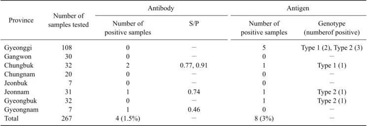
Table 3. Results of PRRSV detection in wild boars from different provinces of Korea
Province Number of samples tested
Antibody Antigen
Number of positive samples
S/P Number of
positive samples
Genotype (numberof positive) Gyeonggi
Gangwon Chungbuk Chungnam Jeonbuk Jeonnam Gyeongbuk Gyeongnam Total
108 30 32 20 7 31 32 7 267
0 0 2 0 0 1 0 1 4 (1.5%)
−
− 0.77, 0.91
−
− 0.74
− 0.46
−
5 0 1 0 0 1 1 0 8 (3%)
Type 1 (2), Type 2 (3)
− Type 1 (1)
−
− Type 2 (1) Type 2 (1)
−
−
S/P: the ratio of sample absorbance to positive control absorbance.
Fig. 1. RT-PCR results for the detection and differentiation of PRRSV in wild boar serum samples. Lane M: 100-bp DNA ladder, Lanes 1, 2, 3, 7, and 8: sample Nos. 49, 77, 167, 193, and 227, respectively (433-bp band), Lanes 4, 5, and 6: sample Nos.
110, 129, and 258, respectively (398-bp band), Lane 9: normal wild boar serum used as a negative control, Lane 10: VR-2332 strain used as a positive control for type 2, Lane 11: Lelystad virus (LV) used as a positive control for type 1.
instructions with primers (Table 2) designed to include the full ORF7 area. The PCR amplicons were purified using a MiniElute gel extraction kit (Qiagen, Germany) and cloned using a pGEMT easy vector system (Promega, USA). Sequencing was then performed using a GenomeLab DTCS-Quick Start Kit (Beckman Coulter, USA) and CEQ8000 automated sequencer (Beckman Coulter, USA). Multiple sequence alignment of the individual sequences was performed using CLUSTALX 1.81, and nucleotide sequence identities among the Korean PRRSV isolates were calculated using BioEdit software (Ibis Biosciences, USA).
Phylogenetic reconstructions were generated with PHYLIP (ver. 3.572c) using the neighbor-joining method based on the Kimura two-parameter model [11,16].
Robustness of the phylogenetic analysis was measured by bootstrap analysis with 1,000 replications. Graphic output was produced by TreeView (ver. 1.6.1) [35]. Evolutionary history was inferred using the neighbor-joining method [36]. An optimal tree in which the sum of branch length was 1.90,543,171 is shown. The percentage of replicate trees in which the associated taxa clustered together in the bootstrap test (1,000 replicates) is shown next to the branches [12]. The tree was drawn to scale with branch lengths in the same units as those of the evolutionary distances used to establish the phylogenetic tree.
Evolutionary distances were calculated using the Kimura two-parameter method [11] and are expressed as units of the number of base substitutions per site. The analysis included 40 nucleotide sequences. All positions containing gaps and missing data were eliminated. There were a total of 363 positions in the final dataset. Evolutionary analyses were conducted with Molecular Evolutionary Genetics Analysis version 5 (MEGA 5) [29].
Results
Out of the 267 sera tested, four (1.5%) were positive for PRRSV antibodies (Table 3). The ELISA S/P ratios for the positive samples were 0.46, 0.74, 0.77, and 0.91. Eight sera samples (3.0%) were positive for PRRSV antigens (Fig. 1).
All PRRSV-positive wild boars were infected with only one genotype. Type 1 virus was detected in three wild boars from two provinces (Gyeonggi and Chungbuk), and type 2 virus was detected in five animals from three provinces (Gyeonggi, Jeonnam, and Gyeongbuk).
Two amplicons (sample No. 49 from Gyeongbuk and sample No. 129 from Chungbuk) from the positive samples were subjected to ORF7 sequencing (Fig. 2).
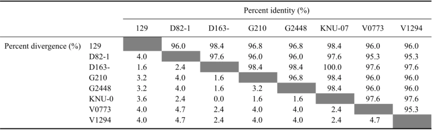
Homology of the deduced amino acid (aa) sequences between the wild boar type 1 virus (sample No. 129) and LV strain was 92.2% (Fig. 2A). Homology between the wild boar type 2 virus (No. 49) and VR-2332 strain was 100% (Fig. 2B). The wild boar type 1 virus had amino acid sequence identities between 96.0% and 98.4% with PRRSVs from domestic pigs in Korea (Table 4).
Phylogenetic analysis revealed that the wild boar type 1