Veterinary Science
http://dx.doi.org/10.4142/jvs.2012.13.2.193
Received: 29 Mar. 2011, Revised: 23 Aug. 2011, Accepted: 17 Mar. 2012
Original Article
*Corresponding author: Tel: +82-2-880-1258; Fax: +82-2-883-8651; E-mail: [email protected]
ⓒ 2012 The Korean Society of Veterinary Science.
This is an Open Access article distributed under the terms of the Creative Commons Attribution Non-Commercial License (http://creativecommons.org/licenses/by-nc/3.0) which permits unrestricted non-commercial use, distribution, and reproduction in any medium, provided the original work is properly cited.
A modified method for inducing periodontitis in dogs using a silk-wire twisted ligature
Se Eun Kim, Eui Ri Lee, Yesran Lee, Manbok Jeong, Young Woo Park, Jae Sang Ahn, Jeong Taek Ahn, Kangmoon Seo*
Department of Veterinary Surgery, College of Veterinary Medicine, Seoul National University, Seoul 151-742, Korea
This study was designed to assess the effectiveness of a modified silk ligature twisted with wire for inducing advanced periodontitis. Periodontitis was induced in five premolars and one molar of 20 healthy dogs over a 60-day period. The dogs were divided into four groups according to the ligature- inducing materials used: soft moistened food only, wire ligature (WL), silk ligature (SL) and twisted ligature with silk and wire (SWL). Periodontal indices were recorded, and dental radiographs were taken before and after 60 days of ligation. The ligatures were checked daily and the day the ligature fell out was noted. The period during which the ligatures were maintained was significantly shorter for the SL group compared to the SWL group (p < 0.05). Results of the clinical examination showed that almost all periodontal status parameters including the plaque index, gingival index, clinical attachment level, and bleeding on probing were significantly exacerbated in the SWL group compared to the other groups (p < 0.05). Radiographic evaluation demonstrated that alveolar bone levels were significantly lower in the SWL group than the other groups on day 60 (p < 0.05). These results suggested that experimental periodontitis induced by SWL could be an effective method for investigating periodontitis in canine models.
Keywords: dog, experimental model, periodontitis
Introduction
Periodontal inflammatory disease is one of the most prevalent disorders affecting both humans and small animals [1,7]. Therefore, several studies have examined the occurrence and treatment of periodontal disease using models of experimentally induced periodontitis. This condition has been induced in rats using a range of methods including pathogen intake, endotoxin injection, high
carbohydrate feeding, or ligature placement around the tooth cervix [12,13,20]. Compared to rat models, ligature induction of periodontitis is used more frequently in dogs [3,17,21].
Ligation materials used to induce periodontitis including silk, cotton, and nylon are placed between the tooth and periodontium to mechanically widen the periodontal pocket and to facilitate plaque accumulation in the dento-gingival region [5]. Several studies have induced experimental periodontitis using a more invasive method in which a full-thickness gingival flap is made and the alveolar bone is destroyed prior to ligation [3,18]. These methods take several months to promote advanced periodontitis, and thread-type ligatures might be lost due to loosening or wearing during this period. In general, ligatures that are lost during the periodontitis induction phase are immediately replaced to maintain the same induction period and environment [3,13].
These reparative procedures require redundant general anesthesia. Furthermore, the induction site can be affected by additional manipulations that may act as variables in the experimental procedure. Therefore, the present study was designed to identify more effective methods for periodontitis induction using modified ligatures, which can promote cases of more advanced periodontitis using a less invasive approach and reduce unnecessary interventions compared to previously established techniques.
Materials and Methods
The protocol used for this study was approved by the
Institutional Animal Care and Use Committee (SNU-
100208-3; Seoul National University, Korea). Twenty
beagle dogs (13 females and 7 males) approximately 1.5
years old were used in this study. All experimental
operations and examinations were performed with dogs
Fig. 1. Ligature materials. (A) 0.012-inch ligature wire, (B) 2-0 braided silk, (C) twisted ligature with silk and wire.
under general anesthesia induced by a combination of medetomidine (0.01 mg/kg; Orion Pharma, Finland), tramadol (2 mg/kg; Samsung Pharm, Korea), and a commercial solution of zolazepam and tiletamine (2.5 mg/kg; Virbac, France) administered via intramuscular injection. To prepare the healthy gingiva, all teeth were scaled and polished using a piezo-type ultrasonic scaler (BonArt, USA), and tooth brushing was performed once daily without anesthesia for the following 2 weeks. During these periods of prophylactic care, the beagles were fed a hard pellet diet to reduce plaque formation [11].
Experimentally induced periodontitis
Two weeks after scaling, experimental periodontitis was induced on the left upper second premolar (PM2), third premolar (PM3), and fourth premolar (PM4) as well as the left lower PM3, PM4, and first molar (M1). Prior to ligation, the gingival attachment was incised slightly by inserting a number 11 scalpel blade (Ailee, Korea), and the periodontal ligaments were undermined until a periodontal pocket depth reached up to 3 mm with a straight elevator (Osung MND, Korea). After undermining, shallow notches for ligature retention were made in the mesial and distal cervical region of each tooth with a round bur (#1; Komet, Germany). An ancillary notch was placed in the mesial surface of the upper PM4.
The dogs were randomly assigned to four groups.
Periodontitis was induced in three groups (Fig. 1) by tying the ligatures around the cervical region of the tooth using dental ligature wire [wire ligature (WL), n = 5; ClassOne Orthodontics, USA)], 2-0 braided silk [silk ligature (SL), n
= 5; Ailee, Korea] or a twisted wire with 2-0 silk [silk-wire twisted ligature (SWL), n = 5]. The remaining group did not undergo any surgical intervention but was fed only soft-moistened food as a control (SF, n = 5). For pain control, tramadol (4 mg/kg, intramuscular injection) was
administered twice a day for 3 days after placing the ligatures. To promote plaque formation, soft-moistened food was given to all groups for the following 60 days. The ligatures were checked daily and the day the ligature fell out was recorded.
Periodontal status evaluation
Before (day 0) and 60 days after (day 60) ligature placement, the clinical periodontal parameters were recorded and digital dental radiographs were taken to evaluate the periodontal status. Clinical parameters included the plaque index (PI), gingival index (GI), periodontal pocket depth (PPD), clinical attachment level (CAL), and bleeding on probing (BoP) [10,16,19]. All measurements were taken at three sites per tooth. If a ligature fell out, the parameters were measured on that day. All measurements were taken by one experienced clinician (S.E. Kim, Seoul National University, Korea) using a Williams periodontal probe (Osung MND, Korea).
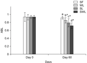
Digital intraoral radiographs (AFP Imaging, USA) were obtained to evaluate the amount of alveolar bone loss after ligature application. An intraoral size 2 sensor (AFP Imaging, USA) was positioned to take dental radiographs using bisecting and parallel techniques for the maxillary and mandibular teeth, respectively, with the same exposure protocol. The alveolar bone level (ABL) was measured using Adobe Systems (USA) software. This measurement was performed at the mesial and distal margin of each tooth except for the upper PM4. ABL of upper PM4 was only measured at the medial margin of the tooth because the ABL at the distal margin was difficult to distinguish from other superimposed dental structures such as the upper M1.
Distances from the alveolar bone margin and cemento- enamel junction to the root apex were measured and the ABL was calculated as a ratio of these two lengths.
Statistical analysis
All data was analyzed using SPSS software (ver. 12.0;
SPSS, USA). The maintenance period and clinical parameters for each group are expressed as the mean ± SD.
Differences in the maintenance periods between the SWL and WL or SL groups were assessed using Student’s t-test.
The clinical parameters measured on day 0 and day 60 in
the same group were analyzed using a paired t-test to
compare the progression of periodontitis in the same tooth
within each group. A one-way analysis of variance was
used to perform an intergroup comparison of the clinical
periodontal parameters and ABL changes on days 0 and 60
with Tukey’s method as a post hoc test. Data within the
95% confidence level were considered significant.
Table 1. Ligature maintenance periods and the ratio of ligature loss in each group
Group Ratio of ligature loss (n / N)
Ligature maintaining period Day*
p-value†WL
SL SWL
4 / 30 5 / 30 0 / 30
54.7 ± 14.6 53.2 ± 16.2 60.0 ± 0.0
0.051 0.046 1.000
*Data are expressed as the mean ± SD. †Statistical significance relative to SWL group. WL: wire ligature, SL: silk ligature, SWL:
silk-wire twisted ligature, n: the number of ligatures that fell out for the group, N: the number of teeth with ligatures in the group.
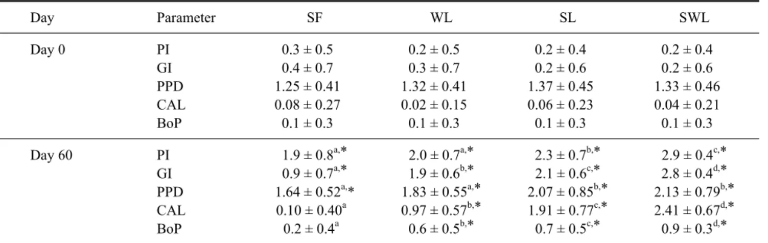
Table 2. Clinical parameters for each group
Day Parameter SF WL SL SWL
Day 0 PI
GI PPD CAL BoP
0.3 ± 0.5 0.4 ± 0.7 1.25 ± 0.41 0.08 ± 0.27 0.1 ± 0.3
0.2 ± 0.5 0.3 ± 0.7 1.32 ± 0.41 0.02 ± 0.15 0.1 ± 0.3
0.2 ± 0.4 0.2 ± 0.6 1.37 ± 0.45 0.06 ± 0.23
0.1 ± 0.3
0.2 ± 0.4 0.2 ± 0.6 1.33 ± 0.46 0.04 ± 0.21 0.1 ± 0.3
Day 60 PI
GI PPD CAL BoP
1.9 ± 0.8
a,* 0.9 ± 0.7
a,* 1.64 ± 0.52
a,* 0.10 ± 0.40
a0.2 ± 0.4
a2.0 ± 0.7
a,* 1.9 ± 0.6
b,* 1.83 ± 0.55
a,* 0.97 ± 0.57
b,* 0.6 ± 0.5
b,*
2.3 ± 0.7
b,* 2.1 ± 0.6
c,* 2.07 ± 0.85
b,* 1.91 ± 0.77
c,* 0.7 ± 0.5
c,*
2.9 ± 0.4
c,* 2.8 ± 0.4
d,* 2.13 ± 0.79
b,* 2.41 ± 0.67
d,* 0.9 ± 0.3
d,*
a,b,c,dValues with different superscript letters across each row are significantly different (p < 0.05). *Significantly increased values within a single group compared to the same parameters measured on day 0 (p < 0.05). PI: plaque index, GI: gingival index, PPD: periodontal pocket depth, CAL: clinical attachment level, BoP: bleeding on probing, SF: negative control, WL: wire ligature, SL: silk ligature, SWL: silk-wire twisted ligature. Data are expressed as the mean ± SD.