Copyright ⓒ the Korean Society for Transplantation, 2018
Successful Treatment of Invasive Gastric Mucormycosis in a Kidney Transplant Recipient
Hyung Nam Kim, M.D.
1, Sun Ae Han, M.D.
1, Ha Yeol Park, M.D.
1, Hyun Woo Kim, M.D.
1Ran Hong, M.D.
2, Nam Gyu Choi, M.D.
3, Min Ho Shin, M.D.
3, Na Ra Yoon, M.D.
1Hyun Lee Kim, M.D.
1, Jong Hoon Chung, M.D.
1, and Byung Chul Shin, M.D.
1Departments of Internal Medicine
1, Pathology
2, and Surgery
3, Chosun University College of Medicine, Gwangju, Korea
Mucormycosis is an extremely rare but potentially life-threatening fungal infection. Gastrointestinal (GI) mucormycosis is very rare and occurs primarily in highly malnourished patients, especially in infants and children. A 55-year-old man with end-stage renal disease due to diabetic nephropathy, who had undergone deceased donor kidney transplantation 2 years prior, complained of abdominal pain and distension with a 3-day duration. Computed tomography revealed diffuse gastric wall thickening, and a huge amount of grey colored necrotic debris surrounded by erythematous erosive mucosa was observed at the antrum to upper body by GI endoscopy. The microscopic examination obtained from a GI endoscopic specimen demonstrated peptic detritus with numerous non-septate mucor hyphae in the mucosa and submucosa. Mucormycosis was diagnosed based on the clinical findings and morphological features. A total gastrectomy was performed and an antifungal agent was administered. A microscopic examination of the surgical specimen demonstrated invasive mucormycosis with numerous fungal hyphae with invasion into the mucosa to subserosa. The patient and graft were treated successfully by total gastrectomy and antifungal therapy.
Key Words: Mucormycosis, Kidney transplantation, Stomach
중심 단어: 털곰팡이증, 신장이식, 위장Received May 17, 2018 Revised October 1, 2018 Accepted October 16, 2018
Corresponding author: Byung Chul Shin
Department of Internal Medicine, Chosun University Hospital, Chosun University College of Medicine, 365 Pilmun-daero, Dong-gu, Gwangju 61453, Korea
Tel: 82-62-220-3967, Fax: 82-62-234-9653 E-mail: [email protected]
Case Report
INTRODUCTION
Invasive fungal infections have been considered as major causes of morbidity and mortality in immunosuppressed pa- tients, such as, kidney or liver transplanted patients, and
Aspergillus
andCandida
species are the commonly isolated fungal pathogens. Recently, fungi of the order Mucorales have been reported to be causative agents of rare and life-threatening opportunistic infections(1). These fungi arecommonly found in decaying food, old bread, and soil(2), usually the upper and lower respiratory tract after in- halation of airborne spores(3). When not treated, mucormy- cosis rapidly progresses by extension to contiguous sites and hematogenous dissemination. Its clinical features are not sig- nificantly different from those of other invasive mycoses and result from hyphal angioinvasion leading to thrombosis and infarction of surrounding tissue and extensive hemor- rhage(3-5). Underlying medical conditions and risk factors determine the presenting clinical syndrome. The rhinocere- bral form is classically observed in poorly controlled dia- betes, especially in patients with ketoacidosis, whereas in- vasive pulmonary disease is more common in neutropenic patients. Cerebral mucormycosis has been reported in drug addicts and AIDS patients(6). Of the several types of mu- cormycosis infections, gastrointestinal (GI) mucormycosis is very rare with manifestations ranging from colonization of
J Korean Soc Transplant 2018;32:104-107
https://doi.org/10.4285/jkstn.2018.32.4.104
105
Hyung Nam Kim, et al: Invasive Gastric Mucormycosis in a KT Recipient
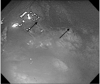
Fig. 1. Gastrointestinal endoscopy showed a huge amount of grey colored elevated necrotic debris surrounded by erythematous erosive mucosa from antrum to upper body (arrows).
peptic ulcers to infiltrative disease with vascular invasion and dissemination(3,6).
Here we report a case of gastric mucormycosis in a kid- ney transplant recipient that was confirmed by histologic findings, which also revealed numerous fungal hyphae had invaded into mucosa to subserosa. This study was approved by the Institutional Review Board of Chosun University Hospital (2018-12-013).
CASE REPORT
A 55-year-old man was admitted to our emergency room because of diffuse abdominal pain and hematemesis. He had undergone deceased donor kidney transplantation 2 years ago because of end-stage renal disease due to diabetic nephr- opathy and maintained immunosuppressive agents with ta- crolimus, mycophenolate mofetil, and methylprednisolone.
The trough level of tacrolimus has been maintained between 4 to 6 ng/mL. The allograft function was kept stable with an estimated glomerular filtration rate of 60 mL/min/1.73 m2. He had also been diagnosed with early gastric carcinoma and was treated by endoscopic submucosal dissection and snare polypectomy some 5 years ago. In the emergency room, he had an acute ill-looking appearance, anemic con- junctiva and an alert mental status. His blood pressure was 90/60 mmHg, body temperature 35oC, pulse rate 104 beats/min, and respiratory rate 20 breaths/min. Abdominal examination revealed decreased bowel sounds and tender- ness in the right upper quadrant with rigidity and rebound tenderness. Laboratory findings were as follows: leukocyte count 11,220/mm3 with 86.7% segment form, hemoglobin 6.7 g/dL, platelet count 174,000/mm3, serum blood urea ni- trogen 68.8 mg/dL, creatinine 3.43 mg/dL, total bilirubin 0.18 mg/dL, albumin 2.18 g/dL, aspartate aminotransferase 2.1 U/L, alanine aminotransferase 2.0 U/L, and C-reactive protein 14.7 mg/dL. Urinalysis results were negative for blood and leukocytes. Computed tomography revealed dif- fuse gastric wall thickening and during GI endoscopy a huge amount of grey colored elevated necrotic debris surrounded by erythematous erosive mucosa was observed from antrum to upper body (Fig. 1). During GI endoscopy the patient’s mental status changed to drowsy. At subsequent emergency total gastrectomy, the stomach was found to have two per-
foration sites and mucosa was partially dissociated from sub- mucosa (Fig. 2A). Microscopic examination of the surgical specimen revealed multifocal transluminal ulcer formation (Fig. 2B), necrotizing vasculitis filled with thrombi (Fig.
2C), a fungal ball (Fig. 2D), and numerous broad-based, non-septate, right angle branched fungal hyphae (Fig. 2E, F).
Mucormycosis was diagnosed based on histologic and clin- ical findings. Initially, intravenous amphotericin B (1 mg/kg/day) was administered for 9 days, but this was later changed liposomal amphotericin B (5 mg/kg/day) because of renal toxicity during 10 days. The patient was discharged on oral posaconazole (400 mg twice daily) for 21 days in good general condition with a healthy transplanted kidney.
He has been maintained immunosuppressive agents with ta- crolimus, mycophenolic acid, and reduced dose of methyl- prednisolone from 4 to 2 mg. The trough level of tacrolimus has been maintained between 2 to 4 ng/mL. The allograft function was slightly decreased with an estimated glomer- ular filtration rate of 50 mL/min/1.73 m2.
DISCUSSION
Mucormycosis is a rare, opportunistic fungal infection that occurs almost exclusively in immunocompromised backgrounds in association with diabetes, leukemia, lympho- ma, renal disease, septicemia, burns, malnutrition, or after
106
J Korean Soc TransplantㆍDecember 2018ㆍVolume 32ㆍIssue 4
Fig. 2. (A) Gross specimen of stomach. The black arrow indicates a perforation lesion. (B) Multifocal transluminal ulcer finding of stomach.
(C) Necrotizing vasculitis finding of filled thrombi and vascular wall destruction. (D) Fungal ball (arrows).
(E, F) PAS stain (E, ×400) and Gomori’s Methenamine silver stain (F, ×400) sections showing broad, non-septated hyphae with right angled branches (B: HE stain, ×200; C, D:
HE stain, ×400).
long-term treatment with immunosuppressive agents(7).
Mucormycosis can manifest as rhinocerebral, pulmonary, disseminated, GI, or cutaneous disease(3). Of these, rhinoc- erebral and pulmonary disease are the most common, and GI involvement is rare, accounting for only 7% of all cases of mucormycosis(3). The GI organs most frequently in- volved in descending order are stomach, colon, small intes- tine, and esophagus(8). The diagnosis of gastric mucormy- cosis is confirmed by presence of aseptate and broad peri- odic acid–Schiff (PAS)-positive fungal hyphae adjacent to necrotic areas by biopsy. Definitive diagnosis by micro- biological culture and identification of the causative organ- ism to the genus and species levels has valuable epidemio-
logical, therapeutic and prognostic implications. Importantly, mucormycosis characteristically invades blood vessels, caus- ing thrombosis, multiple infarctions, and visceral organs hemorrhage(8). Clinically, the lesion mimics an ulcer or carcinoma.
Diagnosis is based on the presence of tissue invasion by biopsy, clinical history taking, radiographic findings, and culture results(3,9). The important aspects of the treatment of mucormycosis are aggressive metabolic support, ampho- tericin B, and surgical debridement of all necrotic involved tissues(10,11).
Our patient had a history of endoscopic mucosal dis- section and deceased donor kidney transplantation, which
107
Hyung Nam Kim, et al: Invasive Gastric Mucormycosis in a KT Recipient
may have led to a systemic predisposition to fungal infection. Gastric mucormycosis was confirmed based on clinical and pathologic findings, and successfully treated by surgical resection and the administration of amphotericin B.
Although the incidence of mucormycosis is increasing slightly, it is still rare in kidney transplantation recipients.
Early diagnosis, rapid medical treatment and appropriate surgical intervention can improve survival in cases of mu- cormycosis in kidney transplant recipients.
CONFLICTS OF INTEREST
No potential conflict of interest relevant to this article was reported.