Fucoidan Attenuates Perfluorooctane Sulfonate-induced Apoptosis of Neuronal Cells
7
0
0
전체 글
(2) J . Mar . Biosci. Biotechnol. 2017,. p.. 01 - 07. Vol.. Among diverse etiologies, environmental factor is. 9,. No.. 1. [Research. Paper]. pollutants through non-provocative approaches.. believed to play as an important risk factor for. Thus, this study attempted to investigate the potential. development of neurodegenerative diseases. While. role of fucoidan as a neuroprotective marine polypeptide. PFOS is mostly accumulated in the liver and blood,. in environmental pollutant-induced apoptosis of neuronal. its levels in the brain gradually increase with time after. cells in culture.. exposure [7]. PFOS is reported to trigger opening of tight junction in brain endothelial cells [8]. Neonatal. Materials and Method. exposure to PFOS can cause neurotoxic effects in adult mice, manifested as changes in spontaneous behavior and habituation [9]. High dose of PFOS leads to neurologic delays and neonatal mortality. Apoptosis of neuronal cells during developmental period of synaptogenesis, known as the brain growth spurt. period,. neurobehavioral. is. a. critical. disturbances. event expressed. to. induce. either. in. childhood or with delayed onset in adulthood [10].. PC-12 cell culture PC12 cells (purchased from Korean cell line bank) were cultured in RPMI 1640 Medium supplemented with 5% heat-inactivated fetal bovine, 10% horse serum and 1% penicillin/streptomycin at 37℃ in a humidified atmosphere of 5% CO2. The cells were used for experiments prior to passage 25.. Many environmental pollutants are known to induce. MTT assay Cell viability was measured using tetrazolium salt. apoptotic neurodegeneration and closely associated with. 3-[4,5-dimethylthiazol -2-yl]-2,5- diphenyltetrazolium. a variety of neuronal diseases. Lee at al. [11] reported. bromide (MTT; Promega, Korea). PC12 cells were. that PFOS induces apoptosis of cerebellar granule cells. seeded in 96-well plate (3 × 10⁴ cells/well) and allowed. via ROS-dependent PKC activation.. to attach overnight. Cells were treated with different. Fucoidan is a main bioactive constituent of the brown. concentrations of PFOS for 24 hr in presence or absence. seaweed, which is widely distributed in East Asia and. of fucoidan (Sigma-Aldrich, St. Louis) (25 or 50 μg/ml). is consumed as a marine vegetable. Fucoidan is also available as food supplement in several countries. While. and then, 10 μl of MTT (5 mg/ml) was added into each. the outstanding physiological functions of fucoidan are well-established, only limited studies are available about. containing MTT was removed, and 100 μl of DMSO was added to dissolve the purple formazan. The. its neuroprotective potentials. It is reported that fucoidan. absorbance was detected at 595 nm by a microplate. induces ROS-dependent apoptosis in 5637 human. reader (Bio-Rad).. bladder cancer cells [12]. Gao et al. [13] reported that fucoidan inhibits hydrogen peroxide-induced apoptosis. well. After 2hr of incubation at 37℃, the medium. of PC12 cells by increasing the Bcl-2/Bax ratio and. LDH Assay Cytotoxicity was evaluated by measurement of. decreasing. lactate dehydrogenase (LDH) activity released from the. caspase-3. activity,. suggesting. the. cytosol of damaged cells. Cells were treated with. neuroprotective role of fucoidan. Potentials of PFOS to elicit neurotoxicity are not only scientifically important but draw a public concern over the long-term health effects. In particular, because. different concentrations of PFOS for 24 hr in presence or absence of fucoidan (25 or 50 μg/ml). The culture medium was collected and a Cytotoxicity Detection Kit. environmental pollutant such as PFOS is exposed to. (Roche, Penzberg, Germany) was used for the assay. The. the general population regardless of age and sex. percentage of cell-mediated lysis was expressed. throughout their entire life span, it is important to. according to the following formula: % cytotoxicity =. identify the ways to alleviate the potential risk of. (exp. value – low control)/ (high control – low control).. - 2 -.
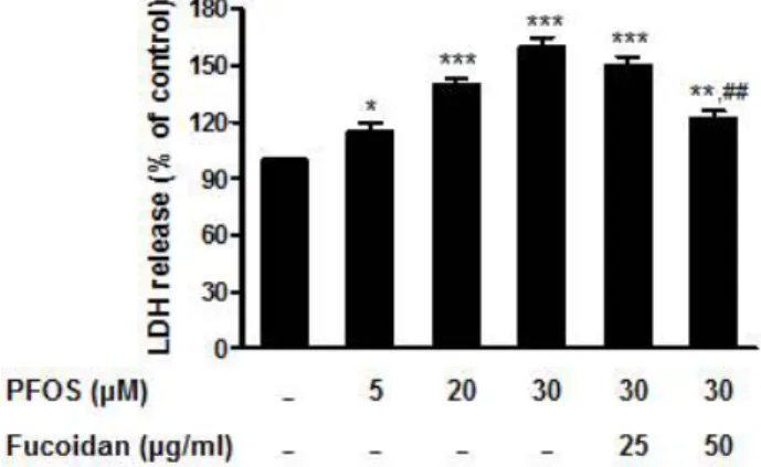
(3) J . Mar . Biosci. Biotechnol. 2017,. p.. 01 - 07. Vol.. 9,. No.. 1. [Research. Paper]. Intracellular ROS measurement Fluorescent probe, 2',7'-dichlorofluorescin diacetate (DCFH-DA), was used to measure the formation of. 50 μg/ml, respectively (Fig 1). In accordance with cell. intracellular ROS as described previously [11]. PC12. was reduced to 150% and 122 % in the presence of. 4. cells were seeded in 96-well black plate (3 × 10. viability, cytotoxicity as measured by LDH release was significantly increased to 160% at 30 μM PFOS but fucoidan 25 and 50 μg/ml, respectively (Fig 2).. cells/well). After 24 hr, cells were treated with PFOS or H2O2 for 4 h and then treated with 5 μM of DCFH-DA for 30 min. After washing cells with PBS, the fluorescence intensity was measured using microplate reader (FLUOstarOPTIMA,BMGLABTECH) with excitation at 485 nm and emission at 520 nm. Caspase-3 activity measurement Cells were grown on 96- well plates (3 × 10⁴ cells/well) and the caspase-3 activity was measured by using commercial assay kits (Promega, Madison, WI). The protocols were provided by the vendor. Briefly, cells were treated with a luminogenic substrate containing the DEVD sequence and the relative light units were measured using a Plus LB96V luminometer (Berthold Detection System, OakRidge, TN). Statistics Data are expressed as means ± SEM. Statistical. Figure 1. Effects of PFOS and fucoidan on cell viability. MTT assay in PC12 cells treated with DMSO as a vehicle control or PFOS (5, 20, and 30 µM), was conducted in the presence or absence of 25 and 50 µg/ml fucoidan as described in materials and method. All values are relative to the control cells. Values represent mean ± SEM of three replicate determinations. *P < 0.05, **P < 0.01, ***P < 0.001 vs. DMSO control. #P < 0.05, ###P < 0.001 vs. PFOS 30 µM-treated cells.. analyses were made by the Student’s t-test to compare values between two groups or by one way ANOVA followed by Tukey’s post hoc test to compare values among more than three groups. A value of P < 0.05 was considered significant. Results. The effects of fucoidan on viability of PC12 cells treated with PFOS Cell viability and cytotoxicity were measured by MTT assay and LDH assay to evaluate the cytotoxic effects of PFOS and the protective effects of fucoidan on PC12 cells. Cells were treated with different concentration (0 ~ 30 μM) of PFOS for 24 h in presence or absence of fucoidan. Cell viability was significantly reduced to 68 % at 30 μM, which was recovered up to 77% and 92 % in the presence of fuoidan 25 and. Figure 2. Effects of PFOS and fucoidan on cytotoxicity. LDH assay in PC12 cells treated with DMSO as a vehicle control or PFOS (5, 20, and 30 µM), was conducted in the presence or absence of 25 and 50 µg/ml fucoidan as described in materials and method. All values are relative to the control cells. Values represent mean ± SEM of three replicate determinations. *P < 0.05, **P < 0.01, ***P < 0.001 vs. DMSO control. ##P < 0.01 vs. PFOS 30 µM-treated cells.. - 3 -.
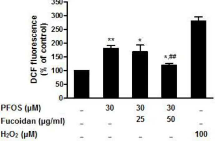
(4) J . Mar . Biosci. Biotechnol. 2017,. p.. 01 - 07. Vol.. The protective effect of fucoidan on PFOS-induced ROS production in PC12 cells ROS plays a pivotal role in environmental. 9,. No.. 1. [Research. Paper]. respectively (Fig 4).. pollutant-induced neuronal stress. The treatment of cells with 30 μM of PFOS for 4 h induced significant increase (170 % of control) in ROS accumulation, which was effectively blocked by pretreatment with fucoidan. While 25 μg/ml fucoidan did not significantly reduce ROS production, 50 μg/ml showed a significant reduction of ROS production (120% of control). H2O2 (100 μM) increased ROS generation about 280% and used as a positive control (Fig3). Figure 4. Effects of PFOS and fucoidan on caspase-3 activity. Caspase-3 activity in PC12 cellse treated with DMSO as a vehicle control or 30 µM PFOS, was measured in the presence or absence of 25 and 50 µg/ml fucoidan as described in materials and method. All values are relative to the control cells. Values represent mean ± SEM of three replicate determinations. **P < 0.01, ***P < 0.001 vs. DMSO control. #P < 0.05, ###P < 0.001 vs. PFOS 30 µM-treated cells. Discussion. PFCs have been found to be relatively high level in human blood compared to other environmental Figure 3. Effects of PFOS and fucoidan on ROS production. ROS production in PC12 cells treated with DMSO as a vehicle control, 30 µM PFOS or 100 µM H2O2 for 4 h, was measured in the presence or absence of 25 and 50 µg/ml fucoidan as described in materials and method. All values are relative to the control cells. Values represent mean ± SEM of three replicate determinations. *P < 0.05, **P < 0.01 vs. DMSO control. vs. PFOS 30 µM-treated cells.. ##P < 0.01. contaminants and of particular concern was their possible neurotoxic effects. PFOS is considered as a neurotoxicant in animal studies. Since there has been a public concern over the long-term health effects of neurotoxic pollutants, development of prophylactic approaches for its prevention is in a high demand. Apoptosis of neuronal cells is a key element in. The effects of fucoidan on PFOS-induced apoptosis of PC12 cells Caspase-3 activity, a hallmark of apoptosis, was measured to determine the cytotoxicity of PFOS and. determining neurotoxicity and apoptotic potential of compound is considered as one of key elements that distinguish neurotoxicants from other toxic materials [14]. While a wide spectrum of biological activities by fucoidan are reported, there is a lack of data that. the cytoprotective effects of fucoidan. PFOS exposure. fucoidan may have a capacity to improve conditions. of cells for 24 h induced a significant increase in caspase-3 activity about 250 % of control at 30 μM.. of environmental pollutant-induced neurotoxicity. Thus, this study attempts to look into PFOS-induced. The increase was dampened to 180 % and 130% in. neurotoxicity with respect to the apoptotic events. μg/ml,. and examine protective effects of readily available. the. presence. of. fucoidan. 25. and. 50. - 4 -.
(5) J . Mar . Biosci. Biotechnol. 2017,. marine. bioactive. p.. 01 - 07. polypeptide,. Vol.. fucoidan,. from. environmental pollutant-induced neurotoxicity.. 9,. No.. 1. [Research. Paper]. associated with a variety of neuronal diseases. However, studies on PFOS-induced apoptosis of. In the present study, dose-dependent increase of. neuronal cells are limited. PFOS induced increase of. cytotoxicity indicates the evidence that PFOS is a. an apoptotic parameter such as caspase-3 activity. An. nuerotoxicant,. effective blocking of apoptosis by fucoidan clearly. as. suggested. in. other. studies.. PFOS-induced cytotoxicity and apoptosis of neuronal cells may play a part in inducing a variety. demonstrates protective effects of fucoidan PFOS-induced neuronal cell death.. on. of neurological conditions. The protective effects of. Fucoidan exhibits various biological activities, such. fucoidan on PFOS-induced cytotoxicity suggest that. as an anti-inflammation in the brain [19] and. this marine polypeptide may be a material useful for. neuroprotectant. the neurological disorders.. injury [20]. Fucoidan has been shown to inhibit β. in. cerebral. ischemic-reperfusion. ROS is a key factor in the regulation of apoptotic. -amyloid protein (Aβ) accumulation within microglia. processes in neurons. Elevated production of ROS. through its effects on multiple scavenger such as. is associated with induction of apoptotic death in. tumor necrosis factor-α, or nitric oxide [21]. Fucoidan. many types of neuronal cells including cerebellar. also interacts with amyloid protein (Aβ) in the rat. granule cells [14]. In accordance with previous. basal forebrain neurons and improves cognitive. studies that demonstrated an increased production of. impairment [22]. Recently, it has been reported that. ROS by PFOS in several different cell types [15-18],. fucoidan attenuates mitochondrial dysfunction [23]. our results also showed a dose-dependent increase. and improves neurological outcome in traumatic brain. of ROS production with exposure to PFOS. While. of aged rat and protects brain microvessel endothelial. antioxidant activity of fucoidan is well established,. cells against diesel exhaust particle exposure- induced. effectiveness. for. disruption [24]. In keeping with the recent series of. environmental pollutants remains to be ascertained.. reports that suggest protective effects of fucoidan in. This study clearly demonstrated that fucoidan is an effective marine bioactive polypeptide for reducing. brain, the current study further supports the potential use of fucoidan in preventing or delaying the. PFOS-induced ROS stress in neuronal cells. The. neurological disorders. Because one third of causes. results further suggest that prophylactic therapy. of diseases is from the environmental origin, it is very. containing fucoidan may be a practical long-term. important to identify the non-provocative therapeutic. approach that may prevent neurological diseases. or prophylactic ways to protect the humans from the. caused by the environmental neurotoxicant exposure. In this study, PFOS generated a dose-dependent. ever-increasing environmental pollutants. Thus, the present study may contribute to establishing the. ROS production, which paralleled with apoptotic. potential role of fucoidan as a neuroprotective. events. The loss of neuronal cells by apoptosis is. polypeptide that protects the risk of neurological. the common final step of most neurodegenerative. disorders from the possible neurotoxic pollutants.. diseases.. of. In. antioxidant. particular,. activity. apoptosis. during. developmental period of synaptogenesis, known as the brain growth spurt (BSG) period, is a critical event. Acknowledgement. to induce neurobehavioral disturbances expressed. This work was supported by Basic Science Research. either in childhood or with delayed onset in adulthood. Program through the National Research Foundation of. [9,10]. Many environmental pollutants are known to. Korea (NRF) funded by the Ministry of Education. induce. (2014R1A1A2056565), Republic of Korea.. apoptotic. neurodegeneration. and. closely. - 5 -.
(6) J . Mar . Biosci. Biotechnol. 2017,. p.. 01 - 07. Vol.. 9,. No.. 1. [Research. Paper]. 659–668.. References. 11.. Lee,. H.G.,. Lee,. Y.J,. and. Yang,. J.H.. 2012.. 1. Giesy, J.P. and Kannan, K. 2002. Perfluorochemical. Perfluorooctane sulfonate induces apoptosis of cerebellar. surfactants in the environment. Environ. Sci. Technol.. granule cells via a ROS-dependent protein kinase C. 36, 146A–152A.. signaling pathway. Neurotoxicology. 33, 314-320.. 2. Harada, K.H., Yang, H.R., Moon, C.S., Hung, N.N., Hitomi, T., Inoue, K., Niisoe, T., Watanabe, T., Kamiyama, S., Takenaka, K., Kim, M.Y., Watanabe, K.,. 12. Han, M.H., Lee, D.S., Jeong, J.W., Hong, S.H., Choi, I.W., Cha, H.J., Kim, S., Kim, H.S., Park, C., Kim, G.Y., Moon, S.K., Kim, W.J., Hyun Choi, Y. 2017. Fucoidan. Takasuga, T. and Koizumi, A. 2010. Levels of. Induces ROS-Dependent Apoptosis in 5637 Human. perfluorooctane sulfonate and perfluorooctanoic acid in. Bladder Cancer Cells by Downregulating Telomerase. female serum samples from Japan in 2008, Korea in. Activity via Inactivation of the PI3K/Akt Signaling. 1994-2008 and Vietnam in 2007-2008. Chemosphere 79,. Pathway. Drug Dev. Res. 78, 37-48. 13. Gao, Y., Dong, C., Yin, J., Shen, J., Tian, J. and Li,. 314–319. 3. Lau, C., Anitole, K., Hodes, C., Lai, D., Pfahles-Hutchens,. C. 2012. Neuroprotective effect of fucoidan on. A. and Seed. J. 2007. Perfluoroalkyl acids: a review of. H2O2-induced apoptosis in PC12 cells via activation of. monitoring and toxicological findings. Toxicol. Sci. 99,. PI3K/Akt pathway. Cell. Mol. Neurobiol. 32, 523-529.. 366–394.. 14. Franklin, J.L. 2011. Redox regulation of the intrinsic. 4. Kannan, K., Tao, L., Sinclair, E., Pastva, S.D., Jude, D.J.. pathway in neuronal apoptosis. Antioxid. Redox. Signal. 14, 1437–1448.. and Giesy, J.P. 2005. Perfluorinated compounds in aquatic organisms at various trophic levels in a Great. 15. Liu, C., Yu, K., Shi, X., Wang, J., Lam, P.K., Wu, R.S.. Lakes food chain. Arch. Environ. Contam. Toxicol. 48,. and Zhou B. 2007. Induction of oxidative stress and. 559–566.. apoptosis by PFOS and PFOA in primary cultured. 5. Fox, N. 2012. When, where, and how does Alzheimer's disease start? Lancet Neurol. 11, 1017-1018. 6. Reverte, I., Klein, A.B., Domingo, J.L. and Colomina, M.T. 2013. Long term effects of murine postnatal. hepatocytes. of. freshwater. tilapia. (Oreochromis. niloticus). Aquat. Toxicol. 82, 135–143. 16. Slotkin, T.A., MacKillop, E.A., Melnick, R.L., Thayer, K.A.. exposure to decabromodiphenylether (BDE-209) on. and. Seidler,. F.J.. 2008.. Developmental. neurotoxicity of perfluorinated chemicals modeled in. learning and memory are dependent upon APOE. vitro. Environ. Health. Perspect. 116, 716–722.. polymorphism and age. Neurotoxicol. Teratol. 40, 17-27.. 17. Eriksen, K.T., Raaschou-Nielsen, O., Sørensen, M.,. 7. Sato, I., Kawamoto, K., Nishikawa, Y., Tsuda, S.,. Roursgaard, M., Loft, S. and Møller, P. 2010. Genotoxic potential of the perfluorinated chemicals PFOA, PFOS,. Yoshida, M., Yaegashi, K., Saito, N., Liu, W. and Jin, Y. 2009. Neurotoxicity of perfluorooctane sulfonate. PFBS, PFNA and PFHxA in human HepG2 cells. Mutat.. (PFOS) in rats and mice after single oral exposure. J. Toxicol. Sci. 34, 569–574.. Res. 700, 39–43. 18. Qian, Y., Ducatman, A., Ward, R., Leonard, S.,. 8. Wang, X., Li, B., Zhao, W.D., Liu, Y.J., Shang, D.S.,. Bukowski, V., Lan Guo, N., Shi, X., Vallyathan, V. and. Fang, W.G. and Chen, Y.H. 2011. Perfluorooctane. Castranova, V. 2010. Perfluorooctane sulfonate (PFOS) induces reactive oxygen species (ROS) production in. sulfonate triggers tight junction "opening" in brain endothelial cells via phosphatidylinositol 3-kinase.. human. Biochem. Biophys. Res. Commun. 410, 258–263. Neonatal exposure to perfluorooctane sulfonate (PFOS) perfluorooctanoic. acid. (PFOA). endothelial. cells:. role. in. endothelial permeability. J. Toxicol. Environ. Health A. 9. Johansson, N., Fredriksson, A. and Eriksson, P. 2008. and. microvascular. 73, 819–836. 19. Del Bigio, M.R., Yan, H.J., Campbell, T.M. and Peeling,. causes. J. 1999. Effect of fucoidan treatment on collagenase-induced intracerebral hemorrhage in rats.. neurobehavioural defects in adult mice. Neurotoxicology 29, 160–169.. Neurol. Res. 21, 415-419.. 10. Olney, J.W. 2002. New insights and new issues in. 20. Che, N., Ma, Y. and Xin, Y. 2016. Protective role of. developmental neurotoxicology. Neurotoxicology 23,. fucoidan in cerebral ischemia-reperfusion injury through. - 6 -.
(7) J . Mar . Biosci. Biotechnol. 2017,. p.. 01 - 07. Vol.. inhibition of MAPK signaling pathway. Biomol.. 23.. Ther.(Seoul). doi:10.4062/biomolther.2016.098.. Wang,. T.,. Zhu,. M.. Low-Molecular-Weight. 9,. No.. and. 1. [Research. He,. fucoidan. Z.Z.. Paper]. 2016.. attenuates. 21. Mitrasinovic, O.M. and Murphy, G.M. Jr. 2002.. mitochondrial dysfunction and improves neurological. Accelerated phagocytosis of amyloid-beta by mouse and. outcome after traumatic brain injury in aged mice:. human. macrophage. Involvement of Sirt3. Cell. Mol. Neurobiol. 36,. colony-stimulating factor receptor. J. Biol. Chem. 277, 29889-29896.. 1257-1268. 24. Choi, Y.S., Eom, S.Y., Kim, I.S., Ali, S.F., Kleinman,. 22. Gao, Y., Li, C., Yin, J., Shen, J., Wang, H., Wu, Y.. M.T., Kim, Y.D. and Kim, H. 2016. Fucoidan extracted. and Jin, H. 2012. Fucoidan, a sulfated polysaccharide. from Hijiki protects brain microvessel endothelial cells. from brown algae, improves cognitive impairment. against. induced by infusion of Aβ peptide in rats. Environ.. disruption. J. Med. Food. 19, 466-471.. microglia. overexpressing. the. Toxicol. Pharmacol. 33, 304-311.. - 7 -. diesel. exhaust. particle. exposure-induced.
(8)
수치
관련 문서
Control- and ASTRIN- depleted HeLa cells were untreated or treated with 5 Gy. IR and were then fixed at
In contrast, cells expressing wild-type form or constitutively active form of RapC (GFP-RapC and GFP-RapC G13V ) showed decreased cell area and cell adhesion, whereas rapC
Sesn2-luciferase transactivation was determined in the lysates of HEK293 cells transfected with an Nrf2 expression construct along with either a Sesn2 (pGL4-phSESN2)
protein levels in control or PIG3 depleted HCT116 and HeLa cells were tested. by
Assessment and recognition in credits or education of experiential learning such as knowledge or skills gained through employment or society, will help expand
Mean arterial plasma concentration-time profiles of 4-hydroxytamoxifen after an oral administration of tamoxifen (10 mg/kg) to rats in the presence or absence
Pharmacokinetic parameters were determined following an oral administration of nateglinide (30 mg/kg) to rabbits in the presence and absence of calcium channel blockers
PI3K inhibition decreased antioxidants/GD-induced apoptosis in A549 cells, and PI3K inhibitor LY294002 had inhibitory effect on antioxidants/GD-induced caspase-3
