Inflammation appears as high Prostate Imaging–
Reporting and Data System scores on prostate magnetic resonance imaging (MRI) leading to false positive MRI fusion biopsy
Elizabeth Rourke1, Abhijit Sunnapwar2, Daniel Mais3, Vishal Kukkar2, John DiGiovanni4, Dharam Kaushik1,5, Michael A. Liss1,4,5
Departments of 1Urology, 2Radiology, and 3Pathology, University of Texas Health San Antonio, San Antonio, TX, 4University of Texas Austin, College of Pharmacy, Austin, TX, 5Mays Cancer Center UT Health San Antonio MD Anderson, San Antonio, TX, USA
Purpose: To investigate if inflammation as a potential cause of false-positive lesions from recent UroNav magnetic resonance im- aging (MRI) fusion prostate biopsy patients.
Materials and Methods: We retrospectively identified 43 men with 61 MRI lesions noted on prostate MRI before MRI ultrasound- guided fusion prostate biopsy. Men underwent MRI with 3T Siemens TIM Trio MRI system (Siemens AG, Germany), and lesions were identified and marked in DynaCAD system (Invivo Corporation, USA) with subsequent biopsy with MRI fusion with UroNav. We ob- tained targeted and standard 12-core needle biopsies. We retrospectively reviewed pathology reports for inflammation.
Results: We noted a total of 43 (70.5%) false-positive lesions with 28 having no cancer on any cores, and 15 lesions with cancer noted on systematic biopsy but not in the target region. Of the men with cancer, 6 of the false positive lesions had inflammation in the location of the targeted region of interest (40.0%, 6/15). However, when we examine the 21/28 lesions with an identified lesion on MRI with no cancer in all cores, 54.5% had inflammation on prostate biopsy pathology (12/22, p=0.024). We noted the highest proportion of inflammation.
Conclusions: Inflammation can confound the interpretation of MRI by mimicking prostate cancer. We suggested focused efforts to differentiate inflammation and cancer on prostate MRI.
Keywords: Inflammation; Magnetic resonance imaging; Prostatic neoplasms; Prostatitis
This is an Open Access article distributed under the terms of the Creative Commons Attribution Non-Commercial License (http://creativecommons.org/licenses/by-nc/4.0) which permits unrestricted non-commercial use, distribution, and reproduction in any medium, provided the original work is properly cited.
Received: 30 December, 2018 • Accepted: 7 May, 2019 Corresponding Author: Michael A. Liss
Department of Urology, University of Texas Health San Antonio, 7703 Floyd Curl Drive, San Antonio, TX 78229, USA TEL: +1-210-567-5676, FAX: +1-210-567-3900, E-mail: liss@uthscsa.edu
ORCID: https://orcid.org/0000-0001-6978-1026
ⓒ The Korean Urological Association
www.icurology.org
Investig Clin Urol 2019;60:388-395.
https://doi.org/10.4111/icu.2019.60.5.388 pISSN 2466-0493 • eISSN 2466-054X
INTRODUCTION
Prostate cancer is the most common non-cutaneous cancer in American men [1]. Since the implementation of
prostate-specific antigen screening in the 1990s, urologists perform on men a non-targeted, template prostate needle biopsy in order to diagnose prostate cancer [2]. Standard template biopsy suffers from sampling error noted by the
30% risk of upgrading at the time of prostatectomy and considering that only 30% to 40% of men who undergo the procedure are diagnosed with prostate cancer [3].
Prostate magnetic resonance imaging (MRI) is an ima- ging modality that may allow more accurate prostate biopsies. Advances in MRI technology have also led to techniques allowing fusion of the MRI images onto the standard ultrasound (US) equipment [4]. Armed with the tools to direct the biopsy to a particular area, urologists have expected improved detection of more aggressive tumors and potentially reduced the number of biopsies with negative MRIs. However, in the recent article by the PRECISION (Prostate Evaluation for Clinically Important Disease:
Sampling Using Image Guidance or Not?) group, randomized men obtained a prostate biopsy based on MRI findings compared to a standard approach without MRI [5]. MRI only improved the detection rate of clinically significant cancer by 12% (95% confidence interval, 4 to 20; e.g., from 26% to 38%). While the result was statistically positive, we argue that a 38% detection rate is still quite weak. We consider other solid organ biopsies which typically reach a detection rate of more than 90% [6]. While MRI does provide incremental benefits to improve cancer detection, in our practice, we have noted a high false-positive rate that could be influencing the accuracy of prostate MRI.
Inflammation is known to mimic prostate cancer lesions on MRI, for example, chronic prostatitis or nodules following bacillus Calmette–Guérin treatment [7-9]. However, it is unknown how commonly inflammation plays a role in MRI fusion ultrasound-guided (MRI-US) prostate needle biopsies.
We investigate inflammation identified on pathology reports from recent UroNav MRI fusion prostate biopsy patients.
MATERIALS AND METHODS
1. Patients
After local IRB approval (approval number: HSC21000480H, University of Texas Health Science Center San Antonio [UTHSCSA]), we retrospectively identified 43 men with 61 MRI lesions noted on prostate MRI before MRI-US fusion prostate biopsy. The patients were consecutive and the only patients in at the UTHSCSA who had a fusion biopsy. There was no specific inclusion or exclusion criteria other than needing to have a fusion biopsy.
2. Multiparametric MRIs
The MRI’s were performed at our local institution, including two different scanners (UT Health: 3T Siemens TIM Trio MRI system [Siemens AG, Erlangen, Germany]
and University Hospital: Phillips 3T [Andover, MA, USA]), with a pelvic phased-array coil, with or without an endo- rectal coil. After MRI, a body MRI radiologist would identify target lesions and outline the region of interest with the DynaCAD system (Invivo Corporation, Gainesville, FL, USA).
We acquired T2-weighted, diffusion-weighted imaging, and dynamic contrast-enhanced sequences with B values of (50, 400, 800, 1,400). Our radiologists categorized lesions according to the Prostate Imaging–Reporting and Data System, version 2 (PI-RADS 2) with a score from one to five [10]. All MRI’s were imported into the DynaCAD system by one radiologist for standard (A.S.) and reviewed by a urologist before biopsy (M.A.L.).
3. Biopsy technique
All patients underwent an enema before the procedure and antibiotics for less than 24 hours starting the morning of the procedure. We performed MRI fusion standard techniques using the UroNav fusion biopsy system. A single surgeon (M.A.L.) performed biopsies at the same location in a surgery center with sedation as needed. We performed standard scanning and segmentation with alignment before prostate biopsy attempt. We performed the biopsy of targeted lesions before a standard 12-core needle biopsy. A target lesion was biopsied three times (two sagittal and one transverse view). If only one lesion were present and if there were more than one lesion, we took two cores of each lesion.
We utilized the UroNav systematic core biopsy guided sys- tem to record cores, and we did not attempt to exclude pre- viously targeted core regions.
4. Pathology
Samples were sent to pathology in formalin as the standard-of-care biopsies. We sent each core in a sep arate container with labels as the target and each syste matic core. We retrospectively reviewed pathology reports for inflammation. After a review of selected cases, a repre- sentative case was utilized to display focal infla mmation- mimicking cancer.
5. Statistical analysis
We utilized IBM SPSS Statistics ver. 22.0 (IBM Corp., New York, USA) for our analysis. We used t-tests for continuous data and the chi-squared test for categorical values.
RESULTS
1. Demographics
We identified a total of 43 patients who underwent MRI fusion biopsy for which we biopsied 61 targeted lesions. We identified cancer on 22 (51.2%) of the biopsies. We display demographics comparing false-positive lesions (no cancer) to true-positive lesions (cancer) in Table 1. The prostate specific antigen (PSA) density was significantly lower in the false-
positive group than the those diagnosed with cancer (median, 0.08 vs. 0.14; p=0.02). Men who have had a previous biopsy were more likely to have a false-positive MRI reading (90.5%
vs. 63.6%, p=0.04). Pathologic inflammation was identified in the pathology reports and all specified chronic inflammation.
We then compared those with any inflammation (n=18) compared to those without inflammation (n=25). Age, body mass index, prostate size, PSA, white blood cells (WBCs) on urine analysis, race, smoking status, previous prostate
Table 1. Cohort demographics comparing false negative MRI lesions to true positive lesions on MRI ultrasound guided fusion prostate biopsy Demographic False positivea no cancer (n=21) True positive cancer (n=22) p-value
Age (y) 64 (60–69) 64.5 (56–70) 0.7
Race 0.4
White, non-Hispanic 10 (47.6) 13 (59.1)
African American 1 (4.8) 2 (9.1)
Hispanic 10 (47.6) 6 (27.3)
Unknown 0 (0.0) 1 (4.5)
Body mass index (kg/m2) 27.7 (25.5–32.8) 28.6 (25.4–31.9) 0.4
Smoking status 0.4
Never 11 (52.4) 13 (59.1)
Current 1 (4.8) 3 (13.6)
Former 9 (42.9) 6 (27.3)
Alcohol status 0.8
Never 5 (23.8) 6 (27.3)
Current 14 (66.7) 15 (68.2)
Former 2 (9.5) 1 (4.5)
Previous biopsy (yes or no) 19 (90.5) 14 (63.6) 0.04
Number of previous biopsies 1 (0–2) 1 (0–2) 0.6
0 4 (19.0) 6 (27.3)
1 10 (47.6) 9 (40.9)
2+ 7 (33.3) 7 (31.8)
Urine white blood cells (>1 HPF) 5/17 (29.4) 6/16 (37.5) 0.8
Finasteride use 6 (28.6) 4 (18.2) 0.4
Prostate size (volume, cm3) 70 (39.7–113.5) 42.8 (25.9–82) 0.1
PSA 5.2 (3.8–9.4) 6.9 (5.8–9.4) 0.2
PSA density 0.08 (0.04–0.14) 0.14 (0.11–0.33) 0.02
PI-RADS score 0.7
2 3 (14.3) 1 (4.5)
3 7 (33.3) 8 (36.4)
4 7 (33.3) 7 (31.8)
5 4 (19.0) 6 (27.3)
Gleason group N/A
1 12 (54.5)
2 5 (22.7)
3 2 (9.1)
4 2 (9.1)
5 1 (4.5)
Values are presented as mean (range) or number (%).
MRI, magnetic resonance imaging; HPF, high-power field; PSA, prostate specific antigen; PI-RADS, Prostate Imaging–Reporting and Data System;
N/A, not applicable.
a:False positive is defined that there was a lesion identified on MRI and no biopsies were identified with cancer.
biopsy, and use of finasteride were all non-significant (p>0.05).
However, PSA density was actually lower in those with any inflammation (mean, 0.11; median [interquartile range, IQR], 0.11 [0.05–0.13]) compared to those without documented inflammation on their biopsy (mean, 0.22; median [IQR], 0.14 [0.9–0.31], p=0.02).
2. False-positive lesions
Of the 61 targeted lesions, 28 (45.9%) did not identify as cancer on any cores from the biopsy, including both the systematic and target lesions for that prostate. The most accurate definition of a false positive is a lesion that was a recognized lesion on MRI, but both biopsy and systematic cores were negative for cancer (false positive biopsy [FP]-, e.g., all cores negative for cancer). Our second definition of a false-positive lesion includes any MRI-detected lesion in which we identified no cancer (negative) on targeted biopsy specifically; however, would include cancer in the systematic cores (FP+, e.g., target negative with and without cancer detection on systematic biopsy). There were four (4/28, 14.3%) lesions negative for cancer yet had positive core biopsy on the systematic biopsy in the region of the target (one PI- RADS 3, and three PI-RADS 4). The targeted biopsies may have missed the lesions; however, due to the subtlety in determining if we genuinely missed the target or not, we included them in the false-positive category for purposes of analysis as this is a difficult determination.
3. Ideal false positive study group (FP-)
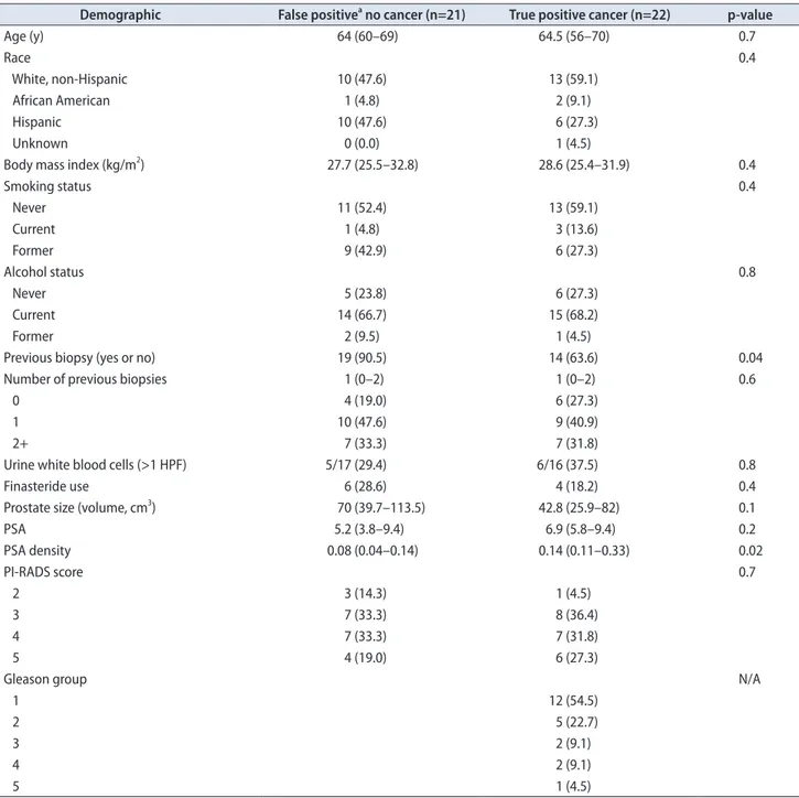
This group of men has entirely negative prostate biopsies and could have potentially avoided biopsy. We display the locations of these false-positive lesions in Fig. 1. In Table 1, we labeled these men as the 21 subjects [48.8% (21/43)] with an actual false-positive targeted biopsy and showed no demographic differences. Inflammation was found in the target lesion in 47.6% (10/21) of lesions identified on MRI in this group.
4. MRI-detection lesions and PI-RADS scores We display the PI-RADS score for each lesion in Table 2.
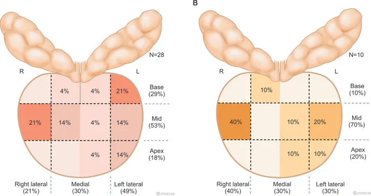
Seven lesions did not have PI-RADS scores and needed re- evaluation, as scans had come from outside facilities in South Texas. In the true-positive lesions, 9/18 (50.0%) were clinically significant cancers (Gleason >3+4, International Society of Urological Pathology [ISUP] group >2). The proportion of clinically significant cancers is higher in those PI-RADS scores >3 (77.8% [7/9] vs. 22.2% [2/9], p=0.02). Taking all 61 lesions into account, 70.5% (43/61) had a false-positive targeted lesion (FP+). More than 50% of the FP+ lesions were PI-RADS >3 (23/43, 53.5%), of which inflammation caused nearly half (16/43 [37.2%] in FP+). We labeled the locations of these lesions in Fig. 2. Only three (7.0%, 3/43) patients had the target lesion alone be the only site of cancer detection.
Moreover, the targeted lesion did not provide a higher Gleason grade than the systematic cores in any patient.
R
N=28
L
Base (29%)
4% 21%
14%
A B
4%
14%
14%
4%
4%
21% Mid
(53%)
Apex (18%)
Right lateral (21%)
Medial (30%)
Left lateral
(49%) CUTHSCSA
R
N=10
L
Base (10%) 10%
20%
40% Mid
(70%)
Apex (20%)
Right lateral (40%)
Medial (30%)
Left lateral
(30%) CUTHSCSA
10%
10% 10%
Fig. 1. (A) Proportion of false positives (no cancer identified) based on location. (B) Proportion of inflammation of false positives (FP-, no cancer identified) based on prostate biopsy location. R, right; L, left.
5. Pathology and inflammation
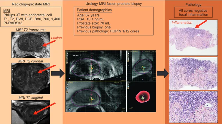
Inflammation was not mentioned in the pathology report (0/33) if any cancer was present on the biopsy. Therefore, inflammation was not mentioned in the presence of cancer and could be underreported in the cancerous prostate, even if the target lesions may have had inflammation. As an example of one of the false-positive lesions caused by inflammation, we provided a summary figure to display the image and pathology (Fig. 3). We show the MRI images, Uronav MRI-US fusion system and pathologic images at 4×, 10×, and 20× magnification. Of the 22 cancerous lesions, 12 (54.5%) were Gleason 3+3 (grade group 1).
DISCUSSION
We identified that our false-positive rate for patients presented with an MRI targetable lesion was 70.5% (43/61).
Despite popular press and improved targeting of lesions, it is evident that improved imaging techniques to reduce false- positive rates are urgently needed. The American Urological Association and the Society of Abdominal Radiology released a joint consensus statement to clarify the utilization of prostate MRI in patients with previous negative biopsies or men on active surveillance (AS). A recent publication from the ASIST trial regarding MRI in patients on AS showed targeted biopsy did not improve upgrading, and systematic biopsies were still necessary [11]. In another context, the use of MRI before the initial biopsy is gaining in popularity [12]. Unfortunately, false-positive lesions continue to pla- gue the accuracy of prostate MRI. Jyoti and colleagues published their experience of in-gantry MRI biopsies from Australia (n=137) noting that PI-RADS 3 and 4 lesions with inflammation accounted for 97% of the false-positive lesions mainly in the transition zone (54%) [9].
Chronic prostatitis shows variable signal intensity and even mimics cancer using MR-spectroscopy with choline peeks and limited citrate values [13]. Nagel et al.
[14] identified significant differences in the value called median apparent diffusion coefficients (ADC) between normal prostate, prostatitis, low- and high-grade cancers (p<0.001). However, the report noted that the ADC values overlapped between the groups and are not likely to be clinically useful. A novel MRI acquisition protocol named restriction spectrum imaging (RSI) may be able to parse the signal of inflammation and cancer [15]. RSI-MRI in the prostate has specifically been shown to be more accurate than ADC values, improve tumor conspicuity, and alter signal between inflammation and cancer [16-19]. Other groups have investigated more efficient use of intravenous Table 2. MRI identified lesions and associated PI-RADS score and pathology
PI-RADS score
False positive True positive (n=61)
Negative for cancer Caused by
inflammation ISUP group 1
(3+3) ISUP group 2
(3+4) ISUP group 3
(4+3) ISUP group 4
(4+4) ISUP group 5 (4+5)
1 1/43 (2.3) 0/1 (0.0) 0 0 0 0 0
2 4/43 (9.3) 0/4 (0.0) 3 0 0 0 0
3 15/43 (34.9) 6/15 (40.0) 4 1 1 0 0
4 18/43 (41.9) 7/18 (38.9) 1 1 0 0 0
5 5/43 (11.6) 3/5 (60.0) 1 4 0 1 1
Total 43/61 (70.5) 16/43 (37.2) 9 (14.8) 6 (9.8) 1 (1.6) 1 (1.6) 1 (1.6)
Values are presented as number (%) or number only.
MRI, magnetic resonance imaging; PI-RADS, Prostate Imaging–Reporting and Data System; ISUP, International Society of Urological Pathology.
R
N=15
L
Base (20%) 7%
20%
13%
27% Mid
(73%)
Apex (7%)
Right lateral (27%)
Medial (60%)
Left lateral
(13%) CUTHSCSA
13% 13%
7%
Fig. 2. Proportion of false positives (FP+, negative target with or with- out cancer on systematic prostate biopsy) based on prostate biopsy location. R, right; L, left.
contrast enhancement techniques to distinguish aggressive tumors [20]. Several other computational measures have been explored to improve tumor conspicuity [21].
As the use of MRI utilization increases for lower risk individuals, such as for prostate cancer screening, we would like to highlight the possibility of inflammation as a potential source for false positive findings. Future studies would ideally focus on improving acquisition protocols to reduce noise from inflammation and potentially combine with prostate inflammation biomarkers. Biomarkers may provide inside into the amount of focal inflammation and allow readers to temper suspicion levels or reduce inflammation and repeat the scan. We did not demonstrate the urinary WBC count on urine analysis to be useful, though other studies have demonstrated this possibility [22].
Despite some interest in the prostate cancer antigen gene 3 (PCA3) urine-based biomarker to determine subsequent MRI and targeted prostate biopsy, studies have shown that PCA3 and other biomarkers may not distinguish between prostatitis and prostate cancer [23]. One study identified SelectMDx biomarker (MDxHealth, Irvine, CA, USA) may be superior to PCA3 in this context [24].
The MRI fusion prostate biopsy is not without its
limitations. There is a significant learning curve for the team over time, which included urologists, pathologists, radiologists and supporting staff [25]. Our data for this study do include our initial biopsy experience and may include missed targeted lesions. Guidelines continue to recommend performing the systematic biopsy along with the targeted approach because an additional 15% of cancers are identified [26]. We retrospectively reviewed our pathology reports, as we did not have adequate funding for the pathology to re- read all previous cores. Our sample size is small and will need larger, prospective targeted studies on this topic to make more definitive statements regarding inflammation and its appearance on multiparametric MRI (mpMRI). We have a low rate of clinically significant prostate cancer detection and is likely due to our population of men with large pro states and negative biopsy and men on AS. When cancer is present, identification of specific inflammation may be minimized, thus reported less. Non-uniformity in the MRI reading of prostate lesions likely leads to more variation and less confidence regarding particular inflammatory lesions. Therefore, artificial intelligence with deep learning of images, among other enhancement processes, may provide more accurate alerts to assist radiologists with false-positive
Radiology-prostate MRI MRI
Phillips 3T with endorectal coil T1, T2, DWI, DCE, B=0, 700, 1,400 PI-RADS=3
MRI T2 transverse
MRI T2 coronal
MRI T2 sagittal
Lesion
All cores negative focal inflammation
Pathology
Inflammation Urology-MRI fusion prostate biopsy
Patient demographics Age: 67 years PSA: 10.1 ng/mL Prostate size: 70 mL Previous biopsy: one
Previous pathology: HGPIN 1/12 cores
Fig. 3. Example of true-positive lesions and false positive caused by inflammation. We present the course of action of a 67-year-old man who presented with elevated prostate specific antigen (PSA) and a previous biopsy with high-grade (HG) PIN in one core of a 12-core biopsy. His magnetic resonance imaging (MRI), which originally was read as Prostate Imaging–Reporting and Data System (PI-RADS) 4, was downgraded to a PI-RADS 3 on re-review. He underwent MRI fusion with UroNav, and the biopsy is displayed. Only inflammation was noted in the pathology specimens displayed on the right side (hematoxylin and eosin stain, top down magnification is 4×, 10×, and 20×). DWI, diffusion-weighted imaging; DCE, dynamic contrast-enhanced.
MRI lesions.
CONCLUSIONS
We could have avoided nearly one-third of our MRI- US fusion biopsies if we could recognize inflammation from cancerous lesions. Concerted effort to minimize the effect of inflammation on MRI should be emphasized to improve cancer detection and improve overall accuracy.
CONFLICTS OF INTEREST
The authors have nothing to disclose.
ACKNOWLEDGMENTS
This work was supported by the NIH (P30CA054174) the Office of the Assistant Secretary of Defense for Health Affairs through the Prostate Cancer Research Program under Award No. W81XWH-15-1-0441. Opinions, interpretations, conclusions, and recommendations are those of the author and are not necessarily endorsed by the Department of Defense.
REFERENCES
1. Siegel RL, Miller KD, Jemal A. Cancer statistics, 2017. CA Can- cer J Clin 2017;67:7-30.
2. Haffty BG, Lawton CA, Sandler H. Watchful waiting-active sur- veillance in low-risk prostate cancer. JAMA Oncol 2015;1:688- 9.
3. Filippou P, Welty CJ, Cowan JE, Perez N, Shinohara K, Carroll PR. Immediate versus delayed radical prostatectomy: updated outcomes following active surveillance of prostate cancer. Eur Urol 2015;68:458-63.
4. Siddiqui MM, Rais-Bahrami S, Turkbey B, George AK, Roth- wax J, Shakir N, et al. Comparison of MR/ultrasound fusion- guided biopsy with ultrasound-guided biopsy for the diagnosis of prostate cancer. JAMA 2015;313:390-7.
5. Kasivisvanathan V, Rannikko AS, Borghi M, Panebianco V, Mynderse LA, Vaarala MH, et al.; PRECISION Study Group Collaborators. MRI-targeted or standard biopsy for prostate- cancer diagnosis. N Engl J Med 2018;378:1767-77.
6. Marconi L, Dabestani S, Lam TB, Hofmann F, Stewart F, Norrie J, et al. Systematic review and meta-analysis of diagnostic accura- cy of percutaneous renal tumour biopsy. Eur Urol 2016;69:660- 73.
7. Sciarra A, Panebianco V, Ciccariello M, Salciccia S, Lisi D, Osi- mani M, et al. Magnetic resonance spectroscopic imaging (1H-
MRSI) and dynamic contrast-enhanced magnetic resonance (DCE-MRI): pattern changes from inflammation to prostate cancer. Cancer Invest 2010;28:424-32.
8. Cheng Y, Zhang X, Ji Q, Shen W. Xanthogranulomatous pro- statitis: multiparametric MRI appearances. Clin Imaging 2014;38:755-7.
9. Jyoti R, Jina NH, Haxhimolla HZ. In-gantry MRI guided pros- tate biopsy diagnosis of prostatitis and its relationship with PIRADS V.2 based score. J Med Imaging Radiat Oncol 2017;
61:212-5.
10. Weinreb JC, Barentsz JO, Choyke PL, Cornud F, Haider MA, Macura KJ, et al. PI-RADS prostate imaging-reporting and data system: 2015, version 2. Eur Urol 2016;69:16-40.
11. Klotz L, Loblaw A, Sugar L, Moussa M, Berman DM, Van der Kwast T, et al. Active Surveillance Magnetic Resonance Imag- ing Study (ASIST): results of a randomized multicenter pro- spective trial. Eur Urol 2019;75:300-9.
12. Rosenkrantz AB, Verma S, Choyke P, Eberhardt SC, Eggener SE, Gaitonde K, et al. Prostate magnetic resonance imaging and magnetic resonance imaging targeted biopsy in patients with a prior negative biopsy: a consensus statement by AUA and SAR.
J Urol 2016;196:1613-8.
13. Shukla-Dave A, Hricak H, Eberhardt SC, Olgac S, Muruga- nandham M, Scardino PT, et al. Chronic prostatitis: MR imag- ing and 1H MR spectroscopic imaging findings--initial obser- vations. Radiology 2004;231:717-24.
14. Nagel KN, Schouten MG, Hambrock T, Litjens GJ, Hoeks CM, ten Haken B, et al. Differentiation of prostatitis and prostate cancer by using diffusion-weighted MR imaging and MR- guided biopsy at 3 T. Radiology 2013;267:164-72.
15. White NS, McDonald C, Farid N, Kuperman J, Karow D, Schenker-Ahmed NM, et al. Diffusion-weighted imaging in cancer: physical foundations and applications of restriction spectrum imaging. Cancer Res 2014;74:4638-52.
16. Liss MA, White NS, Parsons JK, Schenker-Ahmed NM, Rakow- Penner R, Kuperman JM, et al. MRI-derived restriction spec- trum imaging cellularity index is associated with high grade prostate cancer on radical prostatectomy specimens. Front Oncol 2015;5:30.
17. White NS, Leergaard TB, D'Arceuil H, Bjaalie JG, Dale AM.
Probing tissue microstructure with restriction spectrum imag- ing: histological and theoretical validation. Hum Brain Mapp 2013;34:327-46.
18. White NS, McDonald CR, Farid N, Kuperman JM, Kesari S, Dale AM. Improved conspicuity and delineation of high-grade primary and metastatic brain tumors using “restriction spec- trum imaging”: quantitative comparison with high B-value DWI and ADC. AJNR Am J Neuroradiol 2013;34:958-64, S1.
19. Yamin G, Schenker-Ahmed NM, Shabaik A, Adams D, Bartsch
H, Kuperman J, et al. Voxel level radiologic-pathologic validation of restriction spectrum imaging cellularity index with gleason grade in prostate cancer. Clin Cancer Res 2016;22:2668-74.
20. Othman AE, Falkner F, Martirosian P, Schraml C, Schwentner C, Nickel D, et al. Optimized fast dynamic contrast-enhanced magnetic resonance imaging of the prostate: effect of sam- pling duration on pharmacokinetic parameters. Invest Radiol 2016;51:106-12.
21. Mayer R, Simone CB 2nd, Skinner W, Turkbey B, Choykey P.
Pilot study for supervised target detection applied to spatially registered multiparametric MRI in order to non-invasively score prostate cancer. Comput Biol Med 2018;94:65-73.
22. Tekin A, Yuksel A, Tekin S, Gumrukcu G, Aslan AR, Sengor F.
Post-prostatic massage examination for prediction of asymp- tomatic prostatitis in needle biopsies: a prospective study. J Urol 2009;182:564-8; discussion 568-9.
23. De Luca S, Passera R, Fiori C, Bollito E, Cappia S, Mario Scar- pa R, et al. Prostate health index and prostate cancer gene 3
score but not percent-free prostate specific antigen have a pre- dictive role in differentiating histological prostatitis from PCa and other nonneoplastic lesions (BPH and HG-PIN) at repeat biopsy. Urol Oncol 2015;33:424.e17-23.
24. Hendriks RJ, van der Leest MMG, Dijkstra S, Barentsz JO, Van Criekinge W, Hulsbergen-van de Kaa CA, et al. A urinary bio- marker-based risk score correlates with multiparametric MRI for prostate cancer detection. Prostate 2017;77:1401-7.
25. Meng X, Rosenkrantz AB, Huang R, Deng FM, Wysock JS, Bjurlin MA, et al. The institutional learning curve of magnetic resonance imaging-ultrasound fusion targeted prostate biopsy:
temporal improvements in cancer detection in 4 years. J Urol 2018;200:1022-9.
26. Ploussard G, Borgmann H, Briganti A, de Visschere P, Füt- terer JJ, Gandaglia G, et al.; EAU-YAU Prostate Cancer Work- ing Group. Positive pre-biopsy MRI: are systematic biopsies still useful in addition to targeted biopsies? World J Urol 2019;37:243-51.