Received on August 12, 2014. Revised on September 5, 2014. Accepted on September 12, 2014.
CC This is an open access article distributed under the terms of the Creative Commons Attribution Non-Commercial License (http://creativecommons.org/licenses/by-nc/3.0) which permits unrestricted non-commercial use, distribu- tion, and reproduction in any medium, provided the original work is properly cited.
*Corresponding Author. Yeonseok Chung, College of Pharmacy, Seoul National University, 1 Gwanak-ro, Gwanak-gu, Seoul, Korea. Tel: 82-2-880-7874; Fax: 82-2-872-1795; E-mail: Yeonseok@snu.ac.kr
Abbreviations: Th, T helper; Tfh, follicular T helper; CXCR, C-X-C chemokine receptor; Ig, Immunoglobulin; ICOS, in- ducible T cell costimulator; Tfr, follicular regulator T; CTLA-4, cytotoxic T-lymphocyte-associated protein 4; MHC, major histocompatibility complex; HLA, human leukocyte antigen
Regulatory T Cells in B Cell Follicles
Jae-Hoon Chang1 and Yeonseok Chung2*
1College of Pharmacy, Yeungnam University, Gyeongsan 712-749, 2College of Pharmacy, Seoul National University, Seoul 151-742, Korea
Understanding germinal center reactions is crucial not only for the design of effective vaccines against infectious agents and malignant cells but also for the development of ther- apeutic intervention for the treatment of antibody-mediated immune disorders. Recent advances in this field have re- vealed specialized subsets of T cells necessary for the con- trol of B cell responses in the follicle. These cells include fol- licular regulatory T cells and Qa-1-restricted cluster of differ- entiation (CD)8+ regulatory T cells. In this review, we dis- cuss the current knowledge related to the role of regulatory T cells in the B cell follicle.
[Immune Network 2014;14(5):227-236]
Keywords: Germinal center, B cell, Follicular helper T cell, Foxp3+ regulatory T cell, Qa-1 restricted CD8+ regulatory T cell, Autoantibody
INTRODUCTION
Antibodies play an essential role in defending a host organism against infectious agents and mediate this host immunity through multiple mechanisms. Antibodies can neutralize vi- ruses and prevent them from infecting host cells. In addition, they can inactivate toxins derived from infectious microor- ganisms. They can opsonize microorganisms and other for- eign particles and thereby facilitate the recognition of these non-self-antigens by phagocytic cells through Fc receptors.
Furthermore, antibodies can mediate the classical pathway of complement activation that can lead to formation of mem-
brane attack complex which causes target cell lysis. In con- trast to these beneficial roles, antibodies can also mediate det- rimental effects to the host. For instance, binding of IgE ex- pressed on the surface of mast cells to allergens can trigger local or systemic anaphylaxis. Moreover, formation of im- mune complexes as well as binding to self-antigens can in- duce the local or systemic inflammation that often occurs dur- ing autoimmune diseases (1). Therefore, humoral immunity is a double-edged sword that can either protect or damage host tissues, depending on the circumstances. Hence, the production of antibodies by B cells is subject to control by multiple positive and negative regulators.
Th2 cells have been recognized for decades as helper T cells that mediate B cell responses due to the essential role of IL-4 in promoting B cell proliferation and maturation (2-4).
In addition to Th2 cells, a follicular helper T (Tfh) cell pop- ulation has recently been discovered as a distinct lineage of helper T cells that is specialized for facilitating germinal cen- ter reactions (5-7). Tfh cells express CXCR5, which allows their migration into the B cell zone where they facilitate iso- type switching and affinity maturation of Ig as well as the differentiation of B cells to plasma cells or memory B cells.
Multiple types of immune cells have been identified as neg- ative regulators of germinal center reactions. These include IL-10-producing B cells (8-10), Qa-1 restricted cluster of dif- ferentiation (CD)8+ regulatory T cells, and CXCR5+ forkhead box P3 (Foxp3)+ follicular regulatory T (Tfr) cells. The bal- ance between positive and negative regulators of the germinal center reaction therefore determines the magnitude and ki-
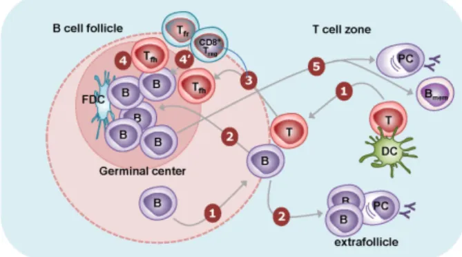
Figure 1. B cell responses to T-dependent antigens. (1) Naïve CD4+ T cells are activated by dendritic cells presenting cognate antigens in the T cell zones. They transiently express CXCR5 and migrate to the T-B border. B cells recognize their cognate antigens through surface Ig and migrate to the T-B border. In the T-B border, activated CD4+ T cells interact with B cells in an antigen-specific manner, termed “linked recognition.” (2) Activated B cells then migrate either to B cell follicles or to the T cell zone. The latter B cells form extrafollicularly and differentiate into short-lived plasma cells with few somatic mutations. (3) Some of the activated CD4+ T cells can acquire characteristics of the Tfh cell lineage and stably express CXCR5. These Tfh cells migrate to the B cell follicle. (4) The activated B cells in B cell follicles are further stimulated by follicular dendritic cells and Tfh cells for multiple rounds. Follicular dendritic cells provide antigens to B cells, leading to the clonal selection of B cells that express high-affinity Ig on their surface. Tfh cells provide IL-21 and costimulation in order to induce the proliferation of B cells, isotype switching, and somatic mutation. The massive B cell expansion and differentiation lead to the formation of germinal centers in the follicle. (4’) Tfr cells and CD8+ Treg cells are thought to suppress this germinal center reaction. (5) The germinal center reaction induces the differentiation of isotype-switched affinity- matured B cells into memory B cells or into long-lived plasma cells.
netics of humoral immunity.
OVERVIEW OF B CELL RESPONSES
Most B cells reside in the B cell follicles of the secondary lymphoid organs, while some B cells travel throughout the body via the circulatory system. B cells in circulation can re- turn to the B cell follicles by following a chemokine gradient of CXCL13 through the CXCR5 expressed on their surface, as dendritic cells in the follicles continuously produce CXCL13.
The first step of antigen-specific humoral immunity is the binding of a cognate antigen to B cells through their surface immunogulobulin (B cell receptor). The binding of antigen to B cell receptors induces the migration of B cells toward the boundary between the B cell and T cell areas (11).
Similarly, antigen-specific CD4+ T cells activated by dendritic cells transiently express CXCR5 on their surface, leading also to their migration toward the boundary between the B cell
and T cell areas (Fig. 1). The migration of antigen-stimulated B and T cells to the same anatomic location increases the chances of antigen-specific interactions between B and T cells. This B:T cell interaction induces the initial proliferation of B cells. Some of these initially activated B cells further mi- grate toward the border of the T cell area (red pulp in the spleen) and form extrafollicular foci, where they can further differentiate into plasmablasts and short-lived plasma cells (Fig. 1). This type of extrafollicular B cell response is critical for the fast production of antibodies during infection (12).
However, due to insufficient help from T cells, the affinity of such antibodies from extrafollicular B cell responses is rela- tively low.
Some of the B cells initially activated after the B:T inter- action in the boundary between the B cell and T cell areas migrate back to the B follicles to form germinal centers.
Germinal centers contain not only activated B cells but also follicular dendritic cells and Tfh cells. Follicular dendritic cells are stromal cells of non-hematopoietic origin. In addition to CXCL13, they express various types of Fc receptors as well as the complement receptors CR1 and CR2, allowing them to trap antigen-antibody-complement complexes for a long time without internalization. Hence, follicular dendritic cells are the main source of antigens that enable B cell proliferation and clonal selection in the germinal center (13). While ini- tially activated CD4+ T cells transiently express CXCR5, some of them can be further differentiated into Tfh cells that stably express CXCR5 (14,15). Tfh cells can therefore migrate into B cell follicles following the CXCL13 gradient, just like B cells, where they further stimulate activated B cells, triggering iso- type-switching, affinity maturation, and differentiation into long-lived plasma cells and memory B cells by providing cyto- kines such as IL-21 as well as costimulatory signals (Fig. 1).
The follicular dendritic cells and Tfh cells are therefore indis- pensable for the generation of high-affinity antibodies and long-lived memory B cells.
TFH CELLS
Tfh cells localized to the germinal center of B cell follicles were first detected in the human tonsil. Further studies re- vealed that CXCR5 expression on these T cells is crucial for their migration into B cell follicles (16). However, these CXCR5+ T cells had been regarded as a subpopulation of Th2 cells for decades until recent studies clearly showed that CXCR5+ CD4+ T cells can be generated in mice lacking Th2
Figure 2. Comparison among Tfh, Tfr, and CD8+ Treg cells. Tfh cells express ICOS, PD-1, CD40L, and CXCR5 on the surface, as well as Bcl-6 and Ascl-2 in the nucleus. Similarly, Tfr cells express PD-1, ICOS, and CXCR5, but not CD40L. Unlike Tfh cells, Tfr cells express glucocorticoid-induced TNF receptor-related protein and CTLA4. They also express Foxp3, Bcl-6, and Blimp-1 in the nucleus. Qa-1-reactive CD8+ Treg cells express ICOSL, CD122, and CXCR5. The transcription factor required for the differentiation of this T cell subset remains to be determined.
cells, such as STAT6−/− and IL-4−/− mice (17,18), indicating that CXCR5+ T cells are distinct from Th2 cells. Indeed, multi- ple groups have shown that the CXCR5+ CD4+ T cell pop- ulation does not belong to Th1, Th2, or Th17 lineages but is instead a distinct subset of CD4+ T cells, termed follicular helper T cells (18). In addition to CXCR5, Tfh cells express ICOS, programmed death (PD)-1, B and T lymphocyte attenu- ator (BTLA) (in mice), CD40L, and CD84 (Fig. 2). Among them, detection of the CXCR5 marker in combination with ei- ther ICOS or PD-1 is the most widely used experimental method for identifying the Tfh cell population in mice as well as in humans.
While the surface phenotype of Tfh cells is well charac- terized, the development of the Tfh lineage from naïve CD4+ T cells is not completely understood. The transcriptional re- pressor Bcl-6 is necessary for the generation of Tfh cells, act- ing in part by repressing the transcription of Tbx21 (encoding T-box expressed in T cells [T-bet]) and Rorc (encoding reti- noic acid-related orphan receptor γt [RORγt]) or direct bind- ing to GATA-bind protein 3 (GATA3) (5-7). Bcl-6 was origi- nally identified as a repressor of B lymphocyte-induced matu- ration protein-1 (Blimp-1), and expression of Blimp-1 is known to suppress the differentiation of Tfh cells. However, enforced expression of Bcl-6 alone in CD4+ T cells is not sufficient to drive Tfh cell differentiation since it cannot in- duce the expression of IL-21 and CXCR5 (16). Of note is a recent study by Liu et al where it was shown that the tran- scription factor achaete scutelike 2 (Ascl2) directly induces the transcription of CXCR5 in Tfh cells (19). In addition to Bcl-6 and Ascl-2, STAT3 (20-22), basic leucine zipper tran- scription factor (BATF) (23,24), and IFN regulatory factor 4
(IRF4) (25,26) are also known to be crucial for Tfh cell development. It is interesting to note that STAT3, BATF, and IRF4 are also needed for differentiation of the Th17 cell lineage.
Interestingly, a cluster of microRNAs known as miR17-92 has been reported to play a pivotal role during Tfh cell differ- entiation, although this role is still controversial. Initially the miR17-92 cluster was proposed to inhibit Tfh cell develop- ment (7); however, more recent studies have demonstrated that these microRNAs promote Th17 cells by facilitating the migration of Tfh cells into the B cell follicles through the sup- pression of the phosphatase pleckstrin homology domain leu- cine-rich repeat protein phosphatase 2, by suppressing the expression of Rora, or through both (27,28).
While the types of signal 3 for the differentiation of Th1, Th2, Th17, and induced regulatory T cells (Treg cells) are well demonstrated, it is still unclear which cytokine signals initiate the differentiation of Tfh cells. Due to its capacity to induce IL-21 expression in T cells, IL-6 has been proposed as a Tfh cell-inducing cytokine (20,29). By contrast, other studies have shown that Tfh cells are unaffected in mice with defective IL-6 signaling (21,30). Moreover, recent studies from multiple researchers convincingly showed that IL-12 can also drive Tfh cell differentiation by inducing IL-21 in a STAT3-dependent manner in mice as well as in humans (22,30-32). In this case, the balance between T-bet and Bcl-6 likely determines whether IL-12 stimulated CD4+ T cells dif- ferentiate into Th1 or Tfh cells. While the identity of the sig- nal 3 cytokine for Tfh cells is unclear, negative regulation of Tfh cell development by IL-2 has been well established.
Mechanistically, IL-2 induces the expression of Blimp-1 in a
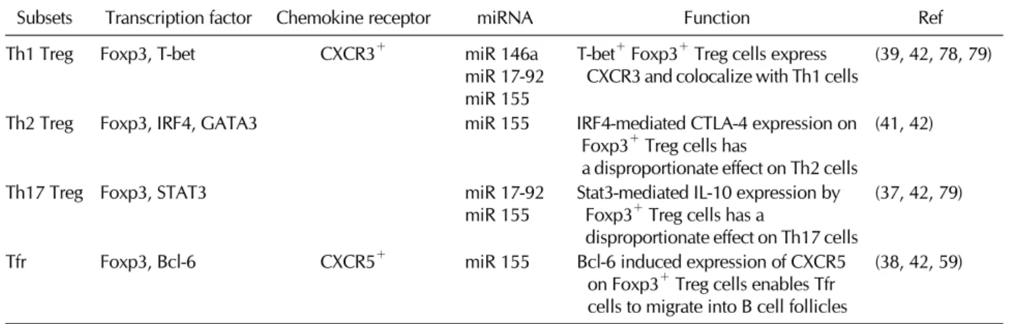
Table I. Diverse subsets of Foxp3+ regulatory T cells
Subsets Transcription factor Chemokine receptor miRNA Function Ref
Th1 Treg Foxp3, T-bet CXCR3+ miR 146a
miR 17-92 miR 155
T-bet+ Foxp3+ Treg cells express CXCR3 and colocalize with Th1 cells
(39, 42, 78, 79)
Th2 Treg Foxp3, IRF4, GATA3 miR 155 IRF4-mediated CTLA-4 expression on
Foxp3+ Treg cells has
a disproportionate effect on Th2 cells
(41, 42)
Th17 Treg Foxp3, STAT3 miR 17-92
miR 155 Stat3-mediated IL-10 expression by Foxp3+ Treg cells has a
disproportionate effect on Th17 cells
(37, 42, 79)
Tfr Foxp3, Bcl-6 CXCR5+ miR 155 Bcl-6 induced expression of CXCR5
on Foxp3+ Treg cells enables Tfr cells to migrate into B cell follicles
(38, 42, 59) STAT5-dependent manner and therefore represses the ex-
pression of Bcl-6 (33,34). Therefore, the key transcription fac- tors and cytokines driving the differentiation of Tfh cells are still obscure. Further studies will be needed to fully define the developmental regulation of the Tfh cell lineage.
TFR CELLS
Foxp3+ Treg cells are a subset of CD4+ T cells with im- munosuppressive properties, including an ability to inhibit the proliferation and cytokine production of effector T cells (35,36).
Notably, a series of recent studies have demonstrated that the suppression of each subset of helper T cell responses by Treg cells requires expression of a certain transcription factor to- gether with Foxp3 in the Treg cells. For instance, Treg cells deficient in Bcl-6, T-bet, IRF4, or Stat3 failed to suppress Tfh, Th1, Th2, or Th17 cells, respectively (37-42) (Table I).
Interestingly, these transcription factors are also indispensable for the differentiation of respective subsets of helper T cells.
Thus, it seems likely that the differentiation of a particular Th subset and a corresponding Treg subset have similar de- velopmental requirements. For instance, the expression of T-bet in conventional CD4+ T cells induces Th1 cells while its expression in Foxp3+ T cells induces Th1-specific Treg cells.
In humoral immune responses, Tfh cells are a unique sub- set of effector T cells capable of migration to the germinal center of B cell follicles. Uncontrolled Tfh responses trigger autoimmunity by inducing excessive autoantibodies (43).
Similar to Tfh cells, Foxp3+ regulatory T cells coexpressing Bcl-6 gain the ability to migrate into B cell follicles through their surface-expressed CXCR5 (Fig. 1). These follicular regu-
latory T cells (Tfr) express CXCR5, ICOS, and PD-1, as well as Bcl-6, but lack CD40L, IL-4, and IL-21 (Fig. 2). They sup- press germinal center reactions, including antibody affinity maturation and differentiation of plasma cells (38,40). These Tfr cells derive from CXCR5− natural Treg cells rather than naïve CD4+ T cells. Like Tfh cells, the differentiation of Tfr cells from CXCR5− Treg cells requires CD28 and ICOS costimulation as well as SAP-dependent stimulation by B cells (38,40).
Tfr cells in humans have also been described in studies of Treg cell migration in human lymphoid tissues. Most of the CD69− Treg cells in humans express CCR7 while only a small population of them express CXCR5. These CXCR5+ Treg cells in humans respond to a CXCL13 gradient in a chemotaxis as- say and suppress antibody production by germinal center B cells and the function of Tfh cells in a coculture experiment in vitro (44,45). Thus, Tfr cells that are present in humans have an immunosuppressive capacity similar to that observed in murine Tfr cells.
Bcl-6 in Tfr cells
Bcl-6+ Treg cells arise from natural Treg cells during active germinal center reactions (40). Since Bcl-6 is required for the expression of CXCR5 on Treg cells and CXCR5-deficient Treg cells are not able to suppress germinal center reactions, the capacity of Tfr to inhibit germinal center B and T cell responses depends on the expression of Bcl-6 in Treg cells (38,40). In addition, isolated Tfr cells have immunosuppressive proper- ties that do not differ in their capacity to inhibit Tfh cells or other effector T cells in vitro. Indeed, the transcriptional pro- file of Tfr cells is similar to that of natural Treg cells with no distinct changes in suppressive function apart from that
of chemotactic localization (38). Thus, the role of Bcl-6 in Treg cells might be the induction of CXCR5 expression on Tfr cells, thus promoting the capacity of Tfr cells to colocalize with activated B cells and Tfh cells in the germinal centers.
Although Bcl-6 is known to repress the expression of Blimp-1, Tfr cell are shown to co-express Bcl-6 and Blimp-1 (40). Blimp-1 is known to mediate the expression of IL-10 in T cells (46,47). It will be interesting if Blimp-1-mediated production of IL-10 plays an important role in suppressing germinal center reactions by Tfr cells in vivo.
TCR and costimulatory signals for Tfr development In addition to Bcl-6, signals from costimulatory molecules ICOS and CD28 are necessary for the development of Tfr cells (48). ICOS is shown to deliver early signals that induce the expression of Bcl-6 and CXCR5 in T cells (49). The phospho- tidylinositol-3-kinase (PI3K) pathway has been reported to conduct the Bcl-6 induction signals further downstream of ICOS during Tfh cell differentiation (50).
Tissue necrosis factor receptor (TNFR)-associated factors (TRAFs) are cytoplasmic adaptor proteins and play a crucial role in the activation of both innate and adaptive immune cells. Molecules in the TRAF family are known to induce the activation of the NF-κB and MAPK pathways in response to various inflammatory stimuli, including TLR and IL-1R signal- ing (51). TRAF3 was the first identified TRAF with the ability to bind the cytoplasmic domain of the TNFR superfamily member CD40. TRAF3 exerts diverse functions depending on the type of receptor (51). In T cells, TRAF3 is crucial for TCR signaling (52). TCR/CD28 mediated early signaling includes phosphorylation of the ZAP70, linker for activation of T cells (LAT), phospholipase Cγ (PLC-γ), and ERK. T cells lacking TRAF3 showed impaired phosphorylation of these early sig- naling molecules, but exhibited normal TCR induced canon- ical NF-κB1 activation (52). Interestingly, we observed that mice with Treg-specific deletion of TRAF3 exhibited sig- nificantly increased IgG production upon immunization with sheep RBCs (53). These mice also showed increased numbers of Tfh cells and germinal center B cells, suggesting ex- aggerated germinal center reactions in the absence of TRAF3 in Treg cells. Mechanistically, the Tfr cell population sig- nificantly decreased in the Treg-specific TRAF3-deficient mice, indicating the crucial role of TRAF3 in the induction of Tfr cells. TRAF3 has little involvement in the induction of Bcl-6.
Instead, TRAF3 seems to be required for the persistent ex- pression of ICOS on Treg cells. Loss of TRAF3 in Treg cells
resulted in decreased activation of the ERK-AP-1 signaling pathway upon the induction of TCR and CD28 signals. Thus, TRAF3 likely regulates ICOS gene induction on Tfr cells via the ERK-AP-1 signaling pathway (53), which is further sup- ported by the fact that the ICOS promoter contains an AP-1 binding site (54). In addition, it has been shown that ERK signaling is essential for the induction of ICOS expression in T cells (55).
Mechanisms underlying Tfr cell-mediated suppression of germinal center reactions
Treg cells regulate all aspects of the immune response. The mechanisms underlying Treg-mediated suppression include the production of suppressor cytokines (e.g., IL-10, TGF-β or IL-35), IL-2 consumption, granzyme-mediated cytolysis, and galectin-1-mediated suppression. In addition, Treg cells are also known to suppress the function of APC and indirectly inhibit the activation of effector T cells (56).
Surface expression of CTLA-4 and PD-1 on Treg cells is crit- ical for controlling humoral immune responses, while the role of cytokines such as IL-10 or TGF-β is incompletely under- stood. CTLA-4 is an inhibitory molecule and exerts its in- hibitory function by limiting the availability of CD80 and CD86 (57). Treg cell-specific deletion of CTLA-4 leads to the induction of fatal autoimmune and lymphoproliferative dis- orders with elevated levels of IgE and IgG in the serum and is also associated with the spontaneous development of Tfh cells and germinal center reactions (57).
PD-1 is another inhibitory receptor belonging to the same superfamily as CD28 and CTLA-4 (58). PD-1 is highly ex- pressed on Tfh and Tfr cells in mice as well as in humans (38,59). PD-1-deficient mice showed dysregulated humoral immunity with increased produciton of autoantibodies (60,61), which is associated with increased expansion of Tfh cells (62). A recent study clearly showed the role of PD-1 in Tfr cells. In a co-transfer study, it was shown that Tfr cells lacking PD-1 exhibited increased expansion compared with wild-type Tfr cells. Moreover, PD-1-deficient Treg cells were more potent in suppressing antibody production from B cells (48), indicating that PD-1 suppresses the generation and func- tion of Tfr cells in vivo. Thus it seems likely that PD-1 ex- pression on Tfr cells places a regulatory check on Tfr cell activity, preventing the excessive suppression of germinal center reactions.
Although Foxp3+ Treg cells are known to produce IL-10 and TGF-β their role in the Tfr cell-mediated regulation of
humoral immunity is unclear. The membrane-bound form of TGF-β was shown to mediate contact-dependent sup- pression of B-cell antibody production (63). IL-10 and TGF-β can induce isotype switching to IgG4 and IgA, respectively (64,65). Further studies will be needed to clarify the cellular and molecular mechanisms underlying Tfr cell-mediated sup- pression of germinal center reactions.
CD8+ TREG CELLS
Although a subpopulation of CD8+ T cells had been long re- garded as suppressor T cells, the CD8+ Treg cells have re- ceived far less attention since the discovery of Foxp3+ CD4+ regulatory T cells. This is at least in part due to the fact that both the identity and the mode of action of such CD8+ Treg cells have been obscure. However, recent advances in this field clearly showed that CD8+ Treg cells are a specialized subset of CD8+ T cells that can suppress activated CD4+ T cells in a Qa-1-dependent manner (66).
Qa-1
In addition to Foxp3+ T cells, Qa-1-reactive CD8+ T (CD8+ Treg) cells are known to suppress autoimmunity. Qa-1 is the murine homolog of a human class Ib MHC molecule, HLA-E.
It can interact with the T cell receptors of CD8+ T cells, and thus mediates positive selection of Qa-1-reactive CD8+ T cells in the thymus (66). In addition, Qa-1 is also a ligand for natu- ral killer group 2 (NKG2) molecules, including inhibitory CD94/NKG2A and CD94/NKG2C receptors on NK cells. Initial studies showed that Qa-1-deficient mice are more susceptible to experimental autoimmune encephalomyelitis, indicating the involvement of this molecule in peripheral tolerance (67-69). The Qa-1-dependent immunosuppression was found to be mediated by CD8+ T cells rather than in an NKG2- mediated manner. Similarly, some CD8+ T cells were also shown to suppress T cell-dependent B cell responses in a Qa-1-dependent manner in multiple animal models (70,71).
Interestingly, the Qa-1 molecule is known to present auto- antigens, including insulin and myelin basic protein. There- fore, it is likely that Qa-1-reactive CD8+ Treg cells are acti- vated by recognizing self-antigens presented by Qa-1 and ac- quire suppressive activity against autoreactive T and B cells.
CD8+ Treg cells suppress Tfh cell responses in vivo More recently, Cantor and colleagues elegantly showed the molecular and cellular mechanisms by which CD8+ Treg cells
suppress adaptive immunity. They generated a Qa-1 knock-in mouse with a single amino acid exchange mutation. The mu- tated Qa-1 was found to be deficient in its ability to bind to T cell receptors and CD8, but maintained its binding to NKG2. Interestingly, these Qa-1 mutant mice exhibited sig- nificantly increased autoantibody levels against nuclear anti- gens as well as Ig deposition in renal glomeruli with in- creased frequency of Tfh cells in the secondary lymphoid or- gans (72). Of note, CXCR5+ CD4+ T cells were found to ex- press Qa-1 on their surface while CXCR5− CD4+ T cells ex- press very low levels of this non-classical MHC molecule, and therefore CD8+ Treg cells are able to specifically suppress Tfh cells by recognizing Qa-1 (72). Qa-1-reactive CD8+ Treg cells express ICOS ligand (ICOSL) and CD122, but unlike Tfr cells, they do not express ICOS and PD-1 (Fig. 2). In addi- tion, the transcription factors driving the CD8+ Treg cell line- age have not yet been identified. Mechanistically, this Qa-1 mediated suppression of Tfh cells by CD8+ Treg cells is de- pendent on perforin. In summary, CD8+ Treg cells suppress Tfh cell-mediated production of autoantibodies by killing Tfh cells in a perforin-dependent manner upon recognizing Qa-1+ Tfh cells through their T cell receptors (72-74).
The immunoregulatory role of CD8+ Treg cells were also shown in murine models of autoimmune diseases, including the B6-Yaa mouse model of lupus and collagen induced ar- thritis (72,75). The IL-15/IL-15 receptor complex induces the expansion of CD8+ Treg cells, and transfer of the expanded CD8+ Treg cells was found to ameliorate the severity of auto- immune arthritis in an animal model by inhibiting autoanti- body production (75).
CD8+ Treg cells in humans
It remains unclear whether Qa-1-reactive CD8+ Treg cells ex- ist in humans. However, a few studies have suggested the existence of HLA-E-mediated immune suppression. For in- stance, the stimulation of CD8+ T cells with dendritic cells that were previously cultured with an HLA-E binding peptide can suppress self-reactive CD4+ T cells in patients with type 1 diabetes (76). Moreover, patients with multiple sclerosis ex- hibit reduced frequency of HLA-E-reactive CD8+ T cells in the peripheral blood (77). Nevertheless, whether the CD8+ Treg cells in humans play any role in Tfh responses remains unexplored. Further studies will be needed to demonstrate the role of these HLA-E-reactive CD8+ Treg cells in the regu- lation of autoimmune diseases in humans.
CONCLUDING REMARKS
Production of high-affinity antibodies is a hallmark of a well-functioning host immune system. However, antibodies produced against self-antigens can destroy host tissues in a number of autoimmune diseases. Therefore, improved knowledge regarding the mechanisms responsible for the suppression of inappropriate antibody production has im- portant implications for our understanding of the immunor- egulatory control of autoimmunity as well as for the develop- ment of effective vaccines against infectious agents and malignancies. With respect to this aspect, it will be important to (i) delineate the underlying cellular and molecular mecha- nisms by which Tfr cells suppress germinal center reactions since it is not yet clear if they directly suppress B cells, Tfh cells, or both; (ii) determine whether adoptive transfer of Tfr cells can ameliorate ongoing autoimmune germinal center re- actions in animal models of diseases; and (iii) determine if Tfr cells and CD8+ Treg cells can suppress autoimmunity in humans.
The use of regulatory T cells in the clinical setting has been largely unsuccessful. This might be due to the low frequen- cies of the specific subsets of Treg cells that are specialized for suppressing particular types of autoimmunity. For in- stance, the use of Tfr cells rather than a broader population of Treg cells could be more efficacious for the suppression of antibody-mediated autoimmunity, while the use of CXCR3+ Treg cells may better suppress unwanted Th1-mediated dis- eases (Table I). Thus, the identification of such a Treg cell subset could open new translational opportunities for the de- velopment of Treg cell-mediated therapies in the era of per- sonalized medicine. In this case, the lower frequency of each Treg subset and the unknown stability of the Treg subset in vivo, as well as the poorly described antigen-specificity of the Treg cells are obvious obstacles. Further studies will be need- ed to further outline the basic biology of regulatory T cells as well as to demonstrate the translational potential of Tfr and CD8+ Treg cells before their use as novel therapeutics for antibody-mediated immune disorders in humans.
ACKNOWLEDGEMENTS
This work was supported by Research Resettlement Fund for the new faculty of Seoul National University (YC), SNU in- vitation for distinguished scholar (YC), and the 2013 Yeungnam University Research Grant (JHC).
CONFLICT OF INTEREST
The authors have no financial conflict of interest.
REFERENCES
1. Wahren-Herlenius, M. and T. Dorner. 2013. Immunopatho- genic mechanisms of systemic autoimmune disease. Lancet 382: 819-831.
2. Paul, W. E. and J. Ohara. 1987. B-cell stimulatory factor-1/in- terleukin 4. Annu. Rev. Immunol. 5: 429-459.
3. Banchereau, J., P. P. de, A. Valle, E. Garcia, and F. Rousset.
1991. Long-term human B cell lines dependent on inter- leukin-4 and antibody to CD40. Science 251: 70-72.
4. Arpin, C., J. Dechanet, K. C. Van, P. Merville, G. Grouard, F. Briere, J. Banchereau, and Y. J. Liu. 1995. Generation of memory B cells and plasma cells in vitro. Science 268:
720-722.
5. Johnston, R. J., A. C. Poholek, D. DiToro, I. Yusuf, D. Eto, B. Barnett, A. L. Dent, J. Craft, and S. Crotty. 2009. Bcl6 and Blimp-1 are reciprocal and antagonistic regulators of T follicular helper cell differentiation. Science 325: 1006-1010.
6. Nurieva, R. I., Y. Chung, G. J. Martinez, X. O. Yang, S.
Tanaka, T. D. Matskevitch, Y. H. Wang, and C. Dong. 2009.
Bcl6 mediates the development of T follicular helper cells.
Science 325: 1001-1005.
7. Yu, D., S. Rao, L. M. Tsai, S. K. Lee, Y. He, E. L. Sutcliffe, M. Srivastava, M. Linterman, L. Zheng, N. Simpson, J. I.
Ellyard, I. A. Parish, C. S. Ma, Q. J. Li, C. R. Parish, C. R.
Mackay, and C. G. Vinuesa. 2009. The transcriptional re- pressor Bcl-6 directs T follicular helper cell lineage commitment. Immunity 31: 457-468.
8. Fillatreau, S., C. H. Sweenie, M. J. McGeachy, D. Gray, and S. M. Anderton. 2002. B cells regulate autoimmunity by pro- vision of IL-10. Nat. Immunol. 3: 944-950.
9. Mizoguchi, A., E. Mizoguchi, H. Takedatsu, R. S. Blumberg, and A. K. Bhan. 2002. Chronic intestinal inflammatory con- dition generates IL-10-producing regulatory B cell subset characterized by CD1d upregulation. Immunity 16: 219-230.
10. Mauri, C., D. Gray, N. Mushtaq, and M. Londei. 2003.
Prevention of arthritis by interleukin 10-producing B cells. J.
Exp. Med. 197: 489-501.
11. Victora, G. D. and M. C. Nussenzweig. 2012. Germinal centers. Annu. Rev. Immunol. 30: 429-457.
12. MacLennan, I. C., K. M. Toellner, A. F. Cunningham, K. Serre, D. M. Sze, E. Zuniga, M. C. Cook, and C. G. Vinuesa. 2003.
Extrafollicular antibody responses. Immunol. Rev. 194: 8-18.
13. Vinuesa, C. G., M. A. Linterman, C. C. Goodnow, and K.
L. Randall. 2010. T cells and follicular dendritic cells in germi- nal center B-cell formation and selection. Immunol. Rev. 237:
72-89.
14. Craft, J. E. 2012. Follicular helper T cells in immunity and systemic autoimmunity. Nat. Rev. Rheumatol. 8: 337-347.
15. Fazilleau, N., L. Mark, L. J. Heyzer-Williams, and M. G.
Heyzer-Williams. 2009. Follicular helper T cells: lineage and location. Immunity 30: 324-335.
16. Crotty, S. 2011. Follicular helper CD4 T cells (TFH). Annu.
Rev. Immunol. 29: 621-663.
17. King, I. L. and M. Mohrs. 2009. IL-4-producing CD4+ T cells in reactive lymph nodes during helminth infection are T fol- licular helper cells. J. Exp. Med. 206: 1001-1007.
18. Nurieva, R. I., Y. Chung, D. Hwang, X. O. Yang, H. S. Kang, L. Ma, Y. H. Wang, S. S. Watowich, A. M. Jetten, Q. Tian, and C. Dong. 2008. Generation of T follicular helper cells is mediated by interleukin-21 but independent of T helper 1, 2, or 17 cell lineages. Immunity 29: 138-149.
19. Liu, X., X. Chen, B. Zhong, A. Wang, X. Wang, F. Chu, R.
I. Nurieva, X. Yan, P. Chen, L. G. van der Flier, H.
Nakatsukasa, S. S. Neelapu, W. Chen, H. Clevers, Q. Tian, H. Qi, L. Wei, and C. Dong. 2014. Transcription factor achaete-scute homologue 2 initiates follicular T-helper-cell development. Nature 507: 513-518.
20. Diehl, S. A., H. Schmidlin, M. Nagasawa, B. Blom, and H.
Spits. 2012. IL-6 triggers IL-21 production by human CD4+ T cells to drive STAT3-dependent plasma cell differentiation in B cells. Immunol. Cell Biol. 90: 802-811.
21. Eddahri, F., S. Denanglaire, F. Bureau, R. Spolski, W. J.
Leonard, O. Leo, and F. Andris. 2009. Interleukin-6/STAT3 signaling regulates the ability of naive T cells to acquire B-cell help capacities. Blood 113: 2426-2433.
22. Ma, C. S., D. T. Avery, A. Chan, M. Batten, J. Bustamante, S. Boisson-Dupuis, P. D. Arkwright, A. Y. Kreins, D.
Averbuch, D. Engelhard, K. Magdorf, S. S. Kilic, Y.
Minegishi, S. Nonoyama, M. A. French, S. Choo, J. M. Smart, J. Peake, M. Wong, P. Gray, M. C. Cook, D. A. Fulcher, J.
L. Casanova, E. K. Deenick, and S. G. Tangye. 2012.
Functional STAT3 deficiency compromises the generation of human T follicular helper cells. Blood 119: 3997-4008.
23. Betz, B. C., K. L. Jordan-Williams, C. Wang, S. G. Kang, J.
Liao, M. R. Logan, C. H. Kim, and E. J. Taparowsky. 2010.
Batf coordinates multiple aspects of B and T cell function re- quired for normal antibody responses. J. Exp. Med. 207:
933-942.
24. Ise, W., M. Kohyama, B. U. Schraml, T. Zhang, B. Schwer, U. Basu, F. W. Alt, J. Tang, E. M. Oltz, T. L. Murphy, and K. M. Murphy. 2011. The transcription factor BATF controls the global regulators of class-switch recombination in both B cells and T cells. Nat. Immunol. 12: 536-543.
25. Biswas, P. S., S. Gupta, R. A. Stirzaker, V. Kumar, R. Jess- berger, T. T. Lu, G. Bhagat, and A. B. Pernis. 2012. Dual regulation of IRF4 function in T and B cells is required for the coordination of T-B cell interactions and the prevention of autoimmunity. J. Exp. Med. 209: 581-596.
26. Bollig, N., A. Brustle, K. Kellner, W. Ackermann, E. Abass, H. Raifer, B. Camara, C. Brendel, G. Giel, E. Bothur, M.
Huber, C. Paul, A. Elli, R. A. Kroczek, R. Nurieva, C. Dong, R. Jacob, T. W. Mak, and M. Lohoff. 2012. Transcription fac- tor IRF4 determines germinal center formation through fol- licular T-helper cell differentiation. Proc. Natl. Acad. Sci. U.
S. A. 109: 8664-8669.
27. Baumjohann, D., R. Kageyama, J. M. Clingan, M. M. Morar, S. Patel, K. D. de, O. Bannard, J. A. Bluestone, M. Matlou- bian, K. M. Ansel, and L. T. Jeker. 2013. The microRNA clus- ter miR-17 approximately 92 promotes TFH cell differentiation and represses subset-inappropriate gene expression. Nat.
Immunol. 14: 840-848.
28. Kang, S. G., W. H. Liu, P. Lu, H. Y. Jin, H. W. Lim, J.
Shepherd, D. Fremgen, E. Verdin, M. B. Oldstone, H. Qi, J. R. Teijaro, and C. Xiao. 2013. MicroRNAs of the miR-17 approximately 92 family are critical regulators of T(FH) differentiation. Nat. Immunol. 14: 849-857.
29. Dienz, O., S. M. Eaton, J. P. Bond, W. Neveu, D. Moquin, R. Noubade, E. M. Briso, C. Charland, W. J. Leonard, G.
Ciliberto, C. Teuscher, L. Haynes, and M. Rincon. 2009. The induction of antibody production by IL-6 is indirectly medi- ated by IL-21 produced by CD4+ T cells. J. Exp. Med. 206:
69-78.
30. Eto, D., C. Lao, D. DiToro, B. Barnett, T. C. Escobar, R.
Kageyama, I. Yusuf, and S. Crotty. 2011. IL-21 and IL-6 are critical for different aspects of B cell immunity and re- dundantly induce optimal follicular helper CD4 T cell (Tfh) differentiation. PLoS One 6: e17739.
31. Nakayamada, S., Y. Kanno, H. Takahashi, D. Jankovic, K.
T. Lu, T. A. Johnson, H. W. Sun, G. Vahedi, O. Hakim, R.
Handon, P. L. Schwartzberg, G. L. Hager, and J. J. O'Shea.
2011. Early Th1 cell differentiation is marked by a Tfh cell-like transition. Immunity 35: 919-931.
32. Schmitt, N., R. Morita, L. Bourdery, S. E. Bentebibel, S. M.
Zurawski, J. Banchereau, and H. Ueno. 2009. Human den- dritic cells induce the differentiation of interleukin-21-produc- ing T follicular helper-like cells through interleukin-12.
Immunity 31: 158-169.
33. Ballesteros-Tato, A., B. Leon, B. A. Graf, A. Moquin, P. S.
Adams, F. E. Lund, and T. D. Randall. 2012. Interleukin-2 inhibits germinal center formation by limiting T follicular helper cell differentiation. Immunity 36: 847-856.
34. Johnston, R. J., Y. S. Choi, J. A. Diamond, J. A. Yang, and S. Crotty. 2012. STAT5 is a potent negative regulator of TFH cell differentiation. J. Exp. Med. 209: 243-250.
35. Sakaguchi, S., N. Sakaguchi, M. Asano, M. Itoh, and M.
Toda. 1995. Immunologic self-tolerance maintained by acti- vated T cells expressing IL-2 receptor alpha-chains (CD25).
Breakdown of a single mechanism of self-tolerance causes various autoimmune diseases. J. Immunol. 155: 1151-1164.
36. Sakaguchi, S., T. Yamaguchi, T. Nomura, and M. Ono. 2008.
Regulatory T cells and immune tolerance. Cell 133: 775-787.
37. Chaudhry, A., D. Rudra, P. Treuting, R. M. Samstein, Y.
Liang, A. Kas, and A. Y. Rudensky. 2009. CD4+ regulatory T cells control TH17 responses in a Stat3-dependent manner.
Science 326: 986-991.
38. Chung, Y., S. Tanaka, F. Chu, R. I. Nurieva, G. J. Martinez, S. Rawal, Y. H. Wang, H. Lim, J. M. Reynolds, X. H. Zhou, H. M. Fan, Z. M. Liu, S. S. Neelapu, and C. Dong. 2011.
Follicular regulatory T cells expressing Foxp3 and Bcl-6 sup- press germinal center reactions. Nat. Med. 17: 983-988.
39. Koch, M. A., G. Tucker-Heard, N. R. Perdue, J. R. Killebrew, K. B. Urdahl, and D. J. Campbell. 2009. The transcription factor T-bet controls regulatory T cell homeostasis and func- tion during type 1 inflammation. Nat. Immunol. 10: 595-602.
40. Linterman, M. A., W. Pierson, S. K. Lee, A. Kallies, S.
Kawamoto, T. F. Rayner, M. Srivastava, D. P. Divekar, L.
Beaton, J. J. Hogan, S. Fagarasan, A. Liston, K. G. Smith, and C. G. Vinuesa. 2011. Foxp3+ follicular regulatory T cells
control the germinal center response. Nat. Med. 17: 975-982.
41. Zheng, Y., A. Chaudhry, A. Kas, P. deRoos, J. M. Kim, T.
T. Chu, L. Corcoran, P. Treuting, U. Klein, and A. Y.
Rudensky. 2009. Regulatory T-cell suppressor program co-opts transcription factor IRF4 to control T(H)2 responses.
Nature 458: 351-356.
42. Lu, L. F., T. H. Thai, D. P. Calado, A. Chaudhry, M. Kubo, K. Tanaka, G. B. Loeb, H. Lee, A. Yoshimura, K. Rajewsky, and A. Y. Rudensky. 2009. Foxp3-dependent microRNA155 confers competitive fitness to regulatory T cells by targeting SOCS1 protein. Immunity 30: 80-91.
43. Vinuesa, C. G., I. Sanz, and M. C. Cook. 2009. Dysregulation of germinal centres in autoimmune disease. Nat. Rev.
Immunol. 9: 845-857.
44. Lim, H. W., P. Hillsamer, and C. H. Kim. 2004. Regulatory T cells can migrate to follicles upon T cell activation and sup- press GC-Th cells and GC-Th cell-driven B cell responses. J.
Clin. Invest. 114: 1640-1649.
45. Lim, H. W., P. Hillsamer, A. H. Banham, and C. H. Kim.
2005. Cutting edge: direct suppression of B cells by CD4+ CD25+ regulatory T cells. J. Immunol. 175: 4180-4183.
46. Neumann, C., F. Heinrich, K. Neumann, V. Junghans, M. F.
Mashreghi, J. Ahlers, M. Janke, C. Rudolph, N. Mockel- Tenbrinck, A. A. Kuhl, M. M. Heimesaat, C. Esser, S. H. Im, A. Radbruch, S. Rutz, and A. Scheffold. 2014. Role of Blimp-1 in programing Th effector cells into IL-10 producers. J. Exp.
Med. 211: 1807-1819
47. Sun, J., H. Dodd, E. K. Moser, R. Sharma, and T. J. Braciale.
2011. CD4+ T cell help and innate-derived IL-27 induce Blimp-1-dependent IL-10 production by antiviral CTLs. Nat.
Immunol. 12: 327-334.
48. Sage, P. T., L. M. Francisco, C. V. Carman, and A. H. Sharpe.
2013. The receptor PD-1 controls follicular regulatory T cells in the lymph nodes and blood. Nat. Immunol. 14: 152-161.
49. Choi, Y. S., R. Kageyama, D. Eto, T. C. Escobar, R. J.
Johnston, L. Monticelli, C. Lao, and S. Crotty. 2011. ICOS re- ceptor instructs T follicular helper cell versus effector cell dif- ferentiation via induction of the transcriptional repressor Bcl6.
Immunity 34: 932-946.
50. Choi, Y. S., J. A. Yang, and S. Crotty. 2013. Dynamic regu- lation of Bcl6 in follicular helper CD4 T (Tfh) cells. Curr.
Opin. Immunol. 25: 366-372.
51. Hildebrand, J. M., Z. Yi, C. M. Buchta, J. Poovassery, L. L.
Stunz, and G. A. Bishop. 2011. Roles of tumor necrosis factor receptor associated factor 3 (TRAF3) and TRAF5 in immune cell functions. Immunol. Rev. 244: 55-74.
52. Xie, P., Z. J. Kraus, L. L. Stunz, Y. Liu, and G. A. Bishop.
2011. TNF receptor-associated factor 3 is required for T cell-mediated immunity and TCR/CD28 signaling. J. Immunol.
186: 143-155.
53. Chang, J. H., H. Hu, J. Jin, N. Puebla-Osorio, Y. Xiao, B.
E. Gilbert, R. Brink, S. E. Ullrich, and S. C. Sun. 2014. TRAF3 regulates the effector function of regulatory T cells and hu- moral immune responses. J. Exp. Med. 211: 137-151.
54. Watanabe, M., S. Nakajima, K. Ohnuki, S. Ogawa, M.
Yamashita, T. Nakayama, Y. Murakami, K. Tanabe, and R.
Abe. 2012. AP-1 is involved in ICOS gene expression down- stream of TCR/CD28 and cytokine receptor signaling. Eur. J.
Immunol. 42: 1850-1862.
55. Tan, A. H., S. C. Wong, and K. P. Lam. 2006. Regulation of mouse inducible costimulator (ICOS) expression by Fyn-NFATc2 and ERK signaling in T cells. J. Biol. Chem. 281:
28666-28678.
56. Shevach, E. M. 2009. Mechanisms of foxp3+ T regulatory cell-mediated suppression. Immunity 30: 636-645.
57. Wing, K., Y. Onishi, P. Prieto-Martin, T. Yamaguchi, M.
Miyara, Z. Fehervari, T. Nomura, and S. Sakaguchi. 2008.
CTLA-4 control over Foxp3+ regulatory T cell function.
Science 322: 271-275.
58. Okazaki, T. and T. Honjo. 2006. The PD-1-PD-L pathway in immunological tolerance. Trends Immunol. 27: 195-201.
59. Linterman, M. A., W. Pierson, S. K. Lee, A. Kallies, S.
Kawamoto, T. F. Rayner, M. Srivastava, D. P. Divekar, L.
Beaton, J. J. Hogan, S. Fagarasan, A. Liston, K. G. Smith, and C. G. Vinuesa. 2011. Foxp3+ follicular regulatory T cells control the germinal center response. Nat. Med. 17: 975-982.
60. Okazaki, T., Y. Tanaka, R. Nishio, T. Mitsuiye, A. Mizoguchi, J. Wang, M. Ishida, H. Hiai, A. Matsumori, N. Minato, and T. Honjo. 2003. Autoantibodies against cardiac troponin I are responsible for dilated cardiomyopathy in PD-1-deficient mice. Nat. Med. 9: 1477-1483.
61. Okazaki, T., Y. Otaka, J. Wang, H. Hiai, T. Takai, J. V.
Ravetch, and T. Honjo. 2005. Hydronephrosis associated with antiurothelial and antinuclear autoantibodies in BALB/
c-Fcgr2b−/−Pdcd1−/− mice. J. Exp. Med. 202: 1643-1648.
62. Good-Jacobson, K. L., C. G. Szumilas, L. Chen, A. H. Sharpe, M. M. Tomayko, and M. J. Shlomchik. 2010. PD-1 regulates germinal center B cell survival and the formation and affinity of long-lived plasma cells. Nat. Immunol. 11: 535-542.
63. Nakamura, K., A. Kitani, and W. Strober. 2001. Cell con- tact-dependent immunosuppression by CD4(+)CD25(+) regulatory T cells is mediated by cell surface-bound trans- forming growth factor beta. J. Exp. Med. 194: 629-644.
64. Jeannin, P., S. Lecoanet, Y. Delneste, J. F. Gauchat, and J.
Y. Bonnefoy. 1998. IgE versus IgG4 production can be differ- entially regulated by IL-10. J. Immunol. 160: 3555-3561.
65. Coffman, R. L., D. A. Lebman, and B. Shrader. 1989. Trans- forming growth factor beta specifically enhances IgA pro- duction by lipopolysaccharide-stimulated murine B lym- phocytes. J. Exp. Med. 170: 1039-1044.
66. Sarantopoulos, S., L. Lu, and H. Cantor. 2004. Qa-1 restriction of CD8+ suppressor T cells. J. Clin. Invest. 114: 1218-1221.
67. Hu, D., K. Ikizawa, L. Lu, M. E. Sanchirico, M. L. Shinohara, and H. Cantor. 2004. Analysis of regulatory CD8 T cells in Qa-1-deficient mice. Nat. Immunol. 5: 516-523.
68. Lu, L., K. Ikizawa, D. Hu, M. B. Werneck, K. W. Wucher- pfennig, and H. Cantor. 2007. Regulation of activated CD4+ T cells by NK cells via the Qa-1-NKG2A inhibitory pathway.
Immunity 26: 593-604.
69. Lu, L., H. J. Kim, M. B. Werneck, and H. Cantor. 2008.
Regulation of CD8+ regulatory T cells: Interruption of the NKG2A-Qa-1 interaction allows robust suppressive activity and resolution of autoimmune disease. Proc. Natl. Acad. Sci.
U. S. A. 105: 19420-19425.
70. Cantor, H. and E. A. Boyse. 1975. Functional subclasses of T-lymphocytes bearing different Ly antigens. I. The gen-
eration of functionally distinct T-cell subclasses is a differ- entiative process independent of antigen. J. Exp. Med. 141:
1376-1389.
71. Cantor, H., F. W. Shen, and E. A. Boyse. 1976. Separation of helper T cells from suppressor T cells expressing different Ly components. II. Activation by antigen: after immunization, antigen-specific suppressor and helper activities are mediated by distinct T-cell subclasses. J. Exp. Med. 143: 1391-1340.
72. Kim, H. J., B. Verbinnen, X. Tang, L. Lu, and H. Cantor.
2010. Inhibition of follicular T-helper cells by CD8(+) regu- latory T cells is essential for self tolerance. Nature 467:
328-332.
73. Kim, H. J. and H. Cantor. 2011. Regulation of self-tolerance by Qa-1-restricted CD8(+) regulatory T cells. Semin. Immunol.
23: 446-452.
74. Kim, H. J., X. Wang, S. Radfar, T. J. Sproule, D. C.
Roopenian, and H. Cantor. 2011. CD8+ T regulatory cells ex- press the Ly49 Class I MHC receptor and are defective in au- toimmune prone B6-Yaa mice. Proc. Natl. Acad. Sci. U. S.
A 108: 2010-2015.
75. Leavenworth, J. W., X. Tang, H. J. Kim, X. Wang, and H.
Cantor. 2013. Amelioration of arthritis through mobilization of peptide-specific CD8+ regulatory T cells. J. Clin. Invest.
123: 1382-1389.
76. Jiang, H., S. M. Canfield, M. P. Gallagher, H. H. Jiang, Y.
Jiang, Z. Zheng, and L. Chess. 2010. HLA-E-restricted regu- latory CD8(+) T cells are involved in development and con- trol of human autoimmune type 1 diabetes. J. Clin. Invest.
120: 3641-3650.
77. Correale, J. and A. Villa. 2008. Isolation and characterization of CD8+ regulatory T cells in multiple sclerosis. J. Neuro- immunol. 195: 121-134.
78. Lu, L. F., M. P. Boldin, A. Chaudhry, L. L. Lin, K. D.
Taganov, T. Hanada, A. Yoshimura, D. Baltimore, and A. Y.
Rudensky. 2010. Function of miR-146a in controlling Treg cell-mediated regulation of Th1 responses. Cell 142: 914-929.
79. de Kouchkovsky, D., J. H. Esensten, W. L. Rosenthal, M. M.
Morar, J. A. Bluestone, and L. T. Jeker. 2013. microRNA- 17-92 regulates IL-10 production by regulatory T cells and control of experimental autoimmune encephalomyelitis. J.
Immunol. 191: 1594-1605.