J. Exp. Biomed. Sci. 2012, 18(3): 218~226 pISSN : 1738-3226
Involvement of D2 Receptor on Dopamine-induced Action in Interstitial Cells of Cajal from Mouse Colonic Intestine
Dong Chuan Zuoa
1, Pawan Kumar Shahia
1, Seok Choia
1, Jae Yeoul Jun
1and Jong-Seong Park
2,†1Department of Physiology, College of Medicine, Chosun University, Gwangju 501-717, Korea
2Department of Physiology, Chonnam National University Medical School, Gwangju 501-746, Korea
Dopamine is an enteric neurotransmitter that regulates gastrointestinal motility. This study was done to investigate whether dopamine modulates spontaneous pacemaker activity in cultured interstitial cells of Cajal (ICCs) from mouse using whole cell patch clamp technique, RT-PCR and live Ca
2+imaging analysis. ICCs generate pacemaker inward currents at a holding potential of -70 mV and generate pacemaker potentials in current-clamp mode. Dopamine did not change the frequency and amplitude of pacemaker activity in small intestinal ICCs. On the contrary dopamine reduced the frequency and amplitude of pacemaker activity in large intestinal ICCs. RT-PCR analysis revealed that Dopamine2 and 4-receptors are expressed in c-Kit positive ICCs. Dopamine2 and 4 receptor agonists inhibited pacemaker activity in large intestinal ICCs mimicked those of dopamine. Domperidone, dopamine2 receptor antagonist, increased the frequency of pacemaker activity of large intestinal ICCs. In Ca
2+-imaging, dopamine inhibited spontaneous intracellular Ca
2+oscillations of ICCs. These results suggest that dopamine can regulate gastrointestinal motility through modulating pacemaker activity of large intestinal ICCs and dopamine effects on ICCs are mediated by dopamine2 receptor and intracellular Ca
2+modulation.
Key Words: Dopamine, Domperidone, Interstitial cells of Cajal, Pacemaker activity
INTRODUCTION
The motility of gastrointestinal (GI) tract is controlled by enteric nervous system, which is part of the autonomic nervous system (Abe et al., 1984). It is well known that sympathetic nerves release norepinephrine (NE) which relaxes smooth muscle (Adler-Graschinsky et al., 1984) and parasympathetic nerves release acetylcholine (ACh) which has an excitatory effect on GI motility (Eaker et al., 1988). Furthermore, GI tract contains a high concentration of the specific dopamine (DA) metabolite 3,4-dihydrioxy-
phenylacetic acid (Gershon, 1967). This observation suggested that DA is an enteric neurotransmitter.
Dopamine relaxes the rat jejunum (Grivegnee et al., 1984), relaxes smooth muscle isolated from dog colon (Hartman and Civelli, 1997), and hyperpolarizes guinea pig submucosal neurons (Hirst and Silinsky, 1975). Thus, these indicate that endogenous dopamine may inhibit in- testinal motility. Furthermore, some studies showed indirect function of dopamine antagonists. Itopride, which has been known to be a gastroprokinetic agent stimulates the release of endogenous ACh release by antagonizing dopamine receptor and has a anticholinesterase activity. Also, this enhances gastric emptying in dogs, rats and humans (Holtmann et al., 2006). Thus dopamine-related agents are used for the treatment of motility disorders of GI tracts.
The GI electrical pacemaker activity originates from the interstitial cells of Cajal (ICCs). The GI pacemaker generates rhythmic depolarizations (also known as slow waves)
Original Article
*Received: July 16, 2012 / Revised: August 2, 2012 Accepted: August 7, 2012
†Corresponding author: Jong-Seong Park. Department of Physiology, Chonnam National University Medical School, 5Hak-dong, Don-gu, Gwangju 501-746, Korea.
Tel: +82-62-220-4264, Fax: +82-62-232-1242 e-mail: [email protected]
○CThe Korean Society for Biomedical Laboratory Sciences. All rights reserved.
(Iwanaga et al., 1994; Huisinga, 2001). Also, ICCs serve as a connection between enteric nerves and smooth muscle cells, participating in neurotransmission and regulating GI motility (Iwanaga et al., 1990; Iwanaga et al., 1996). There- fore, enteric neurotransmitters or endogenous substances regulate GI contractions and electrical activities by modulating ICCs.
However, the role of dopamine in the GI motility has not been clearly investigated now. Especially, the effects of dopamine on ICCs are not reported yet. Therefore, the aim of this study was to investigate dopamine effects on pace- maker activity of ICCs and its related transduction system.
MATERIALS AND METHODS
All experiments were performed according to the Guiding Principles for the Care and Use of Animals approved by the Ethics Committee of Chosun University and the National Institutes of Health Guide for the Care and Use of Laboratory Animals. Every effort was made to minimize both the number of animals used and their suffering.
Preparation of cells
Balb/C mice (8~13 day old) of either sex were anesthetized with diethyl ether and killed by cervical dislocation. The small and large intestines from 1 cm below the pyloric ring to the cecum were removed and were opened along the mesenteric border. The luminal contents were washed away with Krebs-Ringer bicarbonate solution.
The tissues were pinned to the base of a Sylgard dish and the mucosa was removed by sharp dissection. Small strips of intestinal muscle were equilibrated in Ca
2+-free Hank's solution containing KCl 5.36 mM, NaCl 125 mM, NaOH 0.336 mM, Na
2HCO
30.44 mM, glucose 10 mM, sucrose 2.9 mM, and HEPES 11 mM, adjusted to pH 7.4 with Tris for 30 minutes. After incubation for 15 minutes at 37℃
with an enzyme solution containing collagenase 1.3 mg/mL (Worthington Biochemical, Lakewood, NJ, USA), bovine serum albumin 2 mg/mL (Sigma, St. Louis, MO, USA), trypsin inhibitor 2 mg/mL (Sigma), and ATP 0.27 mg/mL, the cells were dispersed. Cells were plated onto sterile glass coverslips coated with Poly-L-lysine solution (Sigma) in
35 mm culture dishes (20). The cells were then cultured at 37℃ in a 5% CO
2incubator in smooth muscle growth medium (Lonza, Cambrex Bio SciencesWalkersville Inc., Walkersville, MD, USA) supplemented with 2% antibiotics/
antimycotics (Gibco, Grand Island, NY, USA) and murine stem cell factor (SCF) (5 ng/mL, Sigma).
Patch-clamp experiments
The patch-clamp experiments of ICCs were performed on cells cultured for 2~3 days. The whole-cell configuration of the patch-clamp technique was used to record membrane currents (voltage clamp) and membrane potentials (current clamp) from cultured ICCs. Currents or potentials were amplified using Axopatch 1-D (Axon Instruments, Foster City, CA, USA). Command pulse was applied using an IBM-compatible personal computer and pClamp software (version 9.2, Axon Instruments). The data were filtered at 5 KHz. All experiments were carried out at 30℃. The results were analyzed using pClamp and Graph Pad Prism (version 2.01, GraphPad Software Inc., San Diego, CA, USA) software.
Measurement of intracellular Ca
2+concentration Changes in intracellular Ca
2+concentration ([Ca
2+]
i) were monitored by using fluo-4/AM, which was initially dissolved in dimethyl sulfoxide and stored at -20℃.
Cultured ICCs on coverslips (25 mm) were rinsed twice with a bath solution (5 mM KCl, 135 mM NaCl, 2 mM CaCl
2, 10 mM glucose, 1.2 mM MgCl
2, and 10 mM HEPES, adjusted to pH 7.4 with Tris). The coverslips were then incubated in the bath solution containing 5 μM fluo-4 with 5% CO
2at 37℃ for 5 min, rinsed two more times with the bath solution, mounted on a perfusion chamber, and scanned every 0.4 seconds with a Nikon Eclipse TE200 inverted microscope equipped with a Perkin-Elmer Ultraview confocal scanner and a Hamamatsu Orca ER 12-bit CCD camera (×200). Fluorescence was excited at a wavelength of 488 nm, and emitted light was observed at 515 nm. During scanning of Ca
2+imaging, the temperature of the perfusion chamber containing the cultured ICC was maintained at 30℃. Variations of the intracellular Ca
2+fluorescence emission intensity were expressed as F1/F0,
where F0 is the intensity of the first imaging.
Cell separation and RT-PCR
The field in the culture dish was selected where there are lots of single cell isolated from each other. The patch pipette with relatively little bigger pore size was used to pick the ICCs. The pipette is moved closer to the single ICCs and collected using applied suction to the pipette that resulted in aspiration of the cell into the patch pipette. The pipette contents were subsequently ejected into a sterile 1 ml eppendorf tube containing sterile and chilled PBS. Cells were rapidly placed in ice and the total RNA was isolated using the Trizol reagent according to the manufacturer's instruction before performing PCR. Real-time polymerase chain reaction (RT-PCR) and cDNA amplification of isolated RNA were performed by using SuperScript one-step RT- PCR with Platinum Taq (Invitrogen, Carlsbad, CA). For amplification, sense and antisense primers were selected according to the mouse sequence for indicated genes:
Dopamine1 receptor (NM_010076), 5'-GCTTTTACATCC- CCGTAGCC-3' and 5'-AGCCAGCAGCACACGAATAC -3' (253 bp); Dopamine2 receptor (NM_010077), 5'- CTTTGCAGACCACCACCAAC-3' and 5'-CATGACA- GTAACTCGGCGCT-3' (275 bp); Dopamine3 receptor (NM_007877), 5'-CATCATCTTTGGCAACGGTC-3' and 5'-AGAGGTTCAGGATGCTGGCT-3' (239 bp); Dopa- mine4 receptor (NM_007878), 5'-GTCTTCCCGCAGAA- GGAGAG-3' and 5'-TGCGGAAGACACTTCGAAAC-3' (257 bp); Dopamine5 receptor (NM_013503), 5'-CAGA- GCGACATGGCTTCAGT-3' and 5'-AGGGTGGGAA- AGCAAAAAGA-3' (237 bp); c-Kit (AY536431), 5'- GCACAGAAGGAGGCACTTATACCT-3' and 5'-TGA- GACAGGAGTGGTACACCTTTG-3' (215 bp); myosin (NM_013607.2), 5'-GAGAAAGGAAACACCAAGGTCA- AGC-3' and 5'-AACAAATGAAGCCTCGTTTCCTCTC- 3' (233 bp); PGP9.5 (NM_011670), 5'-GTACCATCGGG- TTGATCCACGC-3' and 5'-TCGAAACACTTGGCTCT- ATCTTCGG-3' (130 bp).
PCRs with primer pairs for dopamine receptor subtypes were carried out on cDNA templates derived from mRNA isolated from the separated ICCs by performing reverse transcription at 45℃ for 30 min. Cycling parameters
consisted of an initial denaturation at 95℃ for 5 min, then 94℃ for 30 sec, 60℃ for 30 sec, and 72℃ for 30 sec for 35 cycles followed by a 10 min final extension at 72℃. 10 μl of the product was electrophoresed on a 2% agarose gel and stained with ethidium bromide for visualization of DNA fragments.The same PCR protocol was used for c-Kit, myosin and PGP9.5 except the annealing temperature was changed to 60℃.
Solutions and drugs
The cells were bathed in a standard solution containing KCl 5 mM, NaCl 135 mM, CaCl
22 mM, glucose 10 mM, MgCl
21.2 mM, and HEPES 10 mM adjusted to pH 7.4 with Tris. The pipette solution contained K-aspartate 120 mM, KCl 20 mM, MgCl
25 mM, K
2ATP 2.7 mM, Na
2GTP 0.1 mM, creatine phosphate disodium 2.5 mM, EGTA 0.1 mM, and HEPES 5 mM, adjusted to pH 7.4 with Tris.
All drugs in this study were purchased from Sigma Chemical Co. and dissolved in appropriate solvent as mentioned on product information.
Statistical methods
Data are expressed as the mean ± standard errors.
Differences in the data were evaluated using Student's t test.
A P-value less than 0.05 were taken as a statistically significant difference. The n values reported in the text refer to the number of cells used in the patch-clamp experiments.
RESULTS
Effect of dopamine on pacemaker potentials in small and large intestinal ICCs
Cultures of cells contained single cells and networks of cells that had gross morphological properties similar to ICC
in situ. Recordings were made from ICCs with the patchclamp technique as soon as the network-like structures.
Recordings were made from cells within networks that had
morphologies similar to the cells that were immunopositive
for c-Kit. We performed the electrophysiological recording
in cultured ICCs from small and large intestine under
current clamp mode (I=0). After identifying the ICCs under
the microscope and making the whole cell patch, regular
pacemaker potential patterns was observed in small in- testinal ICCs. The resting membrane potential and amplitude recorded from small intestinal ICCs were measured as - 52.6 ± 3.4 mV and 32.74 ± 5.85 mV (n=4; bar graph not shown) respectively in normal condition. Dopamine (10 μM) when administered to bathing solution slightly did not show any influence on pacemaker potentials (Fig. 1A).
However, dopamine (10 μM) inhibited pacemaker potentials in large intestinal ICCs (Fig. 1B). In control condition, the resting membrane potentials, amplitude and frequency recorded from large intestinal ICCs were measured as -60.8
± 3.4 mV, 31.4 ± 4.4 mV and 8.27 ± 2.7 cycles/3 min (n=4) respectively. The treatment of dopamine removed the pacemaker potentials of large intestinal ICCs. The value frequency and amplitude of pacemaker potentials under current clamp mode by dopamaine (10 μM) was significantly different when compared with control value but not resting membrane potential (n=4, Fig. 1C, D and E). So next, we only tested with large intestinal ICCs because of dopamine-induced inhibitory action on large intestinal ICCs.
Effects of dopamine on pacemaker currents in large intestinal ICCs
We examined whether dopamine exposed exogenously to ICCs trigger dose dependent effects on pacemaker currents in voltage clamp mode. Under a voltage clamp at a holding potential of -70 mV, ICCs generated spontaneous inward currents called pacemaker currents. The application of 1 μM dopamine induced the slight inhibition of pacemaker currents frequency without changing of amplitudes (Fig.
2A). However, in the presence of 5 μM and 10 μM dopamine, the pacemaker currents were largely inhibited (Fig. 2B and C). Under control conditions, the frequency generated from the ICCs was recorded as 6.2 ± 1.2 cycles/
3 min respectively. When the ICCs were treated with high concentrations of dopamine, the frequency was changed respectively to 2.5 ± 1.4 cycles/min at 5 μM and 0.8 ± 1.2 at 10 μM (n=4; Fig. 2D).
Involvement of intracellular Ca
2+on dopamine-induced action in large intestinal ICCs
We next examined the effect of dopamine on intra- cellular Ca
2+([Ca
2+]
i) oscillations of ICCs because they are considered the primary mechanism for pacemaker activity
Fig. 1. Effects of dopamine on pacemaker potentials recorded in cultured ICCs from mouse small and large intestine. (A) shows the pacemaker potentials of small intestinal ICCs exposed to dopamine (10 μM) in the current clamping mode (I= 0). (B) shows the pacemaker potentials of large intestinal ICCs exposed to dopamine (10 μM) in the current clamping mode (I= 0). Responses to dopamine in large intestinal ICCs are summarized in (C), (D) and (E). Bars represent means ± SE. *Asterisks mean significantly different from the controls (P < 0.05). RMP: Resting membrane potentials, Con: Control.10 μM 10 μM
A
C D E
B
in gastrointestinal motility. Specifically, we measured spontaneous [Ca
2+]
ioscillations of ICCs using cell cluster preparations. Under control conditions, spontaneous [Ca
2+]
ioscillations were observed in a time series loaded with fluo4-AM (Fig. 3A). In the presence of 10 μM dopamine, [Ca
2+]
ioscillations in ICCs rapidly were declined (Fig. 3A).
The summarized data about inhibitory dopamine-induced action at peak point of [Ca
2+]
ioscillations are shown in Fig.
3B (n=3).
Characterization of dopamine receptor subtypes in large intestinal ICCs
In the present study, we attempted to discern which of dopamine receptors mediates the actions of dopamine on pacemaker currents in ICCs. For finding the dopamine receptors, we performed the RT-PCR assays with c-Kit positive cells. In Fig. 4A lane 3, myosin PCR product was not produced. This mean smooth muscle cells not exist in our collected sample. And RT-PCR using dopamine receptor subtypes-specific primers showed that the dopamine 2
Fig. 2. Effects of dopamine on pacemaker currents recorded in cultured ICCs from mouse large intestine. (A), (B) and (C) show pacemaker currents of ICCs exposed to dopamine (1, 5 or 10 μM respectively) at a holding potential of -70 mV. Vertical solid line scales amplitude of pacemaker current and horizontal solid line scales duration of recording (s) pacemaker currents. Con: Control.Fig. 3. Effects of dopamine on [Ca2+]i oscillation in cultured ICCs from mouse large intestine. (A) shows fluorescence intensity change in absence and presence of dopamine (10 μM). The horizontal solid line scales duration of fluorescence intensity change (s). (B) shows graphic representation of the inhibition of intensity at Ca2+ oscillations by dopamine. Con: Control.
A B
Dopamine 10 μM
A
B
C
D
Dopamine 1 μM
Dopamine 5 μM
Dopamine 10 μM
Dopamine (μM)
(D2) and 4 (D4) PCR product were amplified from c-Kit positive cells (Fig. 4A).
For verifying this result, we tested electrophysiological recording with specific agonists for D2 and D4 receptors.
First, (±)-PPHT hydrochloride (PPHT), a D2 receptor agonist, was used to identify the receptor subtypes of dopamine. Treatment with 10 μM PPHT had inhibitory effect on pacemaker currents in ICCs (Fig. 4B). Under control conditions at a holding potential of -70 mV, the frequency of pacemaker currents was 7.6 ± 0.4 cycles/3 min, respectively (n=4). In the case of PPHT, the frequency of pacemaker currents was 2.7 ± 2.4 cycles/3 min, respectively (n=3, Fig. 4D). Also, the treatment of 10 μM N-[2-(4-(4-Chlorophenyl)piperazin-1-yl)ethyl]-3-methoxy- benzam, a D4 receptor agonist, showed inhibitory effect on pacemaker currents in ICCs (Fig. 4C).
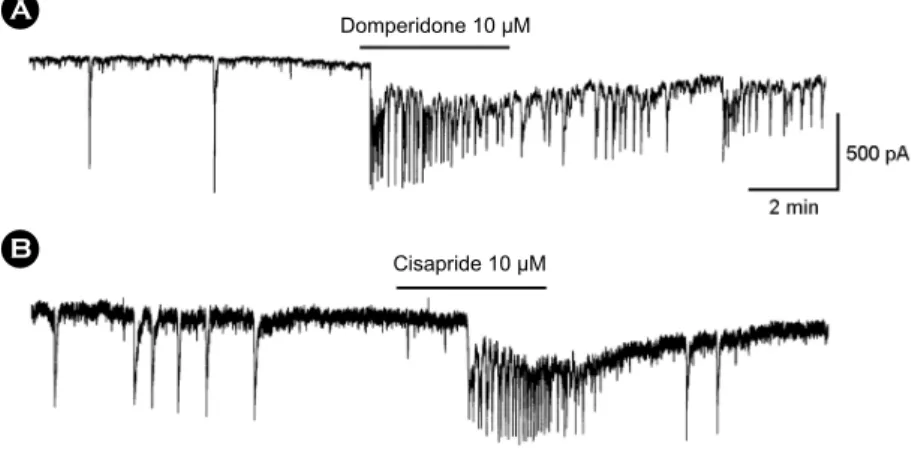
Effects of dopamine 2 receptor antagonists on pacemaker currents in large intestinal ICCs
To investigate the role of dopamine receptor antagonists, we used domperidone, an antagonist of D2 receptor or cisapride, an agonist of serotonin4 receptor. The treatment of ICCs with domperidone (10 μM) increased the freqeuency of pacemaker currents in ICCs compared with normal condition (Fig. 5A). Furthermore, cisapride also evoked the increasing of frequency of pacemaker currents in ICCs (Fig. 5B).
DISCUSSION
Regarding a presumed physiological role for dopamine in the control of GI motility, the present results support that dopamine have inhibitory action on pacemaker activity of
Fig. 4. Effects of D2 or D4 agonists on pacemaker currents recorded in cultured ICCs from mouse large intestine and agarose gels of RT-PCR products of dopamine receptor subtypes in c-Kit positive cells. (A) shows the 1.2% agarose gel which was loaded with 5 μl of PCR product and stained with ethidium bromide. The marker shown in lane 1 indicated bp. M: Marker, Kit: c-Kit, MS: Myosin, PGP:PGP9.5. (B) and (C) show pacemaker currents of ICCs exposed to 10 μM D2 receptor agonist (PPHT) and D4 receptor agonist (Piperazin) at a holding potential of -70 mV. Responses to dopamine in large intestinal ICCs are summarized in (D). Bars represent means ± SE.
*Asterisks mean significantly different from the controls (P < 0.05). Con: Control.
A
C B
D
PPHT 10 μM
Piperazin 10 μM
ICCs. Although many reports have shown the actions of dopamine on GI motility in tissue and smooth muscle cells, this is the first study in which an attempt has been made to determine the effects of dopamine in ICCs on electrical activity.
It is well known that dopamine can activate adrenoceptors (Ji et al., 2003; Jun et al., 2004), which respond to NE and epinephrine. Some reports suggested the nonspecific activation of adrenoceptors is involved on the effects of dopamine in GI tract and denied the existence of enteric receptors for dopamine (Grivegnee et al., 1984; Ji et al., 2003). Also, our previous report showed noradrenaline- induced stimulation of β1-adrenoceptors in ICCs inhibits pacemaker currents (Li et al., 2006). These mean that dopamine may influence on ICCs through stimulation of adrenoceptors. In contrast to these reports, this observation suggests that ICCs is well endowed with specific dopamine receptors. In our result, we showed the transcripts encoding D2 and D4 receptors were expressed in ICCs and specific agonists of D2 and D4 showed the effects on pacemaker activity of ICCs. Thus, our study indicates dopamine may have influence on GI motility by acting D2 and D4 receptors in large intestinal ICCs.
Dopamine generates its physiological effects through five genetically distinct dopamine receptor subtypes: D1, D2, D3, D4 and D5 (Lucchelli et al., 1986; Lucchelli et al., 1990). Whole five dopamine receptor subtypes are present in the GI tract (Mine et al., 1997). D2 and D3 receptor subtypes were found in enteric nervous system (Sakaguchi et al., 1992). However, there are no reports the existence of
dopamine receptor subtypes in ICCs. In this study, we showed the existence of D2 and D4 receptor subtypes.
Interestingly, Li et al., showed the stimulation of D2 receptor in enteric dopaminergic neurons inhibit the intestinal motility and do so physiologically (Sakaguchi et al., 1992).
Our result also showed the inhibitory action of dopamine and existence of D2 receptor subtype. Namely, these are suggested that GI motility is modulated by dopamine through D2 receptor subtype in ICCs.
Many reports showed D2 receptor antagonists are an AChesterase inhibitor (Sanders, 1996; Scheibner et al., 2002). It has been known to enhance gastric motility and is used as a treatment for functional dyspepsia (Scratcherd and Grundy, 1984). Because ACh released from the enteric nerve system stimulates the contraction of smooth muscle, we think D2 receptor antagonists have a prokinetic effect in GI tract. Indeed, many reports showed D2 receptor antagonists had been known to stimulate GI motility (Scheibner et al., 2002). In our study, we found D2 receptor antagonists increase the frequency of pacemaker activity.
This means that ICCs also can be the target for the prokinetic effect of dopamine in GI tract. Although the exact mechanism is not clear how D2 receptor antagonist can act on ICCs, the previous evidences that D2 receptor antagonists stimulate endogenous ACh release by blocking D2 receptor and accumulates endogenous ACh by anti- cholinesterase activity (Iwanaga et al., 1996; So et al., 2009) can be the possible mechanism of dopamine in ICCs. Also we showed carbachol modulates the pacemaker activity of ICCs via muscarinic M3 receptors (Suzuki et al., 2000).
Domperidone 10 μM
Cisapride 10 μM
Fig. 5. Effects of D2 receptor antagonist or serotonin4 receptor agonist on pacemaker currents recorded in cultured ICCs from mouse large intestine. (A) shows pacemaker currents of ICCs exposed to 10 μM dom- peridone (D2 receptor antagonist). (B) shows pacemaker currents of ICCs exposed to 10 μM cisapride (serotonin4 receptor agonist) at a holding potential of -70 mV.
A
B
Furthermore, the effect of D2 antagonists on GI motility was compared with serotonin4 receptor agonists, such as cisapride, mosapride etc. Many selective serotonin4 receptor agonists have been demonstrated to have prokinetic effects on GI motility (Torihashi et al., 1999; Tan et al., 2003). In this study, we also could see the increasing frequency of pacemaker activity by cisapride same as D2 receptor anta- gonist. Commonly, the mechanism of serotonin4 receptor agonists-induced prokinetic effects involves an increase in NE release via presynaptic D2 receptor blockade (Van Nueten and Schuurkes, 1984; Tsai and Cheng, 1992). For finding the mechanism how serotonin4 receptor agonist can increase the frequency of pacemaker activity in ICCs, further studies are needed.
Next with the ground about action of dopamine on pacemaker activity of ICCs, we examined which cellular mechanisms are involved. We focused that the periodic pacemaker activity of ICCs is dependent on [Ca
2+]
ioscil- lations. It is well known that the pacemaker mechanism is initiated by release of Ca
2+from the endoplasmic reticulum and is followed by reuptake of Ca
2+into the mitochondria.
Their results suggested that [Ca
2+]
iplays a crucial role in pacemaking and that Ca
2+imaging at the tissue level is a useful technique to investigate slow wave propagation in GI muscle (Ward, 2000; Ward et al., 2000). In this study, we could see the spontaneous [Ca
2+]
ioscillations using Ca
2+live cell imaging and [Ca
2+]
ioscillations was diminished by dopamine and returned back to normal condition after the washout showing similar effect as electrophysiological studies. The actions of dopamine on [Ca
2+]
iin ICCs are in keeping with the suggestions that intracellular Ca
2+oscil- lations are important actions of pacemaker activity. This means that the inhibitory action of dopamine on pacemaker activity in ICCs is closely related the modulation of [Ca
2+]
i. In this study, it is interesting to observe that the pacemaker current pattern of colonic ICCs and small intestine ICCS is different and dopamine has no action on the pacemaker activity of cultured small intestine ICCs, this suggest that the colonic ICCs are different from the small intestine ICCs in many aspects, such as the pacemaker mechanism, gene expression, etc. In this regard, more comparison studies are required. In conclusion, dopamine has a inhibitory and D2
receptor antagonist has a stimulatory effect on pacemaker activity of ICCs from large intestine and this is mediated through D2 receptor. These data will provide experimental background for the use of dopamine to treat patients with constipation or other functional bowel disorders.
Acknowledgements
This study was financially supported by research fund of Chonnam National University in 2011.
REFERENCES
Abe M, Orita Y, Nakashima Y, Nakamura M. Hypertensive crisis induced by metocloprimed in patient with pheochromocytoma.
Angiology. 1984. 35: 122-128.
Adler-Graschinsky E, Rubio MC, Barontini De Moyano M.
Metoclopramide increases the release of catecholamines from isolated human phaeochromocytomas. J Hypertens. 1984. 2:
127-129.
Eaker EY, Bixler GB, Dunn AJ, Moreshead WV, Mathias JR.
Dopamine and norepinephrine in the gastrointestinal tract of mice and the effects of neurotoxins. J Pharmcol Exp Ther.
1988. 244: 438-442.
Gershon MD. Inhibition of gastrointestinal movement by sympathetic nerve stimulation: the site of action. J Physiol (Lond). 1967. 198: 317-329.
Grivegnee AR, Fontaine J, Reuse J. Effect of dopamine on dog distal colon in-vitro. J Pharm Pharmacol. 1984. 36: 454-457.
Hartman DS, Civelli O. Dopamine receptor diversity: molecular and pharmacological perspectives. Prog Drug Res. 1997. 48:
173-194.
Hirst GD, Silinsky EM. Some effects of 5-hydroxytryptamine, dopamine and noradrenaline on neurons in the submucous plexus of guinea-pig small intestine. J Physiol (Lond). 1975.
251: 817-832.
Holtmann G, Talley NJ, Liebregts T, Adam B, Parow C. A placebo- controlled trial of itopride in functional dyspepsia. N Engl J Med. 2006. 354: 832-840.
Huisinga J. Physiology and pathophysiology of interstitial cells of Cajal: from bench to bedside. II. Gastric motility lessons from mutant mice on slow waves and innervation. Am J Physiol.
2001. 281: G1129-G1134.
Iwanaga Y, Kimura T, Miyashita N, Morikawa K, Nagata O, Itoh Z, Kondo Y. Characterization of acetylcholinesterase-inhibition
by itopride. Jpn J Pharmacol. 1994. 66: 317-322.
Iwanaga Y, Miyashita N, Morikawa K, Mizumoto A, Kondo Y, Itoh Z. A novel water-soluble dopamine-2 antagonist with anticholinesterase activity in gastrointestinal motor activity.
Gastroenterology. 1990. 99: 401-408.
Iwanaga Y, Miyashita N, Saito T, Morikawa K, Itoh Z.
Gastroprokinetic effect of a new benzamide derivative itopride and its action mechanisms in conscious dogs. Jpn J Pharmacol. 1996. 71: 129-137.
Ji SW, Park JH, Cho JS, Lim JH, Lee SI. Investigation into the effects of mosapride on motility of Guinea pig stomach, ileum, and colon. Yonsei Med J. 2003. 44: 653-664.
Jun JY, Choi S, Yeum CH, Chang IY, Park CK, Kim MY, Kong ID, So I, Kim KW, You HJ. Noradrenaline inhibits pacemaker currents through stimulation of b1-adrenoceptors in cultured interstitial cells of Cajal from murine small intestine. Br J Pharmacol. 2004. 141: 670-677.
Li ZS, Schmauss C, Cuenca A, Ratcliffe E, Gershon MD.
Physiological modulation of intestinal motility by enteric dopaminergic neurons and the D2 receptor: analysis of dopamine receptor expression, location, development, and function in wild-type and knock-out mice. J Neurosci. 2006.
26: 2798-2807.
Lucchelli A, Boselli C, Chiari MC, Grana E. Analysis of the relaxing effect of dopamine on the isolated rat jejunum. Arch Int Pharmacodyn Ther. 1986. 279: 234-247.
Lucchelli A, Boselli C, Grana E. Dopamine-induced relaxation of the guinea-pig isolated jejunum is not mediated through dopamine receptors. Pharmacol Res. 1990. 2: 433-444.
Mine Y, Yoshikawa T, Oku S, Nagai R, Yoshida H, Hosoki K.
Comparison of effect of mosapride citrate and existing 5-HT4 receptor agonist on gastrointestinal motility in vivo and in vitro. J Pharmacol Exp Ther. 1997. 283: 1000-1008.
Sakaguchi J, Nishino H, Ogawa N, Iwanaga Y, Yasuda S, Kato H, Ito Y. Synthesis, gastrointestinal prokinetic activity and structure-activity relationships of novel N-[[2-(dialkylamino)- ethoxy]bexzyl]benzamide derivatives. Chem Pharm Bull.
1992. 40: 202-211.
Sanders KM. A case for interstitial cells of Cajal as pacemakers and mediators of neurotransmission in the gastrointestinal tract. Gastroenterology. 1996. 111: 492-515.
Scheibner J, Trendelenburg AU, HeIN L, Starke K, Blandizzi C.
Alpha 2 adrenoceptors in the enteric nervous system: a study in alpha 2A-adrenoceptor-deficient mice. Br J Pharmacol.
2002. 135: 697-704.
Scratcherd T, Grundy D. The physiology of intestinal motility and secretion. Br J Anaesth. 1984. 56: 3-18.
So KY, Kim SH, Sohn HM, Choi SJ, Parajuli SP, Choi S, Yeum CH, Yoon PJ, Jun JY. Carbachol regulates pacemaker activities in cultured interstitial cells of Cajal from the mouse small intestine. Mol Cells. 2009. 27: 525-531.
Suzuki H, Takano H, Yamamoto Y, Komuro T, Saito M, Kato K, Mikoshiba K. Properties of gastric smooth muscles obtained from mice which lack inositol trisphosphate receptor. J Physiol. 2000. 525: 105-111.
Tan S, Hermann B, Borrelli E. Dopaminergic mouse mutants:
investigating the roles of the different dopamine receptor subtypes and the dopamine transporter. Int Rev Neurobiol.
2003. 54: 145-197.
Torihashi S, Horisawa M, Watanabe Y. c-Kit immunoreactive interstitial cells in the human gastrointestinal tract. J Auton Nerv Syst. 1999. 75: 38-50.
Tsai LH, Cheng JT. The effect of exogenous dopamine on ileal smooth muscle of guinea-pigs. Chin J Physiol. 1992. 35: 133 -141.
Van Nueten JM, Schuurkes AJ. Studies on the role of dopamine and dopamine blockers in gastroduodenal motility. Scand Gastroenterol Suppl. 1984. 96: 89-99.
Ward S. Interstitial cells of Cajal in enteric neurotransmission. Gut.
2000. 47: 40-43.
Ward SM, Ordog T, Koh SD, Baker SA, Jun JY, Amberg G, Monaghan K, Sanders KM. Pacemaking in interstitial Cells of Cajal depends upon calcium handling by endoplasmic reticulum and mitochondria. J Physiol. 2000. 525: 355-361.
![Fig. 3. Effects of dopamine on [Ca 2+ ] i oscillation in cultured ICCs from mouse large intestine](https://thumb-ap.123doks.com/thumbv2/123dokinfo/4763982.273321/5.892.123.784.132.473/effects-dopamine-oscillation-cultured-iccs-mouse-large-intestine.webp)