http://dx.doi.org/10.3988/jcn.2014.10.1.37 J Clin Neurol 2014;10:37-41
Introduction
Ocular manifestation is one of the most frequent and impor- tant signs observed during an acute attack of multiple sclero- sis (MS). Common abnormal eye movements include saccad- ic dysmetria, internuclear ophthalmoplegia (INO), impairment of the vestibulo-ocular reflex (VOR), and gaze-evoked nystag- mus (GEN).1 However, primary position upbeat nystagmus (PPUN) is a rare manifestation in MS.
The amplitude of PPUN usually increases during upward gaze, with impaired vertical pursuit, but the amplitude de- creases during downward gaze. It is neither enhanced during lateral gaze nor reduced by fixation, and should be distin- guished from the more common GEN, which is observed only during upward gaze. PPUN can be caused by lesions involv- ing the ventral tegmentum of the rostral medulla and the cau- dal pons, midbrain, brachium conjunctivum (BC), and cere- bellum.2,3 In the lower medulla in particular, it is associated with lesions affecting the prepositus hypoglossi nucleus, ves- tibular nuclei, nucleus intercalatus, and flocculus. Upbeat nys- tagmus (UBN) is caused by damage to the interconnection be- tween the structures controlling the vertical vestibulo-ocular pathways or vertical smooth pursuit,4 and it has been report-
Primary Position Upbeat Nystagmus during an Acute Attack of Multiple Sclerosis
Jee-Ae Kim, In-Hye Jeong, Young-Min Lim, Kwang-Kuk Kim
Department of Neurology, Asan Medical Center, University of Ulsan College of Medicine, Seoul, Korea
Received February 27, 2013 Revised August 18, 2013 Accepted August 18, 2013 Correspondence Kwang-Kuk Kim, MD, PhD Department of Neurology, Asan Medical Center, University of Ulsan College of Medicine,
88 Olympic-ro 43-gil, Songpa-gu, Seoul 138-736, Korea
Tel +82-2-3010-3440 Fax +82-2-474-4691 E-mail [email protected]
Background and PurposezzOcular manifestation is one of the frequent signs of an acute attack in multiple sclerosis (MS), although primary position upbeat nystagmus (PPUN) is rare. The pur- pose of this study is to determine the incidence of PPUN in MS and to determine the lesions that are responsible for this sign.
MethodszzThe medical records of 120 MS patients with acute brain lesions were reviewed over a consecutive period of 9 years; of these, 6 patients were found to have PPUN. Other ocular mo- tor abnormalities were analyzed in combination with upbeat nystagmus, video-oculographic find- ings, and lesions detected on brain MRI.
ResultszzLesions in the pontine tegmentum involving the medial longitudinal fasciculus (MLF) and ventral tegmental tract (VTT) were the most common, being observed in three of the six pa- tients with PPUN. One patient exhibited caudal medullary lesions bilaterally affecting the para- median portion of the posterior tegmentum, and two patients exhibited multiple lesions involving the pons with the cerebral peduncle or medulla. In five patients, other ocular motor dysfunctions, such as gaze-evoked nystagmus (n=3) and internuclear ophthalmoplegia (n=1), were found in combination with upbeat nystagmus.
ConclusionszzPPUN is an infrequent, ocular manifestation noted during an acute attack of MS, and was observed in 5% of the present cases. Brainstem lesions in these cases primarily involved the pontine tegmentum and the caudal medulla. These findings support the theory that upbeat nystagmus is attributable to damage to the upward vestibulo-ocular reflex pathway related to the vestibular nucleus, VTT, and interconnecting pathways. J Clin Neurol 2014;10:37-41 Key Wordszz primary position upbeat nystagmus, multiple sclerosis,
vestibulo-ocular reflex pathway, ventral tegmental tract.
Open Access
cc This is an Open Access article distributed under the terms of the Cre- ative Commons Attribution Non-Commercial License (http://creative- commons.org/licenses/by-nc/3.0) which permits unrestricted non-com- mercial use, distribution, and reproduction in any medium, provided the ori- ginal work is properly cited.
ed in patients with infarctions, demyelination,3 glioma,4 and Wernicke’s encephalopathy.5
The aim of the present study was to determine the incidence of PPUN in acute attacks of MS and describe the anatomic correlation of UBN on brain MRI.
Methods
The medical records of patients who were admitted due to exacerbations of MS at Asan Medical Center between March 2000 and January 2009 were reviewed, and patients with acute
MS lesions on brain MRI and who exhibited PPUN were en- rolled. The diagnosis of MS was based on the 2010 McDonald criteria. PPUN was defined as UBN observed when resting and with stronger upbeat rather than horizontal or torsional components, in addition to increasing amplitude during up- ward gaze. Patients with vertical gaze-evoked UBN observed only during vertical gaze were excluded.
The following clinical details were obtained for each pa- tient: sex, age, other ocular motor abnormalities, video-oculo- graphic findings, and lesions seen on brain MRI. Brain MRI using a 1.5-tesla unit was performed in all patients within 1–28 Table 1. Clinical profiles of the patients with primary position upbeat nystagmus (PPUN) in multiple sclerosis (MS)
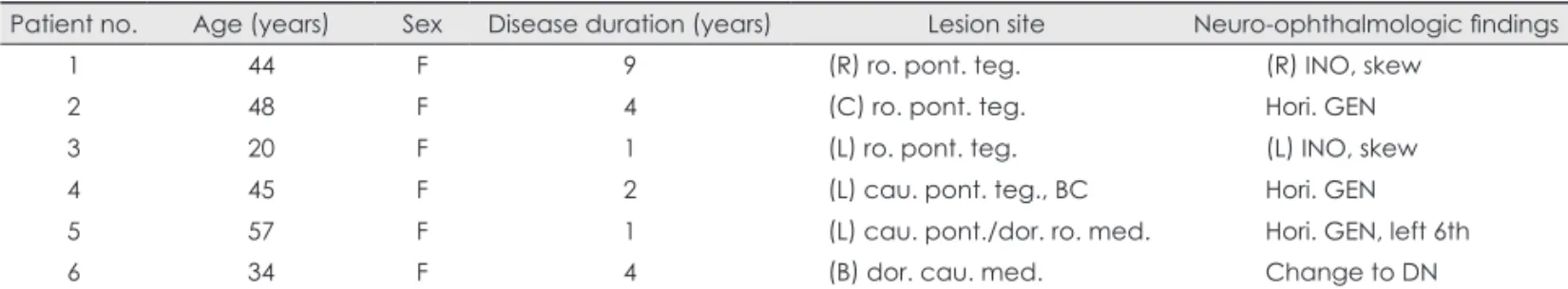
Patient no. Age (years) Sex Disease duration (years) Lesion site Neuro-ophthalmologic findings
1 44 F 9 (R) ro. pont. teg. (R) INO, skew
2 48 F 4 (C) ro. pont. teg. Hori. GEN
3 20 F 1 (L) ro. pont. teg. (L) INO, skew
4 45 F 2 (L) cau. pont. teg., BC Hori. GEN
5 57 F 1 (L) cau. pont./dor. ro. med. Hori. GEN, left 6th
6 34 F 4 (B) dor. cau. med. Change to DN
B: bilateral, BC: brachium conjunctivum, cau.: caudal, DN: downbeat nystagmus, dor.: dorsal, F: female, GEN: gaze-evoked nystag- mus, Hori.: horizontal, INO: internuclear ophthalmoplegia, L: left, med.: medulla, pont.: pontine, R: right, ro.: rostral, teg.: tegmentum.
Table 2. Clinical profiles of 24 patients with the other oculomotor abnormalities
Patient no. Age (years) Sex Lesion site Neuro-ophthalmologic findings
7 59 F (B) pons, med. Hori. GEN
8 55 F (L) pons/(B) med. Hori. GEN
9 34 F PAG of midbrain (L) INO
10 26 F (R) MLF (R) INO
11 32 M (L) ros. to cau. med. (L) Hori. GEN
12 21 F (R) pons (R) 6th nerve palsy
13 26 M (L) midbrain (L) medial gaze limitation
14 25 F (R) MLF (R) INO
15 39 F Negative MRI (R) 3rd nerve palsy
16 33 M (L) midbrain (L) 3rd nerve palsy
17 40 F (B) pont. teg. Hori. GEN
18 20 F (L) pons Hori. GEN
19 19 F PAG. of midbrain DN
20 57 F (B) pont. teg. Hori. GEN
21 41 M (L) midbrain Hori. GEN
22 17 F PAG. of midbrain/pons (L) INO
23 34 F (B) pons/(R) midbrain (L) catch-up saccade
24 36 F (L) midbrain/pons (L) medial gaze limitation
25 24 M (L) SCP/pons Hori. GEN
26 23 M (B) pons (B) 6th nerve palsy
27 25 F (R) MCP Hori. GEN
28 46 F (L) MCP/vermis Hori. GEN/Ver. GEN during upward gaze
29 40 F (B) teg. of midbrain and pons/med. Oscillopsia, Hori. and Ver. GEN
30 45 M (B) pons/(R) cerebellar hemisphere Saccadic hypometria
B: bilateral, DN: downbeat nystagmus, GEN: gaze-evoked nystagmus, Hori: horizontal, ICP: inferior cerebellar peduncle, INO: internu- clear ophthalmoplegia, L: left, MCP: middle cerebellar peduncle, med.: medulla, MLF: longitudinal fasciculus, PAG: periaqueductal gray matter, pont.: pontine, R: right, ro: rostal, SCP: superior cerbellar peduncle, teg.: tegmentum, Ver.: vertical.
days after the symptom onset. An axial T2-weighted scan [repetition time/echo time (TR/TE)=2,500/80 ms] was per- formed in the horizontal plane. T1-weighted (TR/TE=600/20 ms) axial and sagittal images were also obtained. The T2- weighted axial images were used for analysis. Eye movements were observed using video Frenzel goggles and were record- ed using video-oculography (SLMED, Seoul, Korea).
Results
Of the 156 consecutive patients with exacerbations of MS, 120 had acute lesions on brain MRI, of which 6 (5%) exhib- ited PPUN during straight-ahead gaze. All six were women, their mean age was 41 years (range, 20–57 years), and their
mean disease duration was 3.5 years (range, 1–9 years). Table 1 summarizes the clinical and demographic data of these pa- tients. Other oculomotor abnormalities were observed in 24 patients. The demographic and oculomotor findings of these patients are summarized in Table 2.
The most common lesions were pontine lesions involving the ventral tegmental tract (VTT) or medial longitudinal fas- ciculus (MLF); these were observed in three patients (pa- tients 1–3). One patient (patient 6) had MS lesions in the dorsal part of the lower medulla that involved the nucleus intercala- tus. Two patients had multiple brainstem lesions (i.e., pontine tegmentum with superior and middle cerebellar peduncle in patient 4, and diffuse lesions extending from the caudal pons to the dorsal rostral medulla in patient 5). In five patients
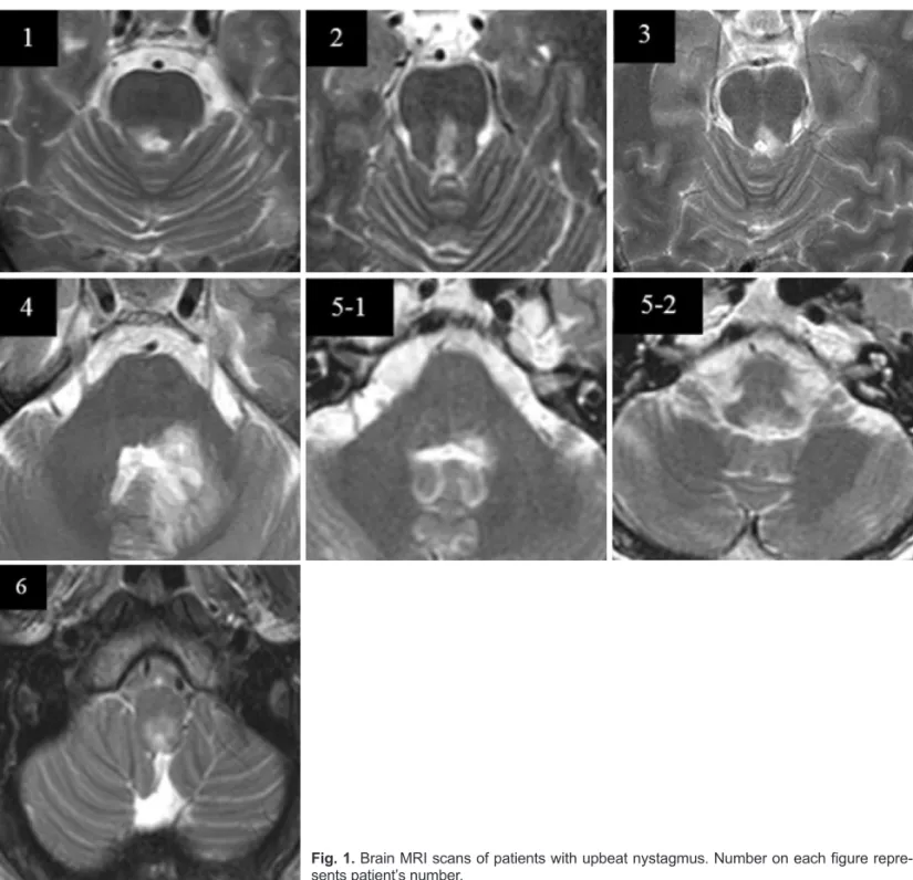
Fig. 1. Brain MRI scans of patients with upbeat nystagmus. Number on each figure repre- sents patient’s number.
(patients 1–5), UBN was accompanied by other ocular ab- normalities such as horizontal GEN (n=3) and INO (n=2). Fig.
1. shows the brain MRI scans of the six MS patients with PPUN.
Illustrative case
A 34-year-old woman presented with dizziness and dyspha- gia. Three years before the onset of her symptoms she had suf- fered from transverse myelitis at the T5-T9 level. A neuro- ophthalmological examination revealed PPUN but without weakness of the extraocular muscles. The amplitude and fre- quency of the UBN increased during upward gaze and de- creased during downward gaze. Video-oculographic record- ing revealed that the nystagmus was upbeating in the primary position of gaze with a mean velocity of 7°/sec, and its slow phase was linear, with a constant velocity. The nystagmus changed to downbeating with a mean velocity of 26.6°/sec during rightward gaze and it disappeared during leftward gaze (Suppl. 1), and reversed direction to downbeat when lying down and during rightward and leftward head turning (Suppl.
2). The nystagmus was also reversed to downbeat during cen- tral head hanging, and right and left head hanging. The gag re- flexes and the power of the tongue were also decreased. Mo- tor and sensory changes were not reported in her extremities.
Brain MRI revealed high T2 signal intensities in the dorsal paramedian area of the caudal medulla, and spinal MRI re- vealed no evidence of new lesions. The patient’s symptoms gradually improved with steroid therapy. However, 20 days later she was seen again because of sudden-onset severe diz- ziness. Video-oculographic findings revealed downbeat nys- tagmus in the primary position, but without other alterations (Suppl. 3). The direction of the nystagmus did not change in any of the head positions. Follow-up brain MRI revealed no definite interval changes of the lower medullary lesion. The downbeat nystagmus persisted to the 1-month follow-up af- ter discharge.
Discussion
Ocular abnormalities can be initial manifestations of MS and may predict additional demyelinating events.6,7 Nevertheless, PPUN is an infrequent ocular manifestation during acute at- tacks of MS, as was observed in 5% of the present cases.
In the present study, the pons was the main anatomical lo- cation responsible for UBN (Fig. 1). In the ascending route of the upward VOR pathway, the signals from the anterior semi- circular canals are sent to the superior vestibular nucleus (SVN) and then through the BC to the oculomotor nucleus in the midbrain via the VTT or MLF.8 In cases of UBN caused by a pontine lesion, the lesion is usually located in the posterior
basis pontis or in the ventral tegmentum at the level of the rostral pons.2 In three of the patients in this study (patients 1–3), the demyelinating lesion was observed in the posterior part of the basis pontis at the level of the rostral pons. In one of these (patient 2), brain MRI revealed involvement of the VTT.
The VTT lies slightly ventral and lateral to the BC in the lower pons, arching medially and decussating above the lev- el of the midpons, in the posterior part of the basis pontis.9 Therefore, UBN may arise from a small, unilateral lesion in the posterior basis pontis of the rostral pons. This patient also harbored midline MLF involvement. The MLF lesion may be another explanation for PPUN. Four patients (patients 1–3, and 5) possessed midline MLF lesions at the level of the rostral to caudal pons. PPUN as a result of MLF lesion is very rare;
damage to the MLF mainly causes INO or skew deviation.
However, a selective lesion in the upward pathway of the VOR with sparing of the downward pathway10 may result in UBN.
One case (patient 4) exhibited involvement of the BC on brain MRI. A few patients with UBN attributed to unilateral BC lesions have also been reported.11,12 The role of the BC could be considered since this structure is generally thought to transmit vertical slow eye movement signals to the oculo- motor nucleus, so that theoretically it is possible for UBN to appear after BC damage.9
Brain MRI in patient 6 demonstrated dorsal caudal medul- lar lesions involving the nucleus intercalatus. The caudal me- dulla is linked to the visual pursuit system. The nucleus inter- calatus, one of the components of the perihypoglossal nuclei, also receives excitatory signals from the SVN and has an in- terconnection with the prepositus hypoglossi nucleus.13 The prepositus hypoglossi nucleus receives afferent inhibitory sig- nals from the cerebellar vermis and the flocculus, and then ex- cites the oculomotor neurons.9 Therefore, UBN may result from lesions in these structures. Some authors have consid- ered a lesion in the nucleus intercalatus of the medulla to be the most reasonable explanation for PPUN.14-16 In patient 6 in the present study, UBN changed to downbeat nystagmus in the primary position 20 days later, without definite changes in the caudal medullar lesions on brain MRI. There have only been a few case reports of UBN spontaneously changing to downbeat nystagmus in patients with Wernicke’s encephalop- athy5,17 and medullary hemorrhage.18 It has been reported that the caudal brainstem, and more specifically the midline cere- bellum, and the nucleus prepositus hypoglossi control vertical smooth pursuit, and that vertical nystagmus can result from damage to these structures.4 Directional changes of vertical nystagmus in our patient are thought to be attributable to in- terruption of tonic balancing smooth pursuit by caudal me- dulla lesions.
As shown by the present illustrative case, UBN could be ob- served transiently, since the latency from the onset was also variable. One limitation of the present retrospective study was the evaluation of delicate and transient neuro-ophthalmolog- ic findings, especially UBN.
In conclusion, the cases described herein support the theory that UBN is caused by damage to the ascending routes of the VOR pathway related to the VTT, SVN, and caudal medulla, and their interconnecting pathways, and are consistent with the results of previous studies. PPUN is not a common ocular manifestation in acute attacks of MS, but it can provide im- portant clues regarding the location of brainstem lesions.
Conflicts of Interest
The authors have no financial conflicts of interest.
REFERENCES
1. Derwenskus J, Rucker JC, Serra A, Stahl JS, Downey DL, Adams NL, et al. Abnormal eye movements predict disability in MS: two-year follow-up. Ann N Y Acad Sci 2005;1039:521-523.
2. Fisher A, Gresty M, Chambers B, Rudge P. Primary position upbeat- ing nystagmus. A variety of central positional nystagmus. Brain 1983;
106:949-964.
3. Hirose G, Kawada J, Tsukada K, Yoshioka A, Sharpe JA. Upbeat nys- tagmus: clinicopathological and pathophysiological considerations. J Neurol Sci 1991;105:159-167.
4. Gilman N, Baloh RW, Tomiyasu U. Primary position upbeat nystag- mus. A clinicopathologic study. Neurology 1977;27:294-298.
5. Cox TA, Corbett JJ, Thompson HS, Lennarson L. Upbeat nystagmus changing to downbeat nystagmus with convergence. Neurology 1981;
31:891-892.
6. Chen L, Gordon LK. Ocular manifestations of multiple sclerosis. Curr Opin Ophthalmol 2005;16:315-320.
7. Solingen LD, Baloh RW, Myers L, Ellison G. Subclinical eye move- ment disorders in patients with multiple sclerosis. Neurology 1977;
27:614-619.
8. Ranalli PJ, Sharpe JA. Upbeat nystagmus and the ventral tegmental pathway of the upward vestibulo-ocular reflex. Neurology 1988;38:
1329-1330.
9. Pierrot-Deseilligny C, Milea D. Vertical nystagmus: clinical facts and hypotheses. Brain 2005;128:1237-1246.
10. Kim JS, Yoon B, Choi KD, Oh SY, Park SH, Kim BK. Upbeat nys- tagmus: clinicoanatomical correlations in 15 patients. J Clin Neurol 2006;2:58-65.
11. Benjamin EE, Zimmerman CF, Troost BT. Lateropulsion and upbeat nystagmus are manifestations of central vestibular dysfunction. Arch Neurol 1986;43:962-964.
12. Kattah JC, Dagi TF. Compensatory head tilt in upbeating nystagmus.
J Clin Neuroophthalmol 1990;10:27-31.
13. Ohkoshi N, Komatsu Y, Mizusawa H, Kanazawa I. Primary position upbeat nystagmus increased on downward gaze: clinicopathologic study of a patient with multiple sclerosis. Neurology 1998;50:551-553.
14. Saito T, Aizawa H, Sawada J, Katayama T, Hasebe N. Lesion of the nucleus intercalatus in primary position upbeat nystagmus. Arch Neu- rol 2010;67:1403-1404.
15. Munro NA, Gaymard B, Rivaud S, Majdalani A, Pierrot-Deseilligny C.
Upbeat nystagmus in a patient with a small medullary infarct. J Neu- rol Neurosurg Psychiatry 1993;56:1126-1128.
16. Adamec I, Gabelić T, Krbot M, Ozretić D, Milivojević I, Habek M.
Primary position upbeat nystagmus. J Clin Neurosci 2012;19:161-162.
17. Kastrup O, Maschke M, Keidel M, Diener HC. Presumed pharmaco- logically induced change from upbeat- to downbeat nystagmus in a patient with Wernicke’s encephalopathy. Clin Neurol Neurosurg 2004;
107:70-72.
18. Janssen JC, Larner AJ, Morris H, Bronstein AM, Farmer SF. Upbeat nystagmus: clinicoanatomical correlation. J Neurol Neurosurg Psychi- atry 1998;65:380-381.
Primary Position Upbeat Nystagmus during an Acute Attack of Multiple Sclerosis
Jee-Ae Kim, In-Hye Jeong, Young-Min Lim, Kwang-Kuk Kim
Department of Neurology, Asan Medical Center, University of Ulsan College of Medicine, Seoul, Korea
Supplemental Video Clip. 1. Video-oculography of patient 6. (a) Upbeating nystagmus in the primary position. (b) During right- ward gaze the nystagmus was changed to downbeating with a mean velocity of 26.6°/sec. (c) During leftward gaze the upbeat nystagmus disappeared.
Supplemental Video Clip. 2. Video-oculography of patient 6. The nystagmus reversed direction to downbeat when lying down (a) and during rightward (b) and leftward (c) head turning.
Supplemental Video Clip. 3. Video-oculography showing the downbeat nystagmus that developed 20 days after the initial symp- toms.