http://dx.doi.org/10.3988/jcn.2011.7.3.115 J Clin Neurol 2011;7:115-127
Introduction
Neuromyelitis optica (NMO, also known as Devic’s disease) is an idiopathic inflammatory syndrome of the central nervous system (CNS) that is characterized by severe attacks of optic neuritis (ON) and myelitis.1 NMO was first described in 1870 by T. Clifford Allbutt, who reported an association between myelitis and unilateral optic nerve disorder.2 In 1894, Eugène Devic coined the term “neuromyelite optique aigue” (acute optic neuromyelitis) to describe 16 patients who had lost vi- sion unilaterally or bilaterally and within weeks developed
severe spastic paraparesis or tetraparesis, loss of sensation, and often loss of sphincter control. These symptoms were recogniz- ed as resulting from inflammation affecting both the optic ner- ve and the spinal cord.3 For the next 100 years, NMO was con- sidered to be a monophasic, devastating disorder that affected the optic nerves and the spinal cord, but spared the remainder of the CNS.
There has long been controversy as to whether NMO is a variant of multiple sclerosis (MS) or a distinct disease. A land- mark advance in this regard was the identification of a disease- specific autoantibody, NMO-IgG, in the serum of patients with NMO.4 NMO-IgG binds selectively to aquaporin (AQP)-4 (AQP4), a water channel that is densely expressed in astrocy- tic foot processes at the blood-brain barrier (BBB).5 This find- ing has prompted revisions of the diagnostic criteria for NMO6 and facilitated an appreciation for the wide spectrum of this
New Insights into Neuromyelitis Optica
Woojun Kim, Su-Hyun Kim, Ho Jin Kim
Department of Neurology, Research Institute and Hospital of National Cancer Center, Goyang, Korea
Received April 6, 2011 Revised June 13, 2011 Accepted June 13, 2011 Correspondence Ho Jin Kim, MD, PhD Department of Neurology, Research Institute and Hospital of National Cancer Center, 323 Ilsan-ro, Ilsandong-gu, Goyang 410-769, Korea Tel +82-31-920-2438 Fax +82-31-905-5524 E-mail hojinkim@ncc.re.kr
Neuromyelitis optica (NMO) is an idiopathic inflammatory disorder of the central nervous sys- tem (CNS) that preferentially affects the optic nerves and spinal cord. In Asia, NMO has long been considered a subtype of multiple sclerosis (MS). However, recent clinical, pathological, immuno- logical, and imaging studies have suggested that NMO is distinct from MS. This reconsideration of NMO was initially prompted by the discovery of a specific antibody for NMO (NMO-IgG) in 2004. NMO-IgG is an autoantibody that targets aquaporin-4 (AQP4), the most abundant water channel in the CNS; hence, it was named anti-AQP4 antibody. Since it demonstrated reasonable sensitivity and high specificity, anti-AQP4 antibody was incorporated into new diagnostic criteria for NMO.The spectrum of NMO is now known to be wider than was previously recognized and in- cludes a proportion of patients with recurrent, isolated, longitudinally extensive myelitis or optic neuritis, and longitudinally extensive myelitis or optic neuritis associated with systemic autoimmune disease or with brain lesions typical of NMO. In this context, a new concept of “NMO spectrum dis- orders” was recently introduced. Furthermore, seropositivity for NMO-IgG predicts future relapses and is recognized as a prognostic marker for NMO spectrum disorders. Humoral immune mech- anisms, including the activation of B-cells and the complement pathway, are considered to play important roles in NMO pathogenesis. This notion is supported by recent studies showing the po- tential pathogenic role of NMO-IgG as an initiator of NMO lesions. However, a demonstration of the involvement of NMO-IgG by the development of active immunization and passive transfer in animal models is still needed. This review focuses on the new concepts of NMO based on its pa- thophysiology and clinical characteristics. Potential management strategies for NMO in light of its pathomechanism are also discussed. J Clin Neurol 2011;7:115-127 Key Wordszz neuromyelitis optica, Devic’s disease, neuromyelitis optica spectrum disorder,
pathogenesis, diagnosis, management.
Open Access
cc This is an Open Access article distributed under the terms of the Cre- ative Commons Attribution Non-Commercial License (http://creative- commons.org/licenses/by-nc/3.0) which permits unrestricted non-com- mercial use, distribution, and reproduction in any medium, provided the ori- ginal work is properly cited.
disorder, which includes a proportion of patients with recur- rent, isolated, longitudinally extensive myelitis (LEM) or ON, and LEM or ON associated with systemic autoimmune disease or with brain lesions typical of NMO. Thus, the concept of
“NMO spectrum disorders” (NMOSDs) was introduced.1 In re- cent years, the clinical spectrum, immunological/pathological profiles, and management issues regarding NMO or NMOSDs have been better characterized. This review focuses on the new concepts of NMO based on its pathophysiology and cli- nical characteristics. Potential management strategies for NMO, in light of its pathomechanism, are also discussed.
Epidemiology and Genetic Predisposition
While there have been few relevant population-based stud- ies, the prevalence of NMO has been estimated by its frequ- ency relative to MS, and has been estimated at 1 per 100,000 population.7-9 In a retrospective, cross-sectional study perform- ed in Italy, the prevalence of NMOSDs was 1.5%, with a ratio of 42 MS cases to 7 NMOSD cases.10 However, the proportion of cases with a non-Caucasian background is significantly greater. NMO is believed to be linked to 30-40% of MS cases in Japanese,11 16.8% in African-Americans,12 and 15% in Afri- can-Brazilians.13 NMO predominantly affects females, with a female-to-male ratio of 9:1. The median age at onset of NMO is the late 30s;14,15 however, there have been several reports of NMO cases in childhood16-18 and in the elderly.19 NMO is not generally considered to have a familial tendency.14 However, genetic susceptibility is possible, since the reported incidence of familial cases of NMO appears to be higher than expected based on its frequency in the general population.20-25
NMO was reported to be associated with the human leu- kocyte antigen DPB1*0501 allele in the Japanese,26,27 but fur- ther analysis of patients with pure opticospinal MS (OSMS) who had consistently normal brain magnetic resonance im- aging (MRI) scans suggested that OSMS is not necessarily associated with DPB1*0501.28 DPB1*0501 was recently re- ported to be associated with NMO in the Han population of southern China.29
Immunopathology
Pathology of NMO lesions
The pathology of NMO lesions was initially described from autopsied cases by Lucchinetti et al.30 Extensive demyelin- ation was found to be present across multiple spinal cord levels and was associated with cavitation, necrosis, and acute axonal pathology (spheroids), in both the gray and white mat- ter. There was a marked loss of oligodendrocytes within the
lesions. The inflammatory infiltrates in active lesions were ch- aracterized by extensive macrophage infiltration associated with large numbers of perivascular granulocytes and eosino- phils, and a few T cells. There was pronounced perivascular deposition of immunoglobulins (mainly IgM) and comple- ment C9neo antigen in active lesions associated with promi- nent vascular fibrosis, and hyalinization in both the active and inactive lesions. These findings support a role for humoral im- munity in the pathogenesis of NMO.
Discovery of NMO-IgG
In 2004, an antibody that reacted specifically with mouse br- ain was identified in the serum of NMO patients.4 This anti- body, named NMO-IgG, was present in the CNS microvessels, pia, subpia, and Virchow-Robin spaces, and was partly colo- calized with laminin. This tissue-based indirect immunofluo- rescence assay (IIFA) was sensitive (73%) and highly specific (91%) to NMO. NMO-IgG was also detected in about half of patients with syndromes that placed them at high risk for NMO, such as idiopathic isolated recurrent ON or LEM. Dur- ing the following year, the same researchers proved that AQP4 is the target antigen of NMO-IgG by showing that NMO-IgG binds selectively to AQP4-transfected human embryonic kid- ney (HEK)-293 cells.5
AQPs and AQP4
AQPs are water-channel proteins that assemble as homotet- ramers in cell membranes and play a major role in fluid ho- meostasis. AQP4 is one of 13 members of the AQP family (AQP0-AQP12) present in mammals and is the predominant AQP in the brain.31 AQP4 is found throughout the brain, but is particularly abundant in the optic nerves and spinal cord.32 Other organs that contain AQP4 include the lung, skeletal muscle, stomach, inner ear, and kidney.33 In rats, AQP4 is pre- sent on astrocytic foot processes along the endothelial tight junctions at the BBB, on the abluminal side of cerebral mi- crovessels, within the cerebellar Purkinje cell layer, and in the hypothalamus.34
AQP4 exists in two isoforms, M1 (34 kDa) and M23 (32 kDa), both of which function as water channels and form st- able tetramers. The relative abundances of the AQP4 isoforms vary with the tissue type, with the rat brain containing appro- ximately three AQP4-M23 proteins per one AQP4-M1 pro- tein. Freeze-fracture electron microscopy of cells transfected with AQP4-M1 versus AQP4-M23 has demonstrated that only the M23 form assembles into large orthogonal arrays of more than 100 particles; AQP4-M1 tetramers were largely dispers- ed and only occasionally formed small arrays of fewer than 12 particles.35 There remains a controversy regarding which iso- form is the major target of the AQP4 antibody (AQP4-Ab). A
study employing Western blotting found AQP-M1 to be the spe- cific target of the AQP4-Ab.36 However, in another study using cell-based IIFA, the sensitivity with AQP4-M1 transfected cells was lower for NMO (70%) and high-risk NMO (39%), suggesting that NMO-IgG targets mainly AQP4-M23.37 Role of AQP4-Ab in the pathogenesis of NMO There is a series of clinical studies showing that AQP4-Ab par- ticipates in the pathogenesis of NMO and that AQP4-Ab may be predictive of relapse and/or later conversion to definite NMO.38,39 Among 23 patients with LEM (14 seronegative and 9 seropositive) followed for 1 year, none of the seronegative patients experienced a relapse or developed ON, whereas 5 of the seropositive patients experienced a second event: 4 (44%) developed recurrent myelitis and 1 (11%) developed ON.38 Si- milarly, among 12 seropositive ON patients for whom long- term follow-up was possible, 6 (50%) experienced an episode of myelitis and fulfilled the diagnostic criteria for NMO. Fur- thermore, the final visual score of seropositive patients was worse than that of seronegative patients.39
AQP4-Ab titers also appear to be correlated with disease activity. In a cross-sectional study of 148 Japanese patients, higher antibody titers measured with a semiquantitative cell- based assay were associated with complete blindness and ex- tensive or large cerebral lesions on MRI.40 The length of spi- nal cord lesions on MRI was positively correlated with the antibody titer at the nadir of exacerbation. AQP4-Ab titers decreased after high-dose methylprednisolone, and a follow- up showed that the antibody titers remained low during re- lapse-free periods under immunosuppression.40,41 In our expe- rience, the AQP4-Ab levels measured by enzyme-linked im- munosorbent assay (ELISA) changed with disease activity.
Of 38 NMOSD patients who were initially seropositive, 21 became seronegative under effective immunosuppressive th- erapy, and 17 continued to be seropositive. During most re- lapses, the serum AQP4-Ab levels were either higher or incre- asing compared with previous levels. However, rising anti- body levels did not always lead to acute exacerbation. Among 13 patients who were initially seronegative, 2 converted to seropositivity following acute exacerbation.42 In a study in- volving eight NMO-IgG positive patients, a fluoroimmuno- precipitation assay revealed that relapses were preceded by an increase in antibody titer of up to threefold, which was pa- ralleled by a rise in other serum autoantibodies in only one patient. Moreover, AQP4-Ab titers correlated with CD19+ cell counts during therapy with rituximab. Treatment with immu- nosuppressants resulted in markedly reduced antibody levels and relapse rates.41 Together these results suggest that NMO and high-risk syndrome are associated with AQP4-Ab titer, and that this antibody titer has significant clinical and immu-
nological implications in NMO.
Pathological data also support the existence of a relation- ship between AQP4-Ab and NMO pathogenesis. In acute and chronic NMO lesions, AQP4 immunoreactivity was lost, and glial fibrillary acidic protein (GFAP) staining was decreased.
Myelin basic protein (MBP)-stained myelinated fibers were relatively preserved, despite the loss of AQP4 and GFAP st- aining. The areas surrounding the lesions in NMO exhibited enhanced expression of AQP4 and GFAP, reflecting reactive gliosis. These features in NMO are distinct from those of MS and suggest that astrocytic impairment is associated with the loss of AQP4.43
In an in vitro study, AQP4-Ab bound to AQP4-expressing cells, activated human and rabbit complement, and caused plasma cell membrane lysis.44-46 In a study using AQP4-ex- pressing HEK-293 cells, serum IgG (predominantly IgG1) from patients with NMO bound to the extracellular domain of AQP4 and initiated two potentially competing outcomes: AQP4 endocytosis/degradation and complement activation.44
NMO-IgG binding to human fetal astrocytes has been found to alter the polarized expression of AQP4 and increase the per- meability of the human BBB. The binding of NMO-IgG to hu- man fetal astrocytes was demonstrated to induce degranula- tion of natural killer cells, astrocyte killing by antibody-de- pendent cellular cytotoxicity, and complement-dependent granulocyte attraction.47 The AQP4-Ab-induced astrocytopa- thy was shown to occur via necrosis rather than apoptosis.46 In contrast, neurons and myelin appear to be preserved at the initiation of the inflammatory process. In line with pathologi- cal findings, Takano et al.48 recently reported a prominent el- evation of cerebrospinal fluid (CSF)-GFAP during the acute phase of NMO, but only a modest elevation of CSF-MBP. De- myelination may occur secondarily by excitotoxicity due to impaired glutamate homeostasis. Hinson et al.49 found that pa- tient serum and active complement compromised the mem- brane integrity of CNS-derived astrocytes. Without comple- ment, astrocytic membranes remained intact, but AQP4 was endocytosed, with the concomitant loss of Na+-dependent glu- tamate transport by excitatory amino acid transporter2,49 re- sulting in the deterioration of glutamate homeostasis.50 Animal studies of NMO
Several studies have employed animal models of NMO.51-55 IgG obtained from NMO patients was injected intraperitone- ally into a rat with experimental autoimmune encephalomy- elitis (EAE), and the passive transfer of NMO-IgG exacerbat- ed the neurologic deficit.51 The active lesions of the rats ex- hibited pathological characteristics similar to those of NMO, including loss of astrocytes and perivascular deposition of im- munoglobulin and complement. Interestingly, GFAP was rel-
atively preserved in lesions completely lacking AQP4, sug- gesting that AQP4 was the primary target in the model. In- tracerebral injection of NMO-IgG and human complement also produced NMO lesions in wild-type mice.52 Bradl et al.53 also demonstrated that NMO-IgG is capable of transforming T-cell-mediated EAE into an NMO-like pathology. However, NMO-IgG injected into naive rats, young rats with a leaky BBB, or rats after the transfer of a nonencephalitogenic T- cell line did not induce disease or pathological alterations in the CNS, suggesting that other factors such as T cells are ne- cessary to trigger active disease in NMO.53
Role of T cells and IgM in the pathogenesis of NMO
A recent study has functionally characterized AQP4-specific T cells.56 Using overlapping 15-residue peptides of AQP4, the immunogenic T-cell epitopes of AQP4 were found to be re- stricted to murine major histocompatibility complex I anti- body. The N-terminal region of AQP4, or more precisely, the intracellular epitope AQP422-36, was detected as a major im- munogenic determinant, along with five more immunogenic epitopes. T cells specific for AQP422-36 and AQP4289-303 were present in the natural T-cell repertoire of wild-type C57BL/6 mice. However, active immunization with these AQP4 pep- tides did not induce signs of spinal cord disease. Rather, sen- sitization with AQP4 peptides resulted in the production of interferon (IFN)-γ as well as interleukin (IL)-5 and IL-10 by antigen-specific T cells. Consistent with this cytokine profile, the antibody response upon immunization with full-length AQP4 included IgG1 and IgG2, which are associated with a mixed T-helper (Th)2/Th1 T-cell response.56
Typically 20-30% of patients with NMO are negative for AQP4-IgG, but it is not known whether AQP4-IgG-negative patients with NMO constitute a pathogenetically distinct dis- ease group. The role of AQP4-IgM may be important in this context. Compared with IgG, IgM antibodies are more po- tent activators of the complement pathway and are detectable at NMO lesional sites. Using a cell-based assay with HEK- 293 cells, anti-AQP4 IgM antibody was detected in 4 of 42 samples from patients with NMOSD, but in none of the 66 control samples. In three patients, antibody titers were higher following the depletion of total IgG from the samples. One sample was positive only after the precipitation of total IgG.
Nevertheless, the role of IgM in the pathogenesis of NMOS- Ds has not yet been determined.57
To summarize what is known about the pathogenesis of NMO, the complement-activating AQP4-Ab, mainly IgG1, is hypothesized to play a critical role in the development of NMO lesions. After the antibody crosses the BBB, it binds to AQP4 molecules on astrocyte foot processes and activates comple-
ment. Activated complement mobilizes neutrophils and eo- sinophils, which then facilitate tissue destruction. The excito- toxicity attributable to impaired glutamate homeostasis cau- ses subsequent secondary demyelination. However, serum AQP4-Ab alone may not be sufficient to induce a clinical out- break; other factors, such as an increase in BBB permeabili- ty, complement activation, and the action of antigen-specific T cells, may also be needed.
Relationship between NMOSDs and Other Autoimmunities
NMO has occasionally been associated with other autoim- mune diseases, including hypothyroidism, Sjögren’s syndrome (SS), systemic lupus erythematosus (SLE), pernicious anemia, ulcerative colitis, primary sclerosing cholangitis, rheumatoid arthritis, mixed connective tissue disorders, and idiopathic thrombocytopenic purpura.14,58 Neuronal and muscle antibo- dies commonly coexist with NMO and its marker, NMO-IgG.59 Muscle acetylcholine receptor antibodies were detected in 13% of NMO patients. Several cases with NMO and myas- thenia gravis were also reported.60-62 Non-organ-specific au- toantibodies such as antinuclear antibodies or SS-A antibodies were often found in patients with NMO or NMOSDs. How- ever, NMO-IgG is highly specific to NMO and does not oc- cur in many other conditions that have been studied as con- trol diseases with alternative causes for severe myelopathy (e.g., documented viral myelitis, vitamin B12 deficiency, sar- coidosis, and tumor) or optic neuropathy (e.g., ischemic or compressive ON).4,63 Therefore, NMOSDs with seropositive findings for NMO-IgG occurring with SS/SLE or non-organ- specific autoantibodies are indications of coexisting NMO, rather than vasculopathic or other complications of SS/SLE.58
Clinical, Imaging, and Laboratory Features
Clinical features
NMO patients can have a monophasic or relapsing course. Al- though a monophasic course of acute transverse myelitis si- multaneously associated with ON is classical, more than 90%
of patients experience a relapsing course.50 ON can be sepa- rated from transverse myelitis by months or years, and episo- des tend to relapse, with significant accumulation of disabili- ty. Among patients who developed a relapsing course, 55%
had experienced their first optic nerve or spinal cord relapse within 1 year, 78% within 3 years, and 90% within 5 years.64
Predictors of a relapsing course are a longer interattack in- terval between the first two clinical events, older age at onset, female gender, and less severe motor impairment with the
sentinel myelitis event. A history of other autoimmune disea- ses, higher attack frequency during the first 2 years of the dis- ease, and better motor recovery following the index myelitis event are associated with mortality due to relapsing NMO.65 Final assessments of visual acuity, motor strength, and sen- sory function were worse in the relapsing group than in the monophasic group. A simple clinical observation that should suggest NMO is the attack severity; individual ON and my- elitis attacks are more severe in NMO compared with MS, and the recovery is less complete.65 Prominent dysesthetic, radicular pain, possibly associated with Lhermitte’s symp- tom, is common and occurs in up to 35% of acute severe my- elitis attacks in relapsing NMO.14 Respiratory failure caused by acute cervical myelitis can also be associated with NMO.
In a study performed in the late 1990s, 33% of relapsing pa- tients and 9% of monophasic patients experienced respirato- ry failure. The 5-year survival rates were 90% in the mono- phasic group and 68% in the relapsing group. All deaths in the relapsing group resulted from respiratory failure.14
NMO has traditionally been regarded as a disease without brain involvement, and a negative brain MRI at disease onset has been considered a major supportive criterion for the diag- nosis of NMO.14 However, NMO is not confined to the spinal cord or optic nerves. In fact, patients who present with brain symptoms at their first manifestation are not rare. We found that 15 of 83 NMOSD patients presented with brain symp- toms.66 Their initial brain manifestations were classified into two groups according to clinical characteristics: 1) encepha- lopathy mimicking acute disseminated encephalomyelitis or posterior reversible encephalopathy syndrome, and 2) char- acteristic brainstem symptoms such as intractable hiccup and vomiting. Most of the brain symptoms and lesions resolved.
Pediatric NMO, in particular, has diverse clinical presenta- tions. Patients can present with cerebral symptoms such as encephalopathy, aphasia, or seizure.67 Intractable hiccup and nausea are symptoms unique to relapsing NMO and are asso- ciated with a linear medullary or medullospinal lesion in- volving the pericanal region, the area postrema, and the nu- cleus tractus solitarius,68 which are sites of high AQP4 ex- pression.69 Since those lesions were not detected in MS cases, a linear medullary lesion causing intractable hiccup and nau- sea may distinguish NMO from MS.68
Imaging characteristics
Brain MRI and orbit MRI often reveal gadolinium enhance- ment of the optic nerve during an acute attack of ON. A spinal cord MRI, when performed within days or weeks after an acu- te myelitis attack, is very likely to show a central cord lesion that extends over three or more vertebral segments. Atrophy and central cavitation are seen in later stages of the disease.
The manifestation of a longitudinally extensive lesion (on at least three vertebral segments) on spinal cord MRI is one of the most specific neuroimaging findings for NMO, with high sensitivity (98%) and specificity (83%).6 However, MRI le- sions are subject to timing issues; a lengthy T2-weighted le- sion might not have developed fully in the first few days after clinical symptom onset, or it might have contracted or resolv- ed over time.6
Several studies have found asymptomatic and symptomatic brain lesions in NMO that were clearly distinct from the le- sions in MS.70-73 Lesions were observed on brain MRI in 60- 88% of patients during follow-up.70,73-75 Asymptomatic lesions are more common, although symptomatic brain involvement is not rare in NMO. In our study analyzing brain MRIs of 78 Korean NMOSD patients who were seropositive for AQP4- Ab, 45% had symptomatic brain involvement. Brain lesions were identified on MRI at first presentation in 24 of 49 pa- tients (49%), and most patients (83%) exhibited abnormali- ties on brain MRI during the disease course.73 The localization and configuration of the lesions were unique; the characteris- tic brain lesions were 1) lesions involving corticospinal tracts such as the posterior limb of internal capsule and cerebral peduncle (44%), 2) extensive hemispheric lesions probably due to vasogenic edema (29%), 3) periependymal lesions sur- rounding the aqueduct and the third and fourth ventricles (22%), 4) periependymal lesions surrounding the lateral ven- tricles (40%), and 5) medullary lesions, often contiguous with cervical lesions (31%).73 The brain lesions adjacent to the ven- tricular system mirror the periventricular and hypothalamic localization of AQP4.32 The extensive hemispheric lesions dif- fered from those observed in regions of high AQP4 expression and were likely related to vasogenic edema; they sometimes looked like “spilled ink” and followed the white-matter tr- acts.73 This assumption was supported by diffusion-weighted images and apparent diffusion coefficient maps, which both ex- hibited a high signal intensity without gadolinium enhance- ment,76 similar to the posterior reversible encephalopathy syn- drome described previously.77 Enhancement on brain MRI is not common and was seen in 13-36% of the cohort.73-75 The most prominent type of enhancement was “cloud-like enhan- cement,” which appears as multiple patches of enhancing le- sions with blurred margins.74
Laboratory findings
Several methods have been developed to test for NMO-IgG or AQP4-Ab; these include tissue-based IIFA4 and cell-based IIFA,40,78-80 radioimmunoprecipitation assays,81 fluoroimmu- noprecipitation assays,45 ELISA,82 flow cytometry,83 and Wes- tern blot analysis.36 All showed good specificity (91-100%) for relapsing NMO, but the sensitivities varied widely (27-91%).
In our experience using ELISA, AQP4-Ab levels and even se- rostatus varied during follow-up. Therefore, negative test re- sults cannot rule out the diagnosis of NMO or an NMOSD, and repeated testing is needed.42
An analysis of the CSF during the acute phase may help to distinguish between NMO and MS. A previous study found pleocytosis [white blood cell count (WBC) >5 cells/mm3] to be present in 11 of 15 monophasic patients (73%) and in 31 of 38 relapsing patients (82%). WBCs higher than 50 cells/
mm3 were observed in 5 of 14 patients (36%) in the mono- phasic group and 13 of 38 patients in the relapsing group (34%).14 Relapsing NMO is characterized by a rarity of oli- goclonal IgG bands and the presence of a normal IgG index, whereas oligoclonal IgG bands are seen in 70-90% of MS patients in Western countries.14,84,85
Diagnosis
Many NMOSD patients develop severe, irreversible impair- ment early in the disease, making early diagnosis critical for effective long-term treatment to prevent attacks. In 1999, Wingerchuck et al.14 proposed the first diagnostic criteria for NMO. The major criteria included three absolute require- ments: ON, acute myelitis, and no symptoms implicating other CNS regions. To enhance the diagnostic specificity, at least one of three minor supportive criteria should be met: 1) brain MRI at disease onset appearing normal or not fulfilling MS imaging criteria, 2) spinal cord MRI showing a lesion ex- tending over at least three vertebral segments, and 3) CSF
with WBC ≥50 cells/mm3 or ≥5 neutrophils/mm3. Alterna- tively, the presence of two of the three minor supportive crite- ria plus bilateral ON, severe residual visual loss, or severe, fix- ed, postattack weakness was sufficient.86
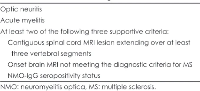
After identifying NMO-IgG, revised diagnostic criteria for definite NMO were proposed. These included ON, myelitis, and at least two of the following three supporting criteria: 1) MRI evidence of a contiguous spinal cord lesion extending over at least vertebral segments, 2) brain MRI at onset non- diagnostic for MS, and 3) NMO-IgG seropositivity (Table 1).6 CNS involvement beyond the optic nerves and spinal cord is compatible with NMO. This diagnostic combination was 99%
sensitive and 90% specific for NMO.6,86 An international task force recently recommended diagnostic criteria for NMO that included more detailed criteria for brain MRI results (Table 2).87
After several reports describing NMO-IgG seropositivity in up to 60% of cases with LEM, necrotic myelopathy, and re- current ON,88,89 the spectrum of disorders classified as NMO was expanded and now includes the variants summarized in Table 3.1
Table 1. Revised criteria for the diagnosis of NMO6 Optic neuritis
Acute myelitis
At least two of the following three supportive criteria:
Contiguous spinal cord MRI lesion extending over at least three vertebral segments
Onset brain MRI not meeting the diagnostic criteria for MS NMO-IgG seropositivity status
NMO: neuromyelitis optica, MS: multiple sclerosis.
Table 2. Diagnostic criteria of NMO*87
Major Criteria (all criteria are required, but may be separated by an unspecified interval) 1) Optic neuritis in one or more eyes
2) Transverse myelitis, clinically complete or incomplete, but associated with radiological evidence of spinal cord lesion extending over three or more spinal segments on T2-weighted MRI images and hypointensity on T1-weighted images when obtained during acute episode of myelitis
3) No evidence for sarcoidosis, vasculitis, clinically manifest systemic lupus erythematosus or Sjögren’s syndrome, or other explanation for the syndrome.
Minor criteria (at least one must be fulfilled)
1. Most recent brain MRI scan of the head must be normal or may show abnormalities not fulfilling Barkhof criteria used for McDonald diagnostic criteria, including†:
1) Non-specific brain T2 signal abnormalities not satisfying Barkhof criteria as outlined in McDonald criteria 2) Lesions in the dorsal medulla, either in contiguity or not in contiguity with a spinal cord lesion
3) Hypothalamic and/or brainstem lesions
4) “Linear” periventricular/corpus callosum signal abnormality, but not ovoid, and not extending into the parenchyma of the cerebral hemispheres in Dawson finger configuration
2. Positive test in serum or CSF for NMO-IgG/aquaporin-4 antibodies
*These criteria exclude limited or inaugural syndromes that may be NMO, such as recurrent transverse myelitis with longitudinally ex- tensive spinal cord lesions, or recurrent ON; further study is warranted to clarify their relationship with NMO, especially in the setting of seropositivity for NMO-IgG/AQP4 antibodies, †Periodic surveillance with brain MRI scanning is necessary to monitor for the emergence of new lesions that may lead to a revised diagnosis.
NMO: neuromyelitis optica, CSF: cerebrospinal fluid.
Management
The optimal treatment for NMO has not yet been established, because no randomized controlled trials have been perform- ed. Thus, treatment recommendations are based on small-siz- ed case series, case reports, or even individual clinical obser- vations.
Acute-phase treatment High-dose corticosteroids
For acute attacks, the traditional treatment is intravenous me- thylprednisolone at 1,000 mg/day for 3-5 consecutive days.
This recommendation is taken from studies of MS and idio- pathic ON; no controlled therapeutic trials have investigated the effectiveness of steroids specifically in NMO.90 Experience has indicated the need to continue steroids in the form of oral prednisone, with very slow tapering over months in the relaps- ing form of the disease. Without tapering, patients tend to re- lapse within weeks after an acute attack.64 Wingerchuck et al.14 reported that 55 of 69 NMO patients with acute attacks show- ed improvement after high-dose corticosteroid treatment. In cases with an unsatisfactory response, a repeated course of cor- ticosteroids may be considered.90
Therapeutic plasmapheresis
A small proportion of patients fail to improve with high-dose corticosteroid therapy, and their prognosis can become grave.91 In such cases, therapeutic plasmapheresis should be consider- ed. The removal of circulating autoantibodies, immune compl- exes, cytokines, and other inflammatory mediators is thought to be the principal mechanism of action for plasmapheresis.92 Plasmapheresis treatments include conventional plasma ex- change using centrifugation, double-filtration plasmapheresis, cryofiltration, and immunoadsorption.93 Most previous studies have used conventional plasma exchange.
Favorable outcomes have been obtained following thera- peutic plasma exchange for severe relapses of conventional MS and other devastating demyelinating disorders such as acu- te myelitis, acute disseminated encephalomyelitis (ADEM), NMO, and recurrent myelitis.94 However, there is a dearth of
data for NMO, because most previous studies have included a broad spectrum of CNS demyelination patients with only a few cases of NMO.95-100 In a retrospective study of 59 con- secutive patients that included 22 MS, 10 NMO, and 10 AD- EM patients, therapeutic plasma exchange was followed by moderate or marked functional improvement in 44.1% of the treated patients, which included 60% of the NMO patients.95 Seven of ten patients with severe isolated ON who were un- responsive to high-dose corticosteroids showed improvement of visual acuity, although their NMO-IgG or AQP4-Ab levels were not measured.96 In a study by Watanabe et al.97 six NMO- IgG-positive patients (three with ON and three with myelitis) who were unresponsive to high-dose intravenous methylpre- dnisolone received plasma exchange (three to five exchanges, each comprising 2-3 L), and three (one with ON and two with myelitis) experienced a definite functional improvement. Cli- nical improvement started to appear after one or two exch- anges.97 A retrospective study by Bonnan et al.98 included 96 severe spinal attacks in 43 Afro-Caribbean patients. As an add- on therapy for 29 attacks, plasma exchange was given daily for 5 days, with the exchange of one volume of plasma with 5%
albumin solution. The plasma-exchange-treated group showed improvements in neurological deficits.
Factors associated with moderate or marked improvement with plasma exchange were male gender, preserved reflexes, and early initiation of treatment.95 In addition, early initiation of plasma exchange and improvement at discharge were sig- nificantly associated with response at 6 months.99 Minor ad- verse events related to treatment in the course of plasma-ex- change sessions were deep hypofibrinogenemia, hematoma at the puncture site, benign vagal reaction, asymptomatic bac- teremia, abdominal syndrome, and extreme bradycardia.98 Pa- tients should be closely monitored for the usual risks associat- ed with long-lasting immobility secondary to severe myelitis attacks, including thromboembolic events and respiratory/uri- nary tract infections.86
In guidelines established by the European Federation of Neurological Societies, early initiation of a rescue therapy with plasmapheresis is indicated in cases that are unresponsive to high-dose corticosteroids, with up to seven treatments every other day.90 Therapeutic plasma exchange with seven exchan- Table 3. NMO spectrum1
NMO
Limited form of NMO
Idiopathic single or recurrent events of longitudinally extensive myelitis (≥3 vertebral segment spinal cord lesions seen on MRI) Optic neuritis: recurrent or simultaneous bilateral
Asian optic-spinal multiple sclerosis
Optic neuritis or longitudinally extensive myelitis associated with systemic autoimmune disease
Optic neuritis or myelitis associated with brain lesions typical of NMO (hypothalamic, corpus callosal, periventricular, or brainstem) NMO: neuromyelitis optica.
ges of approximately 55 mL/kg each, administered every other day for 14 days, should be considered. However, larger-scale studies are needed to determine the optimal number of plasma exchanges.
A trial of double-filtration plasmapheresis for NMO-IgG- positive patients was recently reported. In contrast to conven- tional plasma exchange, which uses one plasma separator fil- ter, double-filtration plasmapheresis uses two types of separa- tor filter. The second filter selectively removes high-mole- cular-weight proteins, including inflammatory cytokines.101 Eight patients were treated with double-filtration plasmaph- eresis for a maximum of seven times per month for 3 months, and 3,000-4,000 mL of plasma were exchanged at each ses- sion. Six of the eight patients (75%) showed therapeutic im- provement after double-filtration plasmapheresis treatment, with improvements of vision (two patients), movement (seven patients), and sensory function (four patients). However, the cli- nical status of the patients, especially whether they were in acute relapse, was not described.
Intravenous immunoglobulin
Intravenous immunoglobulin (IVIg) has been used for the tr- eatment of autoimmune disorders including autoimmune th- rombocytopenia, chronic inflammatory demyelinating poly- radiculoneuropathy, and Guillain-Barré syndrome.102 The best-understood actions of IVIg include 1) modulation of pathogenic autoantibodies, 2) inhibition of complement acti- vation and interception of membranolytic attack complex formation, 3) modulation of the inhibitory or activation Fc receptors on macrophages invading targeted tissues in nerve and muscle, 4) down-regulation of pathogenic cytokines and adhesion molecules, 5) suppression of T-cell functions, and 6) interference with antigen recognition.103 Considering the autoimmune mechanisms in the pathogenesis of NMO, IVIg could be a candidate treatment for NMO. However, IVIg has not yet been evaluated specifically for relapses of NMO and is rarely used for corticosteroid-refractory attacks.104
Prevention of relapses
Many patients with NMO were initially diagnosed as having MS, and treated with approved immunomodulatory therapies such as IFN-β and glatiramer acetate. However, exacerbations of severe ON and myelitis have been reported with IFNβ-1b treatment in Japanese OSMS patients.105,106 IFNβ-1a treatment was recently reported to have increased AQP4-Ab levels in a case of NMO.107
Azathioprine
Azathioprine, a purine antimetabolite, interferes with the pro- liferation of cells, and especially leukocytes,108 and is com-
monly used as an immunosuppressive drug in the treatment of MS.109 Azathioprine (2-3 mg/kg/day), usually in combina- tion with oral prednisone (1 mg/kg/day), is widely employed as a preventative therapy.86 In an observational case series, new- ly diagnosed NMO patients received a maintenance combin- ation treatment of prednisone (1 mg/kg/day) and azathioprine (2-3 mg/kg/day), with a gradual reduction of the prednisone dose after 2 months.110 The patients remained stable, with no attacks, for at least 18 months, and their disability scores im- proved significantly. Gastrointestinal complaints and leukope- nia are the most frequently reported adverse effects of azathio- prine therapy, and occur in more than 10% of patients. Infec- tions, allergy, anemia, thrombocytopenia, and pancytopenia are also common. Most of these adverse effects are easily manag- ed by dosage adjustment or therapy interruption; however, the risk for cancer increases with the treatment duration and cu- mulative dose.111
Low-dose corticosteroids
The results of a retrospective study suggested that there was a beneficial effect of low-dose corticosteroid monotherapy in reducing the incidence of relapse in NMO. The annual re- lapse rate was significantly lower during corticosteroid mo- notherapy than during the period without corticosteroid ther- apy. Relapses occurred significantly more often with corti- costeroid doses of ≤10 mg/day than with doses of >10 mg/
day.112 However, long-term treatment with corticosteroids of- ten causes various complications.
Mycophenolate mofetil
Mycophenolate mofetil (MMF) is a noncompetitive inhibitor of the enzyme inosine 5-monophosphate dehydrogenase. It controls lymphocyte proliferation and T-cell-dependent anti- body responses by inhibiting purine synthesis.113 The lym- phocyte-selective inhibitory effect of MMF presents an ad- vantage over azathioprine. In a case report of a 9-year-old girl who had a severe relapsing-remitting course of NMO de- spite treatment with azathioprine and corticosteroid, a drama- tic and sustained improvement occurred over a 2-year period of treatment with MMF.114 In a retrospective case series with prospective telephone follow-up, 24 patients with NMOSD were treated with MMF (median dose, 2,000 mg/day). The median annualized relapse rate was lower after treatment than before treatment, and disabilities stabilized or decreased in 22 of 24 patients (91%).115 MMF is well tolerated in general, and its typical side effects involve the gastrointestinal (diarrhea, nausea, vomiting, and abdominal cramps), hematologic (leu- kopenia, anemia, and thrombocytopenia), and genitourinary (urgency, frequency, dysuria, and sterile pyuria) systems. In contrast to azathioprine, MMF does not induce nephrotoxici-
ty, hepatotoxicity, or significant bone-marrow suppression.116 Mitoxantrone
Mitoxantrone hydrochloride is an anthracenedione antineo- plastic agent that has been approved as a treatment for sec- ondary progressive and worsening relapsing-remitting MS.
Mitoxantrone potentially suppresses Th lymphocytes and the humoral immune system via both macrophage and B-cell at- tenuation.117 It has been suggested that monthly intravenous infusion of mitoxantrone at 12 mg/m2 for 6 months followed by three additional treatments every 3 months is effective for the prevention of NMO relapses. Among five patients enroll- ed in a study of mitoxantrone treatment, two patients relaps- ed once within the initial 5 months of treatment, and clinical improvement was seen on MRI in four patients.118
In our experience with 20 frequently relapsing NMO pa- tients, the median annualized relapse rate was reduced by 75%, and 50% of patients became relapse-free during mitoxan- trone treatment. Disability improved or stabilized in all pa- tients. No patients had serious adverse effects during the mean follow-up period of 41 months after completing the therapy.
Flow cytometric analysis of cell-surface markers revealed that mitoxantrone treatment preferentially affected CD27+CD19+ memory B cells.119
Common adverse effects of mitoxantrone include leukope- nia, amenorrhea, elevated liver enzyme levels, nausea, alope- cia, bluish discoloration of the urine, and urinary tract infec- tions. The most serious adverse consequences of mitoxan- trone treatment, which occur infrequently, are cardiotoxicity, acute leukemia, and severe infection. The total dose of mito- xantrone should be limited to a cumulative dose of 100 mg/m2, since high doses have the potential to cause cardiotoxicity.120 One study found that therapy-related acute leukemia occurred in 0.3% of patients, and the median onset was at 18.5 months (range, 40-60 months) after the start of mitoxantrone treat- ment; among the 25 patients for which outcomes were re- ported, 6 died (24%). A relationship between total mitoxan- trone dose and acute leukemia is suggested, given that more than 80% of the patients who developed acute leukemia had received >60 mg/m2.121
Intravenous immunoglobulin
There are two reports regarding IVIg for the preventive thera- py of NMO. Two NMO patients with frequent attacks that were unresponsive to daily corticosteroids and azathioprine experienced no further attacks and showed improved neuro- logical status after monthly IVIg infusion; 60 g in one patient and initially 0.4 g/kg/day for 5 days followed by 1.0 g/kg/
day over 2 consecutive days per month for the other patient.122 In the other report, a female NMO patient treated with month-
ly IVIg infusion (0.4 g/kg/day for 1 day) did not experience a relapse for more than 4 years, and her neurologic status also improved during follow-up.123 Further studies assessing the ef- fectiveness and safety of IVIg as a relapse-preventing therapy for NMO are needed.
Rituximab
Since NMO is related to humoral immunity, rituximab may be an effective treatment option. Rituximab is a chimeric, murine/
human monoclonal antibody directed against the CD20 anti- gen expressed on pre-B cells and mature B cells. It causes de- pletion of B cells when administered in vivo. The possibility of rituximab as a therapy for NMO was first suggested by the results of an open-label study in which eight consecutive patients received four intravenous infusions of rituximab at 375 mg/m2, administered once per week.124 When the patients’
peripheral B-cell counts became detectable by fluorescence- activated cell sorting, they were given the option of repeated rituximab treatment. Six of the eight patients became relapse free, and the median attack rate declined dramatically. Seven patients experienced substantial recovery of neurological function during the follow-up period, which averaged 1 year.
In a retrospective analysis of 25 patients with NMO who were treated with rituximab,125 two rituximab regimens were used: 1) 375 mg/m2 infused once per week for 4 weeks (n=
18), and 2) 1,000 mg infused twice, with a 2-week interval be- tween infusions (n=4). The median annualized pretreatment relapse rate was 1.7, and the rate dropped to 0 at a median fol- low-up of 19 months. Disability improved or stabilized in 80%
of the patients. Two patients died during the follow-up period, one from a brainstem relapse and one due to suspected septi- cemia.
We recently completed a study of rituximab treatment in 30 NMOSD patients.126 The treatment protocol consisted of induction therapy (375 mg/m2 once weekly for 4 weeks, or 1,000 mg infused twice, with a 2-week interval between in- fusions), followed by maintenance therapy. The maintenance therapy was a single dose of rituximab at 375 mg/m2 admin- istered whenever re-emerging CD27+ memory B cells repre- sented more than 0.05% of peripheral blood mononuclear cells. Of the 30 NMOSD patients, 29 showed a marked re- duction in relapse rate after 24 months of rituximab treatment.
The relapse rate was reduced significantly by 88%, and 70%
of the patients became relapse-free over 24 months. Disability either improved or stabilized in 97% of the patients. AQP4- Ab levels declined significantly following treatment with rituximab, consistent with the clinical response and the effect on CD27+ memory B cells.
The side effects of rituximab include fever, chills, headache, asthenia, hypotension, bronchospasm, urticaria, and leukope-
nia.127 It has also been associated with progressive multifocal leukoencephalopathy, which was reported in three patients tr- eated for rheumatoid arthritis and two treated for SLE.128 How- ever, these patients had also received other immunosuppres- sive agents prior to or in conjunction with rituximab. The long- term side effects are uncertain.
Intermittent plasma exchange
The therapeutic efficacy of intermittent plasma exchange (dou- ble-filtration plasmapheresis or conventional plasma exch- ange) in combination with immunosuppressants to prevent re- currence was evaluated in two NMO patients, and the recurr- ence rate was significantly reduced after intermittent plasma exchange.129
Conclusion
Recent advances in clinical, neuroimaging, laboratory, and pa- thological hallmarks have established that NMO is a disease that is distinct from MS. Numerous clinical and experimental studies have implicated AQP4-Ab-mediated autoimmunity in the pathogenesis of NMO and provided a strong rationale for the use of therapies targeting humoral immunity in NMO.
The development of the AQP4-Ab test has led to the recog- nition of atypical presentations of NMO that are beyond the traditional view. These new insights into NMO enable us to make an early diagnosis of NMO and to apply appropriate immunotherapies in a wider spectrum of NMO patients.
A future avenue of investigation could include seronega- tive NMO, genetic susceptibility factors, and the development of surrogate markers for clinical efficacy and of promising animal models of NMO. An improved understanding of NMO will lead to the identification of an optimal treatment strategy for this debilitating disease.
Conflicts of Interest
The authors have no financial conflicts of interest.
Acknowledgements
This study was supported by grant A080588-28 from the Korean Health- care Technology R&D Project, Ministry for Health and Welfare, Repub- lid of Korea to HJK.
REFERENCES
1. Wingerchuk DM, Lennon VA, Lucchinetti CF, Pittock SJ, Weinsh- enker BG. The spectrum of neuromyelitis optica. Lancet Neurol 2007;6:805-815.
2. Allbutt T. On the ophthalmoscopic signs of spinal disease. Lancet 1870;1:76-78.
3. Devic E. Myelite subaigue compliquee de nevrite optique. Bull Med (Paris) 1894;8:1033-1034.
4. Lennon VA, Wingerchuk DM, Kryzer TJ, Pittock SJ, Lucchinetti CF, Fujihara K, et al. A serum autoantibody marker of neuromyelitis
optica: distinction from multiple sclerosis. Lancet 2004;364:2106- 2112.
5. Lennon VA, Kryzer TJ, Pittock SJ, Verkman AS, Hinson SR. IgG marker of optic-spinal multiple sclerosis binds to the aquaporin-4 water channel. J Exp Med 2005;202:473-477.
6. Wingerchuk DM, Lennon VA, Pittock SJ, Lucchinetti CF, Weinsh- enker BG. Revised diagnostic criteria for neuromyelitis optica. Neu- rology 2006;66:1485-1489.
7. Matiello M, Jacob A, Wingerchuk DM, Weinshenker BG. Neuro- myelitis optica. Curr Opin Neurol 2007;20:255-260.
8. Cabrera-Gómez JA, Kurtzke JF, González-Quevedo A, Lara-Rodrí- guez R. An epidemiological study of neuromyelitis optica in Cuba. J Neurol 2009;256:35-44.
9. Cabre P. Environmental changes and epidemiology of multiple scle- rosis in the French West Indies. J Neurol Sci 2009;286:58-61.
10. Bizzoco E, Lolli F, Repice AM, Hakiki B, Falcini M, Barilaro A, et al. Prevalence of neuromyelitis optica spectrum disorder and pheno- type distribution. J Neurol 2009;256:1891-1898.
11. Kira J. Multiple sclerosis in the Japanese population. Lancet Neurol 2003;2:117-127.
12. Cree BA, Khan O, Bourdette D, Goodin DS, Cohen JA, Marrie RA, et al. Clinical characteristics of African Americans vs Caucasian Americans with multiple sclerosis. Neurology 2004;63:2039-2045.
13. Papais-Alvarenga RM, Miranda-Santos CM, Puccioni-Sohler M, de Almeida AM, Oliveira S, Basilio De Oliveira CA, et al. Optic neuro- myelitis syndrome in Brazilian patients. J Neurol Neurosurg Psychi- atry 2002;73:429-435.
14. Wingerchuk DM, Hogancamp WF, O’Brien PC, Weinshenker BG.
The clinical course of neuromyelitis optica (Devic’s syndrome). Neu- rology 1999;53:1107-1114.
15. de Seze J, Lebrun C, Stojkovic T, Ferriby D, Chatel M, Vermersch P.
Is Devic’s neuromyelitis optica a separate disease? A comparative study with multiple sclerosis. Mult Scler 2003;9:521-525.
16. Davis R, Thiele E, Barnes P, Riviello JJ Jr. Neuromyelitis optica in childhood: case report with sequential MRI findings. J Child Neurol 1996;11:164-167.
17. Banwell B, Tenembaum S, Lennon VA, Ursell E, Kennedy J, Bar-Or A, et al. Neuromyelitis optica-IgG in childhood inflammatory demy- elinating CNS disorders. Neurology 2008;70:344-352.
18. McKeon A, Lennon VA, Lotze T, Tenenbaum S, Ness JM, Rensel M, et al. CNS aquaporin-4 autoimmunity in children. Neurology 2008;
71:93-100.
19. Filley CM, Sternberg PE, Norenberg MD. Neuromyelitis optica in the elderly. Arch Neurol 1984;41:670-672.
20. McAlpine D. Familial neuromyelitis optica: its occurrence in identi- cal twins. Brain 1938;61:430-438.
21. Ch’ien LT, Medeiros MO, Belluomini JJ, Lemmi H, Whitaker JN.
Neuromyelitis optica (Devic’s syndrome) in two sisters. Clin Elec- troencephalogr 1982;13:36-39.
22. Yamakawa K, Kuroda H, Fujihara K, Sato S, Nakashima I, Takeda A, et al. Familial neuromyelitis optica (Devic’s syndrome) with late onset in Japan. Neurology 2000;55:318-320.
23. Keegan M, Weinshenker B. Familial Devic’s disease. Can J Neurol Sci 2000;27:557-558.
24. Cabrera-Gómez JA, Ramón-Pérez L, Saiz A, Llerena-Fernández P, Fernández-Fernández L, Ercilla G, et al. Neuromyelitis optica and mul- tiple sclerosis in sisters. Mult Scler 2009;15:269-271.
25. Matiello M, Kim HJ, Kim W, Brum DG, Barreira AA, Kingsbury DJ, et al. Familial neuromyelitis optica. Neurology 2010;75:310-315.
26. Yamasaki K, Horiuchi I, Minohara M, Kawano Y, Ohyagi Y, Yamada T, et al. HLA-DPB1*0501-associated opticospinal multiple sclerosis:
clinical, neuroimaging and immunogenetic studies. Brain 1999;122:
1689-1696.
27. Ito H, Yamasaki K, Kawano Y, Horiuchi I, Yun C, Nishimura Y, et al.
HLA-DP-associated susceptibility to the optico-spinal form of mul-
tiple sclerosis in the Japanese. Tissue Antigens 1998;52:179-182.
28. Misu T, Fujihara K, Nakashima I, Miyazawa I, Okita N, Takase S, et al. Pure optic-spinal form of multiple sclerosis in Japan. Brain 2002;
125:2460-2468.
29. Wang H, Dai Y, Qiu W, Zhong X, Wu A, Wang Y, et al. HLA-DPB1 0501 is associated with susceptibility to anti-aquaporin-4 antibodies positive neuromyelitis optica in southern Han Chinese. J Neuroim- munol 2011;233:181-184.
30. Lucchinetti CF, Mandler RN, McGavern D, Bruck W, Gleich G, Ransohoff RM, et al. A role for humoral mechanisms in the patho- genesis of Devic’s neuromyelitis optica. Brain 2002;125:1450-1461.
31. Itoh T, Rai T, Kuwahara M, Ko SB, Uchida S, Sasaki S, et al. Identi- fication of a novel aquaporin, AQP12, expressed in pancreatic acinar cells. Biochem Biophys Res Commun 2005;330:832-838.
32. Pittock SJ, Weinshenker BG, Lucchinetti CF, Wingerchuk DM, Cor- boy JR, Lennon VA. Neuromyelitis optica brain lesions localized at sites of high aquaporin 4 expression. Arch Neurol 2006;63:964-968.
33. Amiry-Moghaddam M, Ottersen OP. The molecular basis of water transport in the brain. Nat Rev Neurosci 2003;4:991-1001.
34. Nielsen S, Nagelhus EA, Amiry-Moghaddam M, Bourque C, Agre P, Ottersen OP. Specialized membrane domains for water transport in glial cells: high-resolution immunogold cytochemistry of aquaporin-4 in rat brain. J Neurosci 1997;17:171-180.
35. Crane JM, Tajima M, Verkman AS. Live-cell imaging of aquaporin-4 diffusion and interactions in orthogonal arrays of particles. Neuro- science 2010;168:892-902.
36. Marnetto F, Hellias B, Granieri L, Frau J, Patanella AK, Nytrova P, et al. Western blot analysis for the detection of serum antibodies rec- ognizing linear Aquaporin-4 epitopes in patients with Neuromyelitis Optica. J Neuroimmunol 2009;217:74-79.
37. Mader S, Lutterotti A, Di Pauli F, Kuenz B, Schanda K, Aboul-Enein F, et al. Patterns of antibody binding to aquaporin-4 isoforms in neu- romyelitis optica. PLoS One 2010;5:e10455.
38. Weinshenker BG, Wingerchuk DM, Vukusic S, Linbo L, Pittock SJ, Lucchinetti CF, et al. Neuromyelitis optica IgG predicts relapse after longitudinally extensive transverse myelitis. Ann Neurol 2006;59:
566-569.
39. Matiello M, Lennon VA, Jacob A, Pittock SJ, Lucchinetti CF, Winger- chuk DM, et al. NMO-IgG predicts the outcome of recurrent optic neuritis. Neurology 2008;70:2197-2200.
40. Takahashi T, Fujihara K, Nakashima I, Misu T, Miyazawa I, Nakamu- ra M, et al. Anti-aquaporin-4 antibody is involved in the pathogenesis of NMO: a study on antibody titre. Brain 2007;130:1235-1243.
41. Jarius S, Aboul-Enein F, Waters P, Kuenz B, Hauser A, Berger T, et al.
Antibody to aquaporin-4 in the long-term course of neuromyelitis op- tica. Brain 2008;131:3072-3080.
42. Kim HJ, Li XF, Kim W, Kim SH, Choi K, Lee JE, et al. Quantitative measurement of anti-aquaporin-4 antibody by enzyme-linked immu- nosorbent assay using purified human aquaporin-4. [abstract P60].
Proceedings of 4th Congress of the Pan-Asian Committee for Treat- ment and Research in Multiple Sclerosis. Singapore, 2010;1290.
43. Misu T, Fujihara K, Kakita A, Konno H, Nakamura M, Watanabe S, et al. Loss of aquaporin 4 in lesions of neuromyelitis optica: distinc- tion from multiple sclerosis. Brain 2007;130:1224-1234.
44. Hinson SR, Pittock SJ, Lucchinetti CF, Roemer SF, Fryer JP, Kryzer TJ, et al. Pathogenic potential of IgG binding to water channel extra- cellular domain in neuromyelitis optica. Neurology 2007;69:2221- 2231.
45. Waters P, Jarius S, Littleton E, Leite MI, Jacob S, Gray B, et al. Aqua- porin-4 antibodies in neuromyelitis optica and longitudinally exten- sive transverse myelitis. Arch Neurol 2008;65:913-919.
46. Kinoshita M, Nakatsuji Y, Moriya M, Okuno T, Kumanogoh A, Na- kano M, et al. Astrocytic necrosis is induced by anti-aquaporin-4 an- tibody-positive serum. Neuroreport 2009;20:508-512.
47. Vincent T, Saikali P, Cayrol R, Roth AD, Bar-Or A, Prat A, et al.
Functional consequences of neuromyelitis optica-IgG astrocyte inter- actions on blood-brain barrier permeability and granulocyte recruit- ment. J Immunol 2008;181:5730-5737.
48. Takano R, Misu T, Takahashi T, Sato S, Fujihara K, Itoyama Y. Astro- cytic damage is far more severe than demyelination in NMO: a clin- ical CSF biomarker study. Neurology 2010;75:208-216.
49. Hinson SR, Roemer SF, Lucchinetti CF, Fryer JP, Kryzer TJ, Cham- berlain JL, et al. Aquaporin-4-binding autoantibodies in patients with neuromyelitis optica impair glutamate transport by down-regulating EAAT2. J Exp Med 2008;205:2473-2481.
50. Marignier R, Giraudon P, Vukusic S, Confavreux C, Honnorat J. An- ti-aquaporin-4 antibodies in Devic’s neuromyelitis optica: therapeutic implications. Ther Adv Neurol Disord 2010;3:311-321.
51. Kinoshita M, Nakatsuji Y, Kimura T, Moriya M, Takata K, Okuno T, et al. Neuromyelitis optica: Passive transfer to rats by human immu- noglobulin. Biochem Biophys Res Commun 2009;386:623-627.
52. Saadoun S, Waters P, Bell BA, Vincent A, Verkman AS, Papadopou- los MC. Intra-cerebral injection of neuromyelitis optica immunoglob- ulin G and human complement produces neuromyelitis optica lesions in mice. Brain 2010;133:349-361.
53. Bradl M, Misu T, Takahashi T, Watanabe M, Mader S, Reindl M, et al.
Neuromyelitis optica: pathogenicity of patient immunoglobulin in vivo. Ann Neurol 2009;66:630-643.
54. Bennett JL, Lam C, Kalluri SR, Saikali P, Bautista K, Dupree C, et al.
Intrathecal pathogenic anti-aquaporin-4 antibodies in early neuromye- litis optica. Ann Neurol 2009;66:617-629.
55. Sharma R, Fischer MT, Bauer J, Felts PA, Smith KJ, Misu T, et al. In- flammation induced by innate immunity in the central nervous system leads to primary astrocyte dysfunction followed by demyelination.
Acta Neuropathol 2010;120:223-236.
56. Kalluri SR, Rothhammer V, Staszewski O, Srivastava R, Petermann F, Prinz M, et al. Functional characterization of aquaporin-4 specific T cells: towards a model for neuromyelitis optica. PLoS One 2011;
6:e16083.
57. Jarius S, Franciotta D, Bergamaschi R, Wildemann B, Wandinger KP.
Immunoglobulin M antibodies to aquaporin-4 in neuromyelitis optica and related disorders. Clin Chem Lab Med 2010;48:659-663.
58. Pittock SJ, Lennon VA, de Seze J, Vermersch P, Homburger HA, Wingerchuk DM, et al. Neuromyelitis optica and non organ-specific autoimmunity. Arch Neurol 2008;65:78-83.
59. Pittock SJ, Weinshenker BG, Wingerchuk D, Lucchinetti CF, Lennon VA. Autoimmune neurological accompaniments of neuromyelitis optica (NMO). Ann Neurol 2006;60:S41.
60. Furukawa Y, Yoshikawa H, Yachie A, Yamada M. Neuromyelitis op- tica associated with myasthenia gravis: characteristic phenotype in Japanese population. Eur J Neurol 2006;13:655-658.
61. Kister I, Gulati S, Boz C, Bergamaschi R, Piccolo G, Oger J, et al. Ne- uromyelitis optica in patients with myasthenia gravis who underwent thymectomy. Arch Neurol 2006;63:851-856.
62. Antoine JC, Camdessanché JP, Absi L, Lassablière F, Féasson L. De- vic disease and thymoma with anti-central nervous system and anti- thymus antibodies. Neurology 2004;62:978-980.
63. Magaña SM, Pittock SJ, Lennon VA, Keegan BM, Weinshenker BG, Lucchinetti CF. NMO-IgG status in fulminant CNS inflammatory de- myelinating disorders (IDD). Neurology 2007;68:A160. Abstract.
64. Mandler RN. Neuromyelitis optica Devic’s syndrome, update. Auto- immun Rev 2006;5:537-543.
65. Wingerchuk DM, Weinshenker BG. Neuromyelitis optica: clinical predictors of a relapsing course and survival. Neurology 2003;60:
848-853.
66. Kim W, Kim SH, Lee SH, Li XF, Kim HJ. Brain abnormalities as an initial manifestation of neuromyelitis optica spectrum disorder. Mult Scler 2011;17:1107-1112.
67. Lotze TE, Northrop JL, Hutton GJ, Ross B, Schiffman JS, Hunter JV.
Spectrum of pediatric neuromyelitis optica. Pediatrics 2008;122: