Introduction
Aortic stenosis (AS) is a highly and increasingly prevalent condition that has become a major health concern.1) Characteris- tically, patients present a long latent asymptomatic period where the risk of sudden death is low, even with severe AS: i.e., peak aortic velocity ≥ 4 m/s, mean aortic gradient ≥ 40 mmHg and aortic valve area ≤ 1.0 cm2 or ≤ 0.60 cm2/m2. However, the risk of sudden death increases dramatically when symptoms ap- pear justifying the European Society of Cardiology/European Association of Cardiothoracic Surgery (ESC/EACTS) to under- line the role of exercise testing to clarify symptomatic status in patients with severe AS.2) On the other side, “truly” asymptom- atic patients undergoing early aortic valve replacement (AVR) may have better outcome compared to medically treated pa- tients.3) Hence, the determination of individual risk factors of rapid clinical deterioration could help identify patients who
may benefit most from early elective surgery. Accordingly, re- cent studies have demonstrated that exercise echocardiography can provide incremental prognostic value over resting echocar- diography and exercise testing.4-6) Exercise echocardiography is useful because it allows assessing clinical, hemodynamic and functional adaptation responses during exercise, which are di- rectly linked to functional status, degree of ventriculo-arterial coupling and left ventricular (LV) myocardial reserve. The pur- pose of this article is to describe the role of exercise testing and echocardiography in the management of asymptomatic patients with severe AS and preserved LV ejection fraction.
Exercise Testing Protocol
A complete clinical evaluation to rule out the presence of REVIEW J Cardiovasc Ultrasound 2014;22(1):1-5
Exercise Echocardiography
in Asymptomatic Patients with Severe Aortic Stenosis and Preserved Left Ventricular Ejection Fraction
Christine Henri, MD and Patrizio Lancellotti, MD, PhD, FESC, FACC
Department of Cardiology and Heart Valve Clinic, GIGA Cardiovascular Sciences, University of Liège, CHU Sart Tilman, Liège, Belgium
The management of asymptomatic patients with severe aortic stenosis (AS) remains controversial. Recent series reported that early aortic valve replacement might be associated with improved clinical outcomes. However, the risk-benefit ratio should be carefully evaluated and early surgery only be proposed to a subset of asymptomatic patients considered at higher risk. Exercise echocardiography can help unmask symptomatic patients combined with assessment of the hemodynamic consequences of AS.
Recent studies have demonstrated that exercise echocardiography can provide incremental prognostic value to identify patients who may benefit most from early surgery. In “truly” asymptomatic patients, an increase in mean aortic gradient ≥ 18–20 mmHg, a limited left ventricular contractile reserve or a pulmonary hypertension during exercise are predictive parameters of adverse cardiac events. Exercise echocardiography is low-cost, safe and available in many referral centers, and does not expose patients to radiation. The purpose of this article is to describe the role of exercise testing and echocardiography in the management of asymptomatic patients with severe AS and preserved left ventricular ejection fraction.
KEY WORDS: Aortic stenosis · Exercise testing · Exercise echocardiography.
• Received: January 29, 2014 • Revised: February 23, 2014 • Accepted: February 23, 2014
• Address for Correspondence: Patrizio Lancellotti, Department of Cardiology, University Hospital, Université de Liège, CHU du Sart Tilman, 4000 Liège, Belgium Tel: +32-4-366-7194, Fax: +32-4-366-7195, E-mail: [email protected]
• This is an Open Access article distributed under the terms of the Creative Commons Attribution Non-Commercial License (http://creativecommons.org/licenses/by-nc/3.0) which permits unrestricted non-commercial use, distribution, and reproduction in any medium, provided the original work is properly cited.
symptoms and to identify potential contraindications is essen- tial before submitting patients to exercise testing. Contraindica- tions to exercise testing include: clear indications for AVR (i.e., symptomatic severe AS), uncontrolled hypertension (systolic pressure > 220 mmHg or diastolic pressure > 110 mmHg), un- controlled or symptomatic arrhythmias, physical or mental dis- ability with the inability to adequately exercise and systemic disease limiting exercise performance.7) A symptom-limited exercise test performed with the goal to reach at least 85% of the age-predicted heart rate is recommended. Treadmill or semi-supine bicycle exercise testing can be used. Safety of both techniques has already been demonstrated and complica- tions remain low under appropriate supervision and monitor- ing.8)9) Treadmill exercise is more commonly used in North America and is realized according to the ACC/AHA practice guidelines using a modified Bruce protocol.10) In contrast, semi- supine ergometer with a tilting table is the preferred approach in Europe reducing the potential risk of hemodynamic col- lapse compared to treadmill test.11) Patients should continue their usual medications, as abnormal results on suboptimal therapy may be confusing for management decisions. The workload should be adjusted for each patient, i.e., beginning at 50 W with an increase of 25 W every 2 minutes for a young patient versus starting at 25 W with an increase of 10 W ev- ery 2 minutes for an older patient. Appearance of symptoms should be assessed regularly, and blood pressure, heart rate and 12-lead electrocardiography should be monitored continuously during the examination. Exercise testing should be interrupted when the target heart rate is reached or if the patient presents typical chest pain, limiting breathlessness, dizziness, hypoten- sion (drop in systolic blood pressure ≥ 20 mmHg), significant ventricular arrhythmia or muscular exhaustion.
Interpretation
Symptomatic status can be difficult to establish because pa- tients may minimize or deny their symptoms or reduce their level of physical activity to avoid them, especially in elderly.
Then, exercise testing can be useful to unmask symptoms in pa- tients with severe AS. Approximately one third of patients who claim to be asymptomatic will develop symptoms on exercise testing.12)13) However, the occurrence of rapidly reversible dys- pnea at high workloads should not be interpreted as abnormal.
Interpretation of exercise testing could be limited in elderly population. In fact, positive predictive value in patients > 70 years old has been shown to be significantly lower than in younger patients (56% compared to 79%) with similar nega- tive predictive values.13) According to this, a negative exercise test should be reassuring, but a positive exercise test may lack of specificity due to frequent comorbidities in elderly patients.
Also, ST segment depression may not improve the positive pre- dictive value of exercise testing, particularly in patients with concomitant coronary artery disease.13) Generally, exercise test- ing is considered positive when the patient presents ≥ 1 of the
following criteria: angina, limiting dyspnea at low workloads, syncope or near-syncope, ≥ 2 mm horizontal or down-slopping ST segment depression, drop or ≥ 20 mmHg rise in systolic blood pressure or complex ventricular arrhythmias.7)
Impact on clinical decision-making
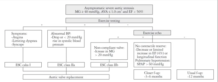
Symptom-limited exercise testing can add important prog- nostic value.14) For this reason, exercise testing is strongly advo- cated by the ESC/EACTS (Class I recommendation) in asymp- tomatic patients with severe AS.2) In fact, a positive exercise test has been shown to predict the rapid onset of symptoms, the oc- currence of cardiac death and the need for AVR. To note, exer- cise-induced dizziness have the highest positive predictive value for the occurrence of symptoms during follow-up.12) In a meta- analysis examining 491 patients, a negative exercise test was as- sociated with no sudden death while 5% of patients with a pos- itive result presented with sudden death during 12-month follow-up. Also, 21% of patients with a negative exercise test had adverse cardiac events compared to 66% of patients with a positive result.14) Therefore, current ESC/EACTS guidelines rec- ommend AVR in asymptomatic patients with severe AS who develop symptoms during exercise testing (ESC/EACTS, Class I) or a fall in systolic blood pressure below baseline value (ESC/
EACTS, Class IIa) (Fig. 1).2)
Exercise Echocardiography Protocol
In valvular heart disease, an experienced sonographer or car- diologist should perform exercise echocardiography. Both types of exercise test can be used. Treadmill allows only post- exercise imaging limiting the accuracy of measurements com- pared to semi-supine cyclo-ergometer permitting optimal im- age acquisitions during each step of exercise testing. Comp- rehensive resting echocardiography should be performed in the same position as during the exercise testing. Echocardio- graphic parameters related to the severity of AS, the conse- quences on the LV and the systolic pulmonary arterial pressure (SPAP) should be recorded throughout the test. To note, the ratio of early diastolic mitral inflow velocity to early diastolic annulus velocity (E/e’ ratio) should be measured before E and A wave fusion appearing at higher heart rates (usually > 100–
110 bpm) (Fig. 2).
Interpretation
Changes in mean aortic gradient
Regardless of resting AS severity, an increase in mean aortic pressure gradient by ≥ 18–20 mmHg during exercise, which occur in about one-third of patients, is associated with an in- creased risk of cardiac-related events.4)6) In fact, an exercise-in- duced increase in mean aortic pressure gradient by ≥ 20 mmHg has been identified as the most powerful predictor of
poor outcomes, even after adjustment for age, exercise LV ejec- tion fraction and resting mean aortic gradient.6) Such an in- crease in mean aortic pressure gradient partly reflects the pres- ence of a rigid and non-compliant aortic valve.15) Moreover, the kinetics of change in mean aortic pressure gradient needs to be assessed as a rapid increase may indicate the presence of a more severe disease.
LV systolic functional reserve
Assessment of LV function during exercise can also provide incremental prognostic information. Limited contractile reserve (i.e., a decrease or a limited increase in LV ejection fraction) has been shown to be associated with an abnormal hemodynamic response to exercise, the development of symptoms and cardio- vascular death.16)17) Limited contractile reserve may represent a more advanced disease process with subclinical intrinsic myo-
cardial damage. The afterload mismatch and the exhausted cor- onary flow reserve with consequent subendocardial ischemia and myocardial fibrosis could contribute to limited functional LV adaptation during test.18) As previously reported, standard LV ejection fraction measurements are relatively insensitive to detect early forms of myocardial dysfunction and assessment of LV longitudinal function seems to be more powerful in predict- ing the occurrence of symptoms, exercise intolerance and out- come in AS.19)20) Longitudinal function can be studied using tis- sue Doppler imaging with pulsed-wave Doppler at the mitral annulus measuring s’ velocities or using 2-dimentional speckle tracking analyzing global longitudinal strain (GLS) (Fig. 3). In a cohort of AS patients, a smaller increase in s’ velocities after exercise was associated with a lower exercise capacity and a low- er exercise-induced increase in systolic blood pressure.21) Accord- ing to strain analysis, Lafitte et al.19) demonstrated that patients
Fig. 1. Impact on clinical decision-making of exercise testing and exercise echocardiography in asymptomatic patients with severe aortic stenosis and preserved left ventricular function. MG: mean aortic pressure gradient, AVA: aortic valve area, BP: blood pressure, EF: ejection fraction, SPAP:
systolic pulmonary artery pressure, ESC: European Society of Cardiology.
Fig. 2. Exercise echocardiography protocol including the sequence of acquisition. *Should be measured at low-level exercise before E and A wave fusion. CW: continuous-wave Doppler, fps: frame per second, LV: left ventricle, PW: pulsed-wave Doppler, SPAP: systolic pulmonary artery pressure, TDI: tissue Doppler imaging, TR: tricuspid regurgitation.
Asymptomatic severe aortic stenosis MG ≥ 40 mmHg, AVA ≤ 1.0 cm2 and EF > 50%
Symptoms:
-Angina -Limiting dyspnea -Syncope
Abnormal BP:
-Drop or < 20 mmHg rise in systolic blood pressure
Non-compliant valve:
-Icrease in MG > 20 mmHg
No contractile reserve:
-Decrease or limited increase in EF (4%) or longitudinal function Pulmonary hypertension:
SPAP > 60 mmHg Closer f-up:
-3–6 months Usual f-up:
-12 months Exercise testing
Exercise echo
ESC calss I
Aortic valve replacement
ESC class IIa ESC class IIb
+ + -
+ + -
Complete resting echocardiography
Mean aortic gradient Aortic valve area CW aortic valve PW LV outflow tract SPAP CW TR jet E/Ea ratio*
PW mitral inflow TDI PW mitral annulus
LV function
2-D grey-scale images in 4, 2, and 3 chambers view (> 60 fps)
Mean aortic gradient Aortic valve area SPAP
Baseline 25 W 50 W 75 W 100 W
with an abnormal exercise response had a significant lower rest- ing GLS and basal longitudinal strain (BLS) compared to pa- tients with a normal exercise test (respectively -14.7% vs.
-19.3% and -10.7% vs. -14.4%). Cutoff values of -18% for GLS and -13% for BLS were able to predict an inadequate exer- cise response with a sensitivity/specificity of 68/75% (GLS) and 77/83% (BLS). Moreover, Donal et al.22) reported that exer- cise-induced changes in LV GLS were lower in AS patients with an abnormal exercise response. In multivariable analysis, a lower resting GLS, a higher exercise-induced increase in mean aortic pressure gradient and a smaller exercise-induced change in GLS were predictors of a positive exercise test.
LV diastolic functional reserve
In patients with severe AS, the chronically increased after-
load is compensated by LV hypertrophy. At a later stage, this adaptive response may be no longer sufficient to mitigate the clinical impact of an increase in LV filling pressure. The E/e’
ratio is recognized as a noninvasive estimate of LV filling pres- sure. At rest, a cutoff value of E/e’ > 13 has been shown to iden- tify LV end-diastolic pressure > 15 mmHg with a sensitivity of 93% and a specificity of 88%.23) At exercise, cutoff values of E/e’
between 13 and 15 were correlated with elevated LV end-dia- stolic pressure during exercise and reduced exercise capacity in patients without valvular disease.24)25) However, these exercise values have not been validated in AS patients.
Changes in systolic pulmonary artery pressure
In asymptomatic patients, exercise echocardiography may unmask dynamic pulmonary hypertension (PHT). In fact, an early exercise-induced increase in SPAP is in favour of a lack of pulmonary vascular adaptation with a high resistance and a low compliance reflecting a more severe disease. In a recent prospective study, exercise-induced PHT, defined as SPAP >
60 mmHg, was associated with a 2-fold increase risk of cardi- ac events at 3-year follow-up in asymptomatic patients with severe AS and preserved LV ejection fraction. Also, in multi- variable model, exercise PHT was identified as an indepen- dent predictor of cardiac events, even when exercise-induced changes in mean aortic gradient were added.5)
Impact on clinical decision-making
Because of its incremental prognostic value, exercise echo- cardiography may be useful to improve risk stratification and identify patients who may benefit from an early surgery. Ac- cording to this, the ESC/EACTS recently added a recommen- dation based on exercise echocardiography. In asymptomatic patients with severe AS and preserved LV ejection fraction, AVR may be considered in patients with an exercise-induced increase in mean aortic pressure gradient > 20 mmHg (Class IIb).2) Otherwise, patients with a significant increase in mean aortic pressure gradient, a limited LV contractile reserve or PHT during exercise could be referred to a dedicated Heart Valve Clinic for a closer follow-up (3–6 months) including re- evaluation of symptoms and repeated exercise echocardiogra- phy.26) Conversely, patients without markers of poor prognosis may be safely reassessed every 12 months (Fig. 1). However, prospective clinical trials are needed to support the widespread use of exercise echocardiography in the routine management of asymptomatic AS.
Conclusion
In AS, exercise echocardiography can help unmask falsely asymptomatic patients as well as patients with abnormal he- modynamic and functional adaptation to exercise who may benefit from early elective AVR. Large-scale studies are need- ed to examine the impact of exercise echocardiography results on post-operative outcome.
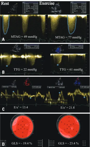
Fig. 3. Example of an asymptomatic patient with severe aortic stenosis presenting a significant exercise-induced increase in mean trans-aortic pressure gradient (A) and exercise pulmonary hypertension (B). In addition, a significant exercise-induced increase in estimated left ventricular filling pressure (C) but a normal contractile reserve assessed by 2-D speckle tracking analyzing global longitudinal strain (D) was observed. MTAG: mean aortic pressure gradient, TTG: trans-tricuspid pressure gradient, GLS: global longitudinal strain.
A
B
C
D
• Acknowledgements
Christine Henri received grants from the Montreal Heart Institute Foundation (Bourse du Bal du Cœur), the Department of Medicine of the University of Montreal and the Association des Cardiologues du Québec P.L. This work was supported by the Belgian National Fund for Scientific Research (F.R.S-FNRS T.0028.14).
References
1. Iung B, Baron G, Butchart EG, Delahaye F, Gohlke-Bärwolf C, Levang OW, Tornos P, Vanoverschelde JL, Vermeer F, Boersma E, Ravaud P, Vahanian A. A prospective survey of patients with valvular heart disease in Europe: The Euro Heart Survey on Valvular Heart Disease.
Eur Heart J 2003;24:1231-43.
2. Joint Task Force on the Management of Valvular Heart Disease of the European Society of Cardiology (ESC); European Association for Cardio-Thoracic Surgery (EACTS), Vahanian A, Alfieri O, Andreot- ti F, Antunes MJ, Barón-Esquivias G, Baumgartner H, Borger MA, Carrel TP, De Bonis M, Evangelista A, Falk V, Iung B, Lancellotti P, Pierard L, Price S, Schäfers HJ, Schuler G, Stepinska J, Swedberg K, Takkenberg J, Von Oppell UO, Windecker S, Zamorano JL, Zem- bala M. Guidelines on the management of valvular heart disease (version 2012). Eur Heart J 2012;33:2451-96.
3. Brown ML, Pellikka PA, Schaff HV, Scott CG, Mullany CJ, Sundt TM, Dearani JA, Daly RC, Orszulak TA. The benefits of early valve re- placement in asymptomatic patients with severe aortic stenosis. J Thorac Cardiovasc Surg 2008;135:308-15.
4. Lancellotti P, Lebois F, Simon M, Tombeux C, Chauvel C, Pierard LA. Prognostic importance of quantitative exercise Doppler echocardiography in asymptomatic valvular aortic stenosis. Circulation 2005;112(9 Suppl):
I377-82.
5. Lancellotti P, Magne J, Donal E, O’Connor K, Dulgheru R, Rosca M, Pierard LA. Determinants and prognostic significance of exercise pulmo- nary hypertension in asymptomatic severe aortic stenosis. Circulation 2012;126:851-9.
6. Maréchaux S, Hachicha Z, Bellouin A, Dumesnil JG, Meimoun P, Pasquet A, Bergeron S, Arsenault M, Le Tourneau T, Ennezat PV, Pibarot P. Usefulness of exercise-stress echocardiography for risk stratifica- tion of true asymptomatic patients with aortic valve stenosis. Eur Heart J 2010;31:1390-7.
7. Lancellotti P, Magne J, Piérard LA. The role of stress testing in evalua- tion of asymptomatic patients with aortic stenosis. Curr Opin Cardiol 2013;28:531-9.
8. Sicari R, Nihoyannopoulos P, Evangelista A, Kasprzak J, Lancellotti P, Poldermans D, Voigt JU, Zamorano JL; European Association of Echocardiography. Stress Echocardiography Expert Consensus Statement-- Executive Summary: European Association of Echocardiography (EAE) (a registered branch of the ESC). Eur Heart J 2009;30:278-89.
9. Pellikka PA, Nagueh SF, Elhendy AA, Kuehl CA, Sawada SG;
American Society of Echocardiography. American Society of Echocar- diography recommendations for performance, interpretation, and application of stress echocardiography. J Am Soc Echocardiogr 2007;20:1021-41.
10. Gibbons RJ, Balady GJ, Bricker JT, Chaitman BR, Fletcher GF, Froelicher VF, Mark DB, McCallister BD, Mooss AN, O’Reilly MG, Winters WL Jr, Gibbons RJ, Antman EM, Alpert JS, Faxon DP, Fuster V, Gregoratos G, Hiratzka LF, Jacobs AK, Russell RO, Smith SC Jr; American College of Cardiology/American Heart As- sociation Task Force on Practice Guidelines (Committee to Update the 1997 Exercise Testing Guidelines). ACC/AHA 2002 guideline update for exercise testing: summary article: a report of the American College of Cardiology/American Heart Association Task Force on Practice Guide-
lines (Committee to Update the 1997 Exercise Testing Guidelines). Circu- lation 2002;106:1883-92.
11. Picano E, Pibarot P, Lancellotti P, Monin JL, Bonow RO. The emerg- ing role of exercise testing and stress echocardiography in valvular heart dis- ease. J Am Coll Cardiol 2009;54:2251-60.
12. Amato MC, Moffa PJ, Werner KE, Ramires JA. Treatment decision in asymptomatic aortic valve stenosis: role of exercise testing. Heart 2001;86:
381-6.
13. Das P, Rimington H, Chambers J. Exercise testing to stratify risk in aortic stenosis. Eur Heart J 2005;26:1309-13.
14. Rafique AM, Biner S, Ray I, Forrester JS, Tolstrup K, Siegel RJ.
Meta-analysis of prognostic value of stress testing in patients with asymp- tomatic severe aortic stenosis. Am J Cardiol 2009;104:972-7.
15. Leurent G, Donal E, de Place C, Chabanne C, Gervais R, Fougerou C, le Helloco A, Daubert JC, Mabo P, Laurent M. Argument for a Doppler echocardiography during exercise in assessing asymptomatic patients with severe aortic stenosis. Eur J Echocardiogr 2009;10:69-73.
16. Lancellotti P, Karsera D, Tumminello G, Lebois F, Piérard LA. De- terminants of an abnormal response to exercise in patients with asymptomatic valvular aortic stenosis. Eur J Echocardiogr 2008;9:338-43.
17. Maréchaux S, Ennezat PV, LeJemtel TH, Polge AS, de Groote P, Asseman P, Nevière R, Le Tourneau T, Deklunder G. Left ventricular response to exercise in aortic stenosis: an exercise echocardiographic study.
Echocardiography 2007;24:955-9.
18. Lancellotti P, Donal E, Magne J, Moonen M, O’Connor K, Daubert JC, Pierard LA. Risk stratification in asymptomatic moderate to severe aor- tic stenosis: the importance of the valvular, arterial and ventricular inter- play. Heart 2010;96:1364-71.
19. Lafitte S, Perlant M, Reant P, Serri K, Douard H, DeMaria A, Rou- daut R. Impact of impaired myocardial deformations on exercise tolerance and prognosis in patients with asymptomatic aortic stenosis. Eur J Echocar- diogr 2009;10:414-9.
20. Lancellotti P, Moonen M, Magne J, O’Connor K, Cosyns B, Attena E, Donal E, Pierard L. Prognostic effect of long-axis left ventricular dys- function and B-type natriuretic peptide levels in asymptomatic aortic stenosis.
Am J Cardiol 2010;105:383-8.
21. Van Pelt NC, Stewart RA, Legget ME, Whalley GA, Wong SP, Zeng I, Oldfield M, Kerr AJ. Longitudinal left ventricular contractile dysfunction after exercise in aortic stenosis. Heart 2007;93:732-8.
22. Donal E, Thebault C, O’Connor K, Veillard D, Rosca M, Pierard L, Lancellotti P. Impact of aortic stenosis on longitudinal myocardial deforma- tion during exercise. Eur J Echocardiogr 2011;12:235-41.
23. Bruch C, Stypmann J, Grude M, Gradaus R, Breithardt G, Wichter T. Tissue Doppler imaging in patients with moderate to severe aortic valve stenosis: clinical usefulness and diagnostic accuracy. Am Heart J 2004;148:
696-702.
24. Burgess MI, Jenkins C, Sharman JE, Marwick TH. Diastolic stress echocardiography: hemodynamic validation and clinical significance of esti- mation of ventricular filling pressure with exercise. J Am Coll Cardiol 2006;47:1891-900.
25. Grewal J, McCully RB, Kane GC, Lam C, Pellikka PA. Left ventric- ular function and exercise capacity. JAMA 2009;301:286-94.
26. Lancellotti P, Rosenhek R, Pibarot P, Iung B, Otto CM, Tornos P, Donal E, Prendergast B, Magne J, La Canna G, Piérard LA, Maurer G. ESC Working Group on Valvular Heart Disease position paper--heart valve clinics: organization, structure, and experiences. Eur Heart J 2013;
34:1597-606.