* Corresponding author: Jae-Ho Shin
Department of Biomedical Laboratory Science, Eulji University, 553 Sanseong-daero, Sujeong-gu, Sungnam 13135, Korea
E-mail: [email protected]
* ORCID: https://orcid.org/0000-0002-2141-1090
ORIGINAL ARTICLE
A Study on the Protective Effect of Antioxidants on
Damage Induced by Liver Ischemia/Repefusion in a Rat Model
Yong Ho Ahn 1 , Pu Reum Seok 2 , Su Jin Oh 2 , Jin Woo Choi 2 , Jae-Ho Shin 2
1
Department of Clinical Laboratory Science, Dongnam Health University, Suwon, Korea
2
Department of Biomedical Laboratory Science, Eulji University, Sungnam, Korea
모델 랫드에 간 허혈/재관류로 유발된 손상에 대한 항산화제의 보호 효과에 관한 연구
안용호 1 , 석푸름 2 , 오수진 2 , 최진우 2 , 신재호 2
1
동남보건대학교 임상병리과,
2을지대학교 임상병리학과
ARTICLE INFO ABSTRACT
Received
June 26, 2019Revised
July 22, 2019Accepted
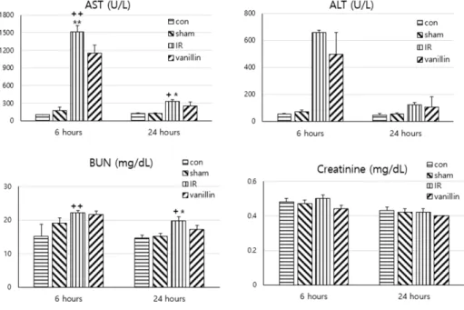
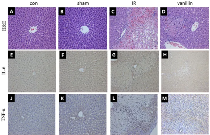
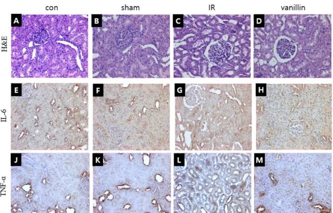
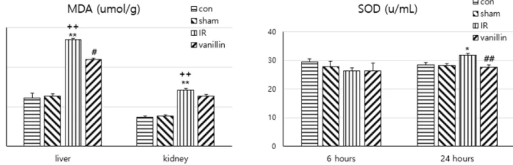
August 4, 2019The hepatic ischemic model has recently been widely used for the epidemiological study of ischemic reperfusion injury. This study was carried out to investigate the protective effect of vanillin, which is known to have antioxidant and anti-inflammatory effects, against hepatic and renal injury using an ischemia-reperfusion rat model, and we also investigated the mechanism related to vanillins’ protective effect. The test material was administered at a concentration of 100 mg/kg for 3 days, followed by ligation of the liver for 60 minutes to induce ischemia reperfusion. As control groups, there was a negative control, sham control and ischemia-reperfusion-only ischemia reperfusion control, and the controls groups were compared with the drug administration group. In the vanillin group, aspartate aminotransferase (AST) and alanine aminotransferase (ALT) activities were significantly inhibited compared with the AST and ALT activities of the ischemia-reperfusion group, and histopathological examination showed significant reduction of both inflammation and necrosis. The malondialdehyde (MDA) and superoxide dismutase (SOD) levels were significantly different from the ischemia-reperfusion group. In conclusion, vanillin showed a hepatocyte protective action by alleviating the cellular inflammation and cell necrosis caused by hepatic ischemia-reperfusion, and vanillin mitigated inflammatory changes in the kidney glomeruli and distal tubules. The protective effect is considered to be caused by vanillin’s antioxidant function.
Further studies such as on cell death and possibly vanillin’s same effect on damaged tissue will be necessary for clinical applications such as organ transplantation.
Copyright © 2019 The Korean Society for Clinical Laboratory Science. All rights reserved.
Key words Ischemia-reperfusion Liver
Kidney Rat Vanillin
INTRODUCTION
Postoperative dysfunction and death are still serious problems, and the main cause is ischemia-reperfusion injury during surgery [1, 2]. It is reported that massive bleeding during surgical resection of hepatocellular carcinoma results in unstable cardiovascular events, and
Korean Society for Clinical Laboratory Science