Patients’ Satisfaction after Reverse Total Shoulder Arthroplasty Is Affected by Preoperative Functional Status
Jong Pil Yoon, Dong-Hyun Kim, Seok Won Chung1
Department of Orthopedic Surgery, Kyungpook University School of Medicine, Daegu, 1Department of Orthopedic Surgery, Konkuk University School of Medicine, Seoul, Korea
Background: The purpose of this study is to evaluate the functional outcomes of reverse total shoulder arthroplasty (RTSA) and to assess factors affecting the patients’ subjective satisfaction after RTSA.
Methods: Forty-three patients (mean age, 75.0 ± 5.2 years) who underwent RTSA for cuff tear arthropathy or irreparable cuff tears with preoperative magnetic resonance imaging and pre- and postoperative radiographs at 1 year, and whose various functional outcomes in- cluding pain visual analogue scale (VAS), simple shoulder test, Constant score, American Shoulder and Elbow Surgeons score, and active range of motion were evaluated preoperatively and at the last follow-up (>12 months) were enrolled. The outcome parameter was set as a satisfaction scale. Various clinical and radiographic factors were analyzed, and their correlations with postoperative satisfaction were evaluated.
Results: All functional scores, VAS pain score, and active forward flexion showed significant improvement after surgery (all p<0.001).
Twenty-nine patients were satisfied with the results and 14 were dissatisfied. The presence of pseudoparalysis (p=0.028) and worse preoperative function (all p<0.05) were related with higher satisfaction. Any radiologic parameters did not affect patients’ postoperative satisfaction.
Conclusions: All patients showed a good functional outcome after RTSA, however the patients’ subjective postoperative satisfaction was affected by preoperative functional status (higher satisfaction in poor preoperative function), not by radiological findings.
(Clin Shoulder Elbow 2016;19(3):119-124)
Key Words: Reverse total shoulder arthroplasty; Satisfaction; Preoperative function
Copyright © 2016 Korean Shoulder and Elbow Society. All Rights Reserved. pISSN 2383-8337
Clinics in Shoulder and Elbow Vol. 19, No. 3, September, 2016 http://dx.doi.org/10.5397/cise.2016.19.3.119
Received September 17, 2015. Revised November 26, 2015. Accepted November 30, 2015.
Correspondence to: Seok Won Chung
Department of Orthopedic Surgery, Konkuk University School of Medicine, 120-1 Neungdong-ro, Gwangjin-gu, Seoul 05030, Korea Tel: +82-2-2030-7604, Fax: +82-2-2030-7748, E-mail: [email protected]
IRB approval (No. KUH1060101).
Financial support: This work was supported by Institute for Information & Communications Technology Promotion (IITP) grant funded by the Korea government (MSIP) (B0101-15-1081). Conflict of interests: None.
Introduction
Reverse total shoulder arthroplasty (RTSA) is a treatment op- tion for patients with irreparable massive rotator cuff tears and cuff tear arthropathy.1) The modern RTSA is based on the design described by Grammont and Baulot,1) in which the center of joint rotation is moved medially and distally to maximize the lever arm and tension of the deltoid as well as the recruited portion of the deltoid for compensation of the dysfunctional rotator cuff, thus permitting elevation of the arm above shoulder height.2)
The RTSA was developed to improve and modify implant design to reduce complications and enhance function, and recent clinical studies have reported promising results after RTSA.2-4) In addition, efforts have been made to investigate the factors that affect surgical outcomes, and several factors includ- ing age,5) obesity,6) fatty infiltration (FI) of the teres minor,7) scapu- lar notching,8) and lengthening of the lever arm9) have been sug- gested as prognostic factors for clinical outcomes. However, no study has focused on the subjective satisfaction after surgery.
Thus, the aim of this study was to evaluate the functional outcomes of RTSA, and the correlation of various clinical and
radiographic factors including preoperative function with post- operative satisfaction in patients who underwent RTSA for cuff tear arthropathy or irreparable massive rotator cuff tears. We hy- pothesized that the functional outcome of RTSA would be good, and the subjective satisfaction after RTSA might be affected by preoperative function.
Methods
Demographic Data
The study protocol was approved by the Institutional Review Board of Konkuk University Medical Center. Forty-nine consecu- tive patients who underwent RTSA for cuff tear arthropathy or irreparable cuff tears between March 2009 and December 2012 at the author’s institution were evaluated. Six patients were lost to follow-up; the remaining 43 patients with a complete follow- up (cuff tear arthropathy=30, irreparable cuff tear=13) were included in the analysis. Patients with massive rotator cuff tears, who had persistent severe pain, difficulty in performance of daily functions with a consistent reduction of shoulder motion in physical examinations, and were not responding to conserva- tive treatment such as medications, physical therapy, and steroid injection, were considered candidates for RTSA.
The patients underwent preoperative magnetic resonance imaging (MRI) and pre- and postoperative anteroposterior (AP) and axial plain radiographs in a neutral position at 1 year, and were evaluated with satisfaction at least 1 year after surgery as well as functional outcome scores such as visual analogue scale (VAS) for pain, simple shoulder test (SST), Constant score, American Shoulder and Elbow Surgeons (ASES) score, and active range of motion (ROM) preoperatively and at least 1 year post- operatively. All patients had massive rotator cuff tears and those with Hamada criteria grade10) 4 or 5 were considered to have cuff tear arthropathy. Among the 43 patients, 4 patients were Hamada criteria grade 1, 6 were grade 2, 3 were grade 3, 13 were grade 4a, 10 were grade 4b, and 7 were grade 5 massive rotator cuff tears. Exclusion criteria included revision procedures for prior failed arthroplasty or proximal humerus open reduction and internal fixation or deep space infection, acute proximal humerus fracture or fracture dislocation, inflammatory arthropa- thies, and less than 1 year of follow-up. None of the patients had undergone previous rotator cuff surgery. The patient group included 14 men (32.6%) and 29 women (67.4%) with a mean age of 75.0 ± 5.2 years (range, 64–86) at the time of surgery and a mean follow-up period of 19.3 ± 7.1 months (range, 12–
36). The dominant shoulder was affected in 36 patients (83.7%) and the non-dominant shoulder in 7 patients (16.3%).
Evaluation of Factors Associated with the Surgical Outcomes
Information on factors that can affect RTSA outcomes, includ-
ing age, sex, symptom duration, etiology, side of involvement, bone mineral density, body mass index, concomitant medical disease, shoulder usage level, subscapularis integrity, pseudopa- ralysis, and various radiological parameters of FI of each rotator cuff muscle, Hamada criteria grade,10) postoperative scapular notching,11) acromion-deltoid tuberosity distance,12) and center of rotation (COR) distance13) was evaluated (Table 1).
A high level of shoulder usage was defined as participating in manual labor or enjoying dynamic sports (e.g., tennis, table tennis, and badminton), medium level as work with less activity or enjoying static sports (e.g., golf, yoga, and running), and low
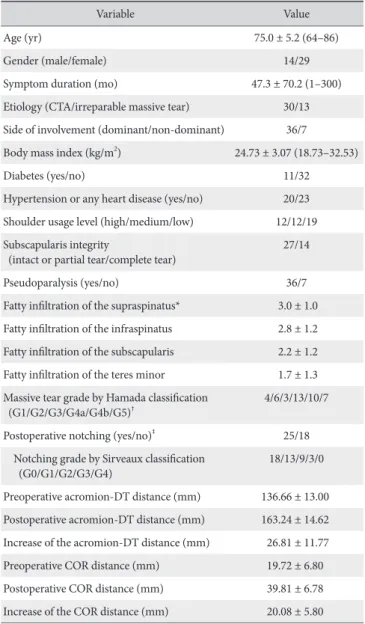
Table 1. Clinical and Radiographic Data
Variable Value
Age (yr) 75.0 ± 5.2 (64–86)
Gender (male/female) 14/29
Symptom duration (mo) 47.3 ± 70.2 (1–300)
Etiology (CTA/irreparable massive tear) 30/13 Side of involvement (dominant/non-dominant) 36/7 Body mass index (kg/m2) 24.73 ± 3.07 (18.73–32.53)
Diabetes (yes/no) 11/32
Hypertension or any heart disease (yes/no) 20/23 Shoulder usage level (high/medium/low) 12/12/19 Subscapularis integrity
(intact or partial tear/complete tear) 27/14
Pseudoparalysis (yes/no) 36/7
Fatty infiltration of the supraspinatus* 3.0 ± 1.0 Fatty infiltration of the infraspinatus 2.8 ± 1.2 Fatty infiltration of the subscapularis 2.2 ± 1.2 Fatty infiltration of the teres minor 1.7 ± 1.3 Massive tear grade by Hamada classification
(G1/G2/G3/G4a/G4b/G5)† 4/6/3/13/10/7
Postoperative notching (yes/no)‡ 25/18
Notching grade by Sirveaux classification
(G0/G1/G2/G3/G4) 18/13/9/3/0
Preoperative acromion-DT distance (mm) 136.66 ± 13.00 Postoperative acromion-DT distance (mm) 163.24 ± 14.62 Increase of the acromion-DT distance (mm) 26.81 ± 11.77 Preoperative COR distance (mm) 19.72 ± 6.80 Postoperative COR distance (mm) 39.81 ± 6.78 Increase of the COR distance (mm) 20.08 ± 5.80 Values are presented as mean ± standard deviation (range), number only, or mean ± standard deviation only.
CTA: cuff tear arthropathy, DT: deltoid tuberosity, COR: center of rotation.
*Fatty infiltration was graded according to the criteria by Goutallier et al.16)
†Massive rotator cuff tear was graded according to the criteria by Hamada et al.10)
‡Scapular notching was graded according to the criteria by Sirveaux et al.11)
level as retired or rarely participating sports.14) Pseudoparalysis was defined as active shoulder elevation <90o in the pres- ence of full passive forward elevation.15) FI of each rotator cuff muscle (supraspinatus, infraspinatus, and subscapularis) was evaluated using MRI (3-T scanner Signa® HDxt MRI scanner/
Discovery® MR750w system; General Electric, Milwaukee, WI, USA), according to the criteria established by Goutallier et al.16) and modified by Fuchs et al.,17) at the most lateral section of the oblique sagittal image, at the point where the scapular spine was still in continuity with the body of the scapula forming a Y-shape.
Radiographic evaluations were performed using standard AP radiographs with the arm in neutral rotation and 0o abduction, and axial radiographs, at the final follow-up (>1 year) for scapu- lar notching, postoperative increase in the acromion-deltoid tuberosity distance, and COR medialization. Scapular notch- ing was classified on AP radiographs according to the Nerot- Sirveaux classification.11) In this classification, a grade 1 defect is contained within the inferior pillar, a grade 2 defect is erosion up to the level of the inferior screw, a grade 3 defect extends over the inferior screw, and a grade 4 defect extends to the baseplate.
The acromion-deltoid tuberosity distance was defined as the distance between the inferolateral tip of the acromion and the deltoid tuberosity on the standard AP radiographs with the arm in neutral rotation and 0o abduction.12) In addition, the COR of the glenohumeral joint was the center of the ‘best fit’ circle over- lay on the articular surface of the humeral head, and the COR distance was defined as the perpendicular distance from the acromion-deltoid tuberosity line to the COR.13) The postopera- tive increased or medialized length of each measurement was calculated.
Surgical Procedures
All procedures were performed using a delto-pectoral ap- proach with the patient in the beach-chair position. The pros- theses implanted were Aequalis reverse shoulder system (Tornier, Montbonnot, France) in 33 patients and Comprehensive reverse shoulder system (Biomet, Warsaw, IN, USA) in 10 patients. The subscapularis tendon, when present, was cut from the lesser tuberosity, and was repaired later in a transosseous manner. The long head of the biceps tendon was tenotomized at its origin and sutured to the surrounding tissue later. The glenohumeral joint was approached, and the labrum was removed completely and glenoid was fully exposed. The prostheses were implanted according to the instructions provided by the manufacturer of the respective implant. The articular cartilage and sclerotic bone of the glenoid were removed using a flat reamer, and the base- plate was carefully positioned approximately 2–3 mm inferior to the center of the glenoid with a 10o inferior tilt in order to re- duce notching.18) In addition, humeral head osteotomy was per- formed with a targeted retroversion of 0o to 20o after removing all osteophytes from the humeral head-neck junction, and the
humeral implant was implanted with gentamicin-impregnated cement in all cases. Before the final implantation, the soft tissue tension, implant stability, and impingement-free shoulder mo- tion was checked with a trial implant. No other concomitant procedures such as bone grafting or latissimus dorsi transfer were performed in any cases. One or two suction drains were inserted before wound closure.
Postoperatively, all patients underwent the same rehabilita- tion protocol. Immobilization was maintained with an abduction sling for 4 weeks. Passive motion was initiated the day after sur- gery, with limited external rotation allowed to protect the sub- scapularis repair. Active-assisted ROM exercise was encouraged after patients were weaned off the sling. Muscle strengthening exercises were initiated at 9 to 12 weeks postoperatively.
Outcome Assessment
Various functional outcomes and satisfaction were evalu- ated in all patients. The functional outcome evaluation was performed using the VAS for pain, ASES score, Constant score, SST score, and active shoulder ROM pre- and postoperatively, and the satisfaction scale postoperatively. On the VAS, pain was scored on a scale of 0–10, with 10 indicating the highest level of pain; the ASES score consisted of a score summation using a 100-point system (50 points for daily function and 50 points for pain). Forward elevation was measured in degrees between the arm and the thorax with the elbow held straight. External rota- tion at the side was measured in degrees between the thorax and forearm with the arm held in an adducted position and the elbow in 90o flexion. Internal rotation of the shoulder was mea- sured by the vertebral level reached in the back with the tip of the thumb and numbered serially as 1 to 12 for the 1st to 12th thoracic vertebra, 13 to 17 for the 1st to 5th lumbar vertebra, and 18 for any level below the sacral region.
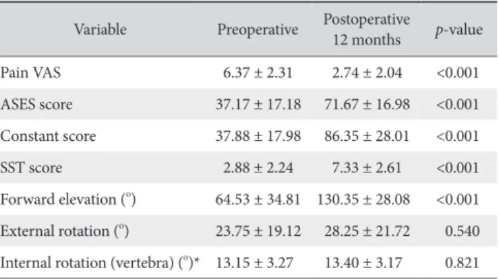
Table 2. Functional Outcomes after Reverse Total Shoulder Arthroplasty Variable Preoperative Postoperative
12 months p-value
Pain VAS 6.37 ± 2.31 2.74 ± 2.04 <0.001
ASES score 37.17 ± 17.18 71.67 ± 16.98 <0.001 Constant score 37.88 ± 17.98 86.35 ± 28.01 <0.001
SST score 2.88 ± 2.24 7.33 ± 2.61 <0.001
Forward elevation (o) 64.53 ± 34.81 130.35 ± 28.08 <0.001 External rotation (o) 23.75 ± 19.12 28.25 ± 21.72 0.540 Internal rotation (vertebra) (o)* 13.15 ± 3.27 13.40 ± 3.17 0.821 Values are presented as mean ± standard deviation.
VAS: visual analogue scale, ASES: American Shoulder and Elbow Surgeons, SST: simple shoulder test.
*The vertebral level of internal rotation was numbered serially as follows: 1–12 for the 1st to 12th thoracic vertebra, 13–17 for the 1st to 5th lumbar vertebra, and 18 for any level below the sacral region.
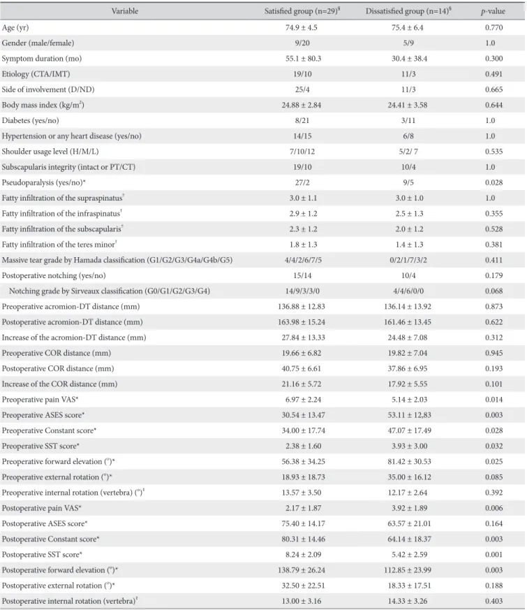
Table 3. Comparison between the Satisfied and Dissatisfied Groups
Variable Satisfied group (n=29)§ Dissatisfied group (n=14)§ p-value
Age (yr) 74.9 ± 4.5 75.4 ± 6.4 0.770
Gender (male/female) 9/20 5/9 1.0
Symptom duration (mo) 55.1 ± 80.3 30.4 ± 38.4 0.300
Etiology (CTA/IMT) 19/10 11/3 0.491
Side of involvement (D/ND) 25/4 11/3 0.665
Body mass index (kg/m2) 24.88 ± 2.84 24.41 ± 3.58 0.644
Diabetes (yes/no) 8/21 3/11 1.0
Hypertension or any heart disease (yes/no) 14/15 6/8 1.0
Shoulder usage level (H/M/L) 7/10/12 5/2/ 7 0.535
Subscapularis integrity (intact or PT/CT) 19/10 10/4 1.0
Pseudoparalysis (yes/no)* 27/2 9/5 0.028
Fatty infiltration of the supraspinatus† 3.0 ± 1.1 3.0 ± 1.0 1.0
Fatty infiltration of the infraspinatus† 2.9 ± 1.2 2.5 ± 1.3 0.355
Fatty infiltration of the subscapularis† 2.3 ± 1.2 2.0 ± 1.2 0.528
Fatty infiltration of the teres minor† 1.8 ± 1.3 1.4 ± 1.3 0.381
Massive tear grade by Hamada classification (G1/G2/G3/G4a/G4b/G5) 4/4/2/6/7/5 0/2/1/7/3/2 0.411
Postoperative notching (yes/no) 15/14 10/4 0.179
Notching grade by Sirveaux classification (G0/G1/G2/G3/G4) 14/9/3/3/0 4/4/6/0/0 0.068
Preoperative acromion-DT distance (mm) 136.88 ± 12.83 136.14 ± 13.92 0.873
Postoperative acromion-DT distance (mm) 163.98 ± 15.24 161.46 ± 13.45 0.622
Increase of the acromion-DT distance (mm) 27.84 ± 13.33 24.48 ± 7.08 0.312
Preoperative COR distance (mm) 19.66 ± 6.82 19.82 ± 7.04 0.945
Postoperative COR distance (mm) 40.75 ± 6.61 37.86 ± 6.95 0.193
Increase of the COR distance (mm) 21.16 ± 5.72 17.92 ± 5.55 0.101
Preoperative pain VAS* 6.97 ± 2.24 5.14 ± 2.03 0.014
Preoperative ASES score* 30.54 ± 13.47 53.11 ± 12,83 0.003
Preoperative Constant score* 34.00 ± 17.74 47.07 ± 17.49 0.028
Preoperative SST score* 2.38 ± 1.60 3.93 ± 3.00 0.032
Preoperative forward elevation (o)* 56.38 ± 34.25 81.42 ± 30.53 0.025
Preoperative external rotation (o)* 18.93 ± 18.73 35.00 ± 16.12 0.085
Preoperative internal rotation (vertebra) (o)‡ 13.57 ± 3.50 12.17 ± 2.64 0.392
Postoperative pain VAS* 2.17 ± 1.87 3.92 ± 1.89 0.006
Postoperative ASES score* 75.40 ± 14.17 63.57 ± 21.01 0.164
Postoperative Constant score* 80.31 ± 14.46 64.14 ± 18.37 0.003
Postoperative SST score* 8.24 ± 2.09 5.42 ± 2.59 0.001
Postoperative forward elevation (o)* 138.79 ± 26.24 112.85 ± 23.99 0.003
Postoperative external rotation (o)* 32.50 ± 22.51 18.33 ± 17.51 0.188
Postoperative internal rotation (vertebra)‡ 13.00 ± 3.16 14.33 ± 3.26 0.403
Values are presented as mean ± standard deviation or number only.
CTA: cuff tear arthropathy, IMT: irreparable massive tear, D: dominant, ND: non-dominant, H: high level, M: middle level, L: low level, PT: partial tear, CT: com- plete tear, DT: deltoid tuberosity, COR: center of rotation, VAS: visual analogue scale, ASES: American Shoulder and Elbow Surgeons, SST: simple shoulder test.
*Statistically significant. †Fatty infiltration was graded according to the criteria by Goutallier et al.16)‡The vertebral level of internal rotation was numbered serially as follows: 1–12 for the 1st to 12th thoracic vertebra, 13–17 for the 1st to 5th lumbar vertebra, and 18 for any level below the sacral region. §Satisfied group includ- ed those who responded with the choices ‘very satisfied’ or ‘satisfied’, and dissatisfied group included those who responded with ‘neither satisfied nor dissatisfied’,
‘dissatisfied’, or ‘very dissatisfied’.
Satisfaction was assessed by asking the patients to rate their overall experience with the surgery as very satisfied, satisfied, neither satisfied nor dissatisfied, dissatisfied, or very dissatisfied.
The outcome parameter was set as a satisfaction scale (very satis- fied or satisfied vs. neither satisfied nor dissatisfied, dissatisfied, or very dissatisfied).19) The patients who responded with the choices ‘very satisfied’ or ‘satisfied’ were classified as the satisfied group, and those who responded with the choices ‘neither satis- fied nor dissatisfied’, or ‘dissatisfied’, or ‘very dissatisfied’ were classified as the dissatisfied group.
Statistics
A paired t-test was used for comparison of the pre- and postoperative results of pain using the VAS, ROM, and func- tional scores. The mean values were compared using the Mann- Whitney U test for continuous variables and χ2 test or Fisher’s exact test for categorical variables to determine the differences between the satisfied and dissatisfied groups. SPSS ver. 13.0 (SPSS Inc., Chicago, IL, USA) was used for statistical analyses and p<0.05 indicated statistical significance.
Results
All functional scores, VAS pain score, and active forward flex- ion improved significantly after surgery (all p<0.001) (Table 2).
Overall, 29 patients were satisfied with the results and 14 were dissatisfied. In the postoperative satisfaction outcome parameter, the presence of pseudoparalysis (p=0.028) and worse preop- erative function with regard to pain VAS, ASES score, Constant score, SST score, and forward flexion were related to higher satisfaction (all p<0.05) (Table 3). Other radiological factors including the grade of the massive cuff tear, FI of rotator cuffs, postoperative scapular notching, increase of acromion-deltoid tuberosity distance, and increase of the COR were not associ- ated with the postoperative satisfaction (all p>0.05).
Twenty-five (58.1%) patients showed postoperative scapular notching, with no influence on the functional outcome. One patient had acromio-clavicular separation at 6 months postop- eratively, and 1 had an acromial fracture at 3 months postopera- tively. Other complications such as dislocation or infection did not occur.
Discussion
The aim of this study was to comprehensively examine the correlation of various clinical and radiographic factors including preoperative function with postoperative satisfaction in patients with RTSA, and demonstrated that the presence of pseudopa- ralysis and worse preoperative function are prognostic factors for patient satisfaction after RTSA. It is interesting to note that subjective satisfaction was not affected by the reported demo-
graphic or radiographic factors such as age,5) obesity,6) FI of the teres minor,7) scapular notching,8) and lengthening of the lever arm,9) rather by the preoperative functional status of the patients.
In this study, the worse preoperative condition of the patients, as shown by the presence of preoperative pseudoparalysis, severe pain, and poor functional disabilities, rather resulted in higher satisfaction after RTSA. We do not know the exact reason why patients who showed poor preoperative function were more sat- isfied with the results; however we think that this is due in part to the unique mechanism of RTSA, which restores the functional deficit of the rotator cuff deficient shoulder. Patients who had greater disability preoperatively due to limited shoulder motion and weakness (worse preoperative functional status) appear to be more satisfied with their restoration of active shoulder motion and strength after surgery, even though the actual level of func- tional outcome would not be very high. It might be reasonable that a patient who initially could not easily perform daily activi- ties such as washing face or changing clothes who is able to per- form those activities more easily will have far greater satisfaction than those whose initial function was relatively good. That is, we think that the restoration of active shoulder ROM and strength deficit by RTSA1) therefore appears to be a major determinant of patients’ subjective satisfaction after RTSA.
To the best of our knowledge, this was the first study to evalu- ate the relationship between various preoperative factors includ- ing preoperative function and patients’ postoperative subjective satisfaction. However, several limitations should be noted when interpreting our findings. First, the number of patients was rela- tively small, thus it is possible that the lack of statistical signifi- cance in some factors may be due to the relatively small number of cases. Further studies including more cases may be needed to exclude the possibility of type 2 error and to confirm our results.
Second, the follow-up period was relatively short (mean, 19.3 ± 7.1 months), thus the satisfaction and functional outcomes may change further with a longer follow-up period. This study should be interpreted as a result of early to mid-term follow-up. Third, this was not a prospective cohort study. Even though most vari- ables were gathered prospectively, we cannot deny the possibil- ity of a selection bias and a confounding effect that we were not aware of. In addition, the drop-out rate (6/49, 12.2%) may also have caused selection bias. Therefore, a well-organized prospec- tive cohort study might be needed to confirm our results.
Conclusion
All patients showed a good functional outcome after RTSA, however the patients’ subjective postoperative satisfaction was affected by preoperative functional status (higher satisfaction in poor preoperative function), not by radiological findings, in a given follow-up period. Further longer term follow-up may be needed to confirm this result.
References
1. Grammont PM, Baulot E. Delta shoulder prosthesis for rotator cuff rupture. Orthopedics. 1993;16(1):65-8.
2. Boileau P, Watkinson DJ, Hatzidakis AM, Balg F. Grammont re- verse prosthesis: design, rationale, and biomechanics. J Shoul- der Elbow Surg. 2005;14(1 Suppl S):147S-61S.
3. Werner CM, Steinmann PA, Gilbart M, Gerber C. Treatment of painful pseudoparesis due to irreparable rotator cuff dysfunc- tion with the Delta III reverse-ball-and-socket total shoulder prosthesis. J Bone Joint Surg Am. 2005;87(7):1476-86.
4. Molé D, Favard L. Excentered scapulohumeral osteoarthritis.
Rev Chir Orthop Reparatrice Appar Mot. 2007;93(6 Suppl):
37-94.
5. Muh SJ, Streit JJ, Wanner JP, et al. Early follow-up of reverse total shoulder arthroplasty in patients sixty years of age or younger. J Bone Joint Surg Am. 2013;95(20):1877-83.
6. Beck JD, Irgit KS, Andreychik CM, Maloney PJ, Tang X, Harter GD. Reverse total shoulder arthroplasty in obese patients. J Hand Surg Am. 2013;38(5):965-70.
7. Simovitch RW, Helmy N, Zumstein MA, Gerber C. Impact of fatty infiltration of the teres minor muscle on the outcome of reverse total shoulder arthroplasty. J Bone Joint Surg Am.
2007;89(5):934-9.
8. Ek ET, Neukom L, Catanzaro S, Gerber C. Reverse total shoul- der arthroplasty for massive irreparable rotator cuff tears in patients younger than 65 years old: results after five to fifteen years. J Shoulder Elbow Surg. 2013;22(9):1199-208.
9. Kasten P, Maier M, Rettig O, Raiss P, Wolf S, Loew M. Proprio- ception in total, hemi- and reverse shoulder arthroplasty in 3D motion analyses: a prospective study. Int Orthop. 2009;33(6):
1641-7.
10. Hamada K, Fukuda H, Mikasa M, Kobayashi Y. Roentgeno- graphic findings in massive rotator cuff tears. A long-term ob- servation. Clin Orthop Relat Res. 1990;(254):92-6.
11. Sirveaux F, Favard L, Oudet D, Huquet D, Walch G, Molé D.
Grammont inverted total shoulder arthroplasty in the treat- ment of glenohumeral osteoarthritis with massive rupture of the cuff. Results of a multicentre study of 80 shoulders. J Bone Joint Surg Br. 2004;86(3):388-95.
12. De Wilde L, Audenaert E, Barbaix E, Audenaert A, Soudan K.
Consequences of deltoid muscle elongation on deltoid muscle performance: a computerised study. Clin Biomech (Bristol, Avon). 2002;17(7):499-505.
13. Jobin CM, Brown GD, Bahu MJ, et al. Reverse total shoulder arthroplasty for cuff tear arthropathy: the clinical effect of del- toid lengthening and center of rotation medialization. J Shoul- der Elbow Surg. 2012;21(10):1269-77.
14. Chung SW, Kim JY, Kim MH, Kim SH, Oh JH. Arthroscopic repair of massive rotator cuff tears: outcome and analysis of factors associated with healing failure or poor postoperative function. Am J Sports Med. 2013;41(7):1674-83.
15. Oh JH, Kim SH, Shin SH, et al. Outcome of rotator cuff repair in large-to-massive tear with pseudoparalysis: a comparative study with propensity score matching. Am J Sports Med. 2011;
39(7):1413-20.
16. Goutallier D, Postel JM, Bernageau J, Lavau L, Voisin MC. Fatty muscle degeneration in cuff ruptures. Pre- and postoperative evaluation by CT scan. Clin Orthop Relat Res. 1994;(304):78- 83.
17. Fuchs B, Weishaupt D, Zanetti M, Hodler J, Gerber C. Fatty degeneration of the muscles of the rotator cuff: assessment by computed tomography versus magnetic resonance imaging. J Shoulder Elbow Surg. 1999;8(6):599-605.
18. Nyffeler RW, Werner CM, Gerber C. Biomechanical relevance of glenoid component positioning in the reverse Delta III total shoulder prosthesis. J Shoulder Elbow Surg. 2005;14(5):524-8.
19. Young AA, Smith MM, Bacle G, Moraga C, Walch G. Early results of reverse shoulder arthroplasty in patients with rheu- matoid arthritis. J Bone Joint Surg Am. 2011;93(20):1915-23.