Corresponding author:
Lorenzo Alibardi
Department of Bigea, University of Bologna, via Selmi 3, 40126, Bologna, Italy
Tel: +39-051-2094138, Fax: +39-051-2094286, E-mail: lorenzo.alibardi@
unibo.it
Copyright © 2013. Anatomy & Cell Biology
Ultrastructural immunolocalization of beta- defensin-27 in granulocytes of the dermis
and wound epidermis of lizard suggests they contribute to the anti-microbial skin barrier
Lorenzo Alibardi
Department of Bigea, University of Bologna, Bologna, Italy
Abstract: The high resistance to infections in lizard wounds suggests that these reptiles possess effective antimicrobial peptides in their tissues. The present immunocytochemical study shows the cellular localization of beta-defensin 27 in tail tissues and in the blood, a defensin previously identified in the lizard Anolis carolinensis through biomolecular methods. Beta-defensin-27 immunoreactivity is only observed in some large granules mainly contained in heterophilic granulocytes that are sparse within the dermis of the skin or in the isolated blood. This peptide is absent in other cell types of the skin, in keratinocytes and in subdermal muscle tissue of the tail in normal conditions. Pre-corneous keratinocytes of the regenerating tail epidermis are unlabeled or show a weak labeling for the peptide only in sparse cytoplasmic areas or in the extracellular spaces among corneocytes of the wound and regenerating epidermis. The study suggests that beta-defensin 27 is normally stored in granulocytes present in the blood or in connective tissues while in the epidermis keratinocytes do not show the presence of this peptide unless these cells are stimulated from injury to produce and likely release beta-defensins.
Key words: Lizards, Skin, Blood, Beta-defensin-27, Immunolocalization
Received July 15, 2013; Revised September 5, 2013; Accepted September 27, 2013
antiseptic treatment, an ability indicating that they possess an effective and rapid innate immunity.
Previous studies have suggested that part of the strong innate immunity in injured lizards derives from the intense activity of granulocytes that destroy bacteria and likely release antimicrobial agents in injured connective tissues present beneath the scab [3-5]. Recent molecular biology studies have indeed found the presence of two types of antimicrobial peptides in lizard wounds and in regenerating tissues, beta- defensins and cathelicidins [6, 7].
Beta-defensins are included in the large family of anti- microbial peptides, with sequences of 18‒40 amino acids and containing a characteristic cysteines motif comprising three disulphide bonds [8, 9]. These peptides have been discovered in numerous plants, invertebrates and vertebrates, including reptiles, where they elicit a potent anti-microbial
Introduction
The capability of lizards to resist infections after large wounds or organ amputation in the tail or limb is in part due to the anti-microbial barrier represented by the epidermis, and to circulating or tissue granulocytes in the dermis [1-3].
Numerous bacteria are found over the exposed tissues of an amputated limb or tail that are in contact with the microbial- rich ground substratum. Lizards are however little affected from these bacteria and heal their tissues with no need for
activity (Fig. 1) [6, 10-13]. Beta-defensin peptides derive from a larger protein precursor of 90‒110 amino acids, made of 19‒20 amino acid signal sequence, a pro-peptide sequence of 45‒50 amino acids, and a 32‒36 amino acid peptide at the C-terminal, that is cleaved from the remaining protein to produce the mature and active peptide.
Another largely characterized group of antimicrobial peptides present in lizard tissues includes the cathelicidins [7], cathionic peptides of 12‒80 amino acids rich in lysine, phenylalanine, trypthophane and proline, and initially synthesized in the cytoplasm of granulocytes as precursors proteins [14, 15]. These peptides also derive from a larger protein precursor. The larger protein contain a pro-peptide sequence and a conserved cathelin domain containing about 100 amino acids that is joined to a terminal amino acid sequence of 12‒100 amino acids, the latter corresponding to the mature peptide (cathelicidin). In mammalian cells, the mature peptide derives from the cleavage from the cathelin domain by an intracellular or an extracellular elastase-like protein [16].
In previous molecular studies on lizard anti-microbial peptides we found an expression for both beta-defensins (Ac-BD-15, -25, and -27) and cathelicidins (Ac-CATH-1, -2, -3) in different tissues, including normal and regenerating skin, but the specific cell source for these molecules was not known [6, 7]. These studies have indicated that lizard beta- defensin-27 (Ac-BD-27), together 15, 25, 29, and 30 are the beta-defensins with the larger spectrum of expression in different tissues. Using reverse transcription polymerase chain reaction (RT-PCR) expression analysis of Ac-BD-25 and -27, the study showed that the mRNAs for these two peptides were present in the skin, but only during wound healing and skin regeneration (Dalla Valle et al., unpublished observations).
The localization of the more widespread beta-defensin, Ac- BD-15, essentially in granulocytes and phagocytes of Anolis carolinensis [5, 17] suggested that also other beta-defensins could be present in similar cells and become active during wound healing and tissue regeneration. In order to detect the presence and specific localization of other beta-defensins other than Ac-BD-15 the present qualitative study aims to detect the cell localization of Ac-BD-27 within the connective and epidermal tissues of the tail in the lizard A. carolinensis in normal and regenerating conditions.
Materials and Methods
AnimalsThe study was conducted on adult lizards of the green anolis (A. carolinensis, n=4) maintained in a terrarium at 25‒33oC as previously indicated [5]. The tail was amputated by twisting it to exploit the natural fracture plains present in the tail (autotomous planes) [1]. Despite the little bleeding, some blood was aspirated from the amputation surface of the stump with a Pasteur pipette and fixed into an eppendhorf test tube. Also, pieces of the detached normal tail were sampled for comparison with regenerated tissues (see later).
The animals were left to regenerate their tail at room temperature (25‒30oC) in cages. In the first sampling, stumps of healing tails were collected after 4 days post-amputation (wound healing stage), and the same animals were re-sampled after 4 weeks when regenerating tails of 3‒5 mm in length were formed. The tissues were immediately fixed for the following immunocytochemical study. Also the blood derived from the second amputation was collected in the fixative solution as indicated above.
Immunocytochemistry and electron microscopy
Tissues were fixed at 0‒4oC in fresh 4% paraformaldehyde in 0.1 M phosphate buffer at pH 7.4 for 7‒8 hours, rinsed in buffer, dehydrated in alcohol, and embedded in the resins Lowicryl or LR-White. In three cases also the blood from normal and regenerating lizards (3‒4 days after tail amputation) was fixed for 1 hour and then centrifuged in an eppendhorf test tube for 3 minutes at 10,000 g. The super- natant was discharged and the pellet was dehydrated and embedded as indicated above.
The embedded tissues or blood were sectioned using an ultramicrotome, and semithin sections (2‒3 µm thick) and thin sections (70‒90 nm thick) were collected, stained in Fig. 1. Amino acid sequences of beta-defensins from the lizard Anolis
carolinensis (AcBD15 and -27 [6]), the soft-shelled turtle Apalone spinifera (TuBD-1 [13]), the turtles Caretta caretta [10], Pelodiscus sinensis [11] and Emys orbicularis [12]. Note the characteristics positions of the 6 cysteines ( ) and the glycines ( ) localized two positions before the second (arrowhead) and the fourth (double arrowhead) cysteine.
1% toluidine blue for the histological examination or were utilized for the immunohistochemical study. The antibody against BD-27 was raised in a rabbit immunized with a 35 amino acid long peptide (DLECQKAKGLCLHQRCRRPWR SVGTCNNLQHHCCQ) for A. carolinensis beta-defensin-27 (Fig. 1) [6]. The peptides were conjugated to Key Limpet Haemocyanin before injection into rabbits, and the derived antibodies present in the immune serum were purified by affinity chromatography (Davids Biotechnologie, Regensburg, Germany). The reactivity of the immune serum to the anti- genic peptides was tested by the manufacturer using the enzyme-linked immunosorbent assay-test.
Light microscope immunocytochemistry was performed incubating the sections overnight at 0‒4oC with the beta- defensin-27 or cathelicidins antibodies diluted 1:200 in buffer (Tris 0.05 M at pH 7.6 containing 1% bovine serum albumin).
In control sections, the primary antibody was omitted or pre-absorbed for 24 hours at 4oC mixing 3:1 of the solution containing the peptide antigen with the purified antibody
solution. After rinsing in buffer, the sections were incubated for 60 minutes at room temperature in a fluorescein- conjugated anti-rabbit antibody (1:100, Sigma, St. Louis, MO, USA), rinsed in buffer, mounted in 10% glycerol in phosphate buffered saline, and observed under a fluorescence microscope. Pictures were taken by a digital camera and digitalized into a computer using the Adobe Photoshop Program.
Thin sections of 40‒80 nm in thickness were collected on Nickel grids, and the sections were incubated for 10 minutes in the Tris buffer containing 1% Cold Water Fish Gelatin to block non-specific binding sites, then the grids were incubated overnight at 0‒4oC in the primary antibodies (1:200). After rinsing in the Buffer, a 5 or 10 nm gold conjugated anti- rabbit secondary antibody was applied for one hour at room temperature, the grids were rinsed in buffer and stained for 6 minutes in 2% aqueous uranyl acetate, rinsed and observed with a Zeiss 10C/10 CR electron microscope (Zeiss, Jena, Germany). In control sections, the primary antibodies were
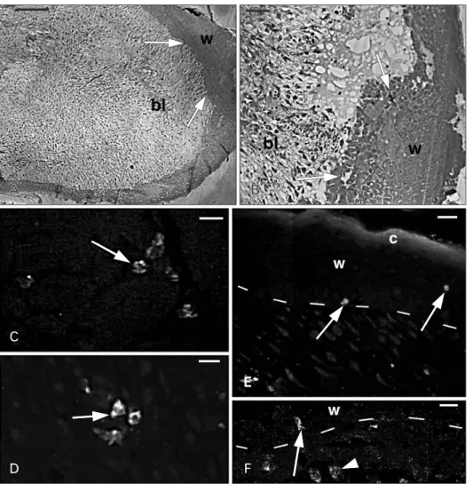
Fig. 2. Histology of the apical region of a regenerating tail (A, B) and Ac- BD-27 immunoreactivity in sparse cells (C‒F). (A) The blastema (bl) consists in a mesenchymal tissue surrounded by a thick wound epidermis (w).
The arrows indicate the areas where granulocytes were seen using the elec- tron microscope. (B) Detail of the wound epidermis (w) and the under- lying blastema mesenchyme (bl). The arrows indicate the areas were granu- locytes were later identified under the electron microscope. (C) Positive gra nular cells (arrow) in the inter- muscle connective of the tail stump.
(D) Positive cells (arrow) located in the mesenchyme of the regenerating tail.
(E) Immunofluorescent cells (arrows) in the wound epithelium (w) with its stratum corneum (c). Dashes indicate the dermal-epidermal boundary. (F) Detail of immunofluorescent granular cells localized in the dermis (arrowhead) and at the base (arrow) of the wound epidermis (w). Dashes indicate the dermal-epidermal boundary. Scale bars=100 µm (A), 20 µm (B), 10 µm (C−
F).
omitted or pre-absorbed for 24 hours at 4°C mixing 3:1 the solution containing the antigen with the purified antibody solution. The grids containing the immunolabeled sections were completely scanned checking for the localization of gold particles in all cell types present in the tissues.
Results
Histology and light immunocytochemistry
The tissues of the skin, dermis and epidermis, in the normal tail and the mesenchyme and epidermis present in the regenerating tail were studied under the light microscope.
The regenerating tail was formed by a loose mesenchyme at the tip, called the blastema, in contact with a multilayered epidermis, the wound epidermis (Fig. 2A). Although most cells were mesenchymal, sparse blood cells and phagocytes
were present in the blastema, and these cells were also localized at the base and sparsely within the wound epidermis (Fig. 2B). The electron microscope study allowed a conclusive identification of cells sparsely located among the keratinocytes of the wound epidermis (arrows in Fig. 2B) as granulocytes (see later description).
The study under the fluorescent microscope of sections of normal and regenerating tail after immunostaining with the Ac-BD-27 antibody showed very few and sparse immunofluorescent cells, more frequently encountered in regenerating then in normal tail tissues. In sections of the blood in normal or regenerating condition, sparse granular cells were also seen (see ultrastructural description). Im- mu no fluorescent cells appeared isolated or, more occa sio- nally, forming small clusters in the inter-muscular con- nectives, dermis, and especially in the regenerating tail
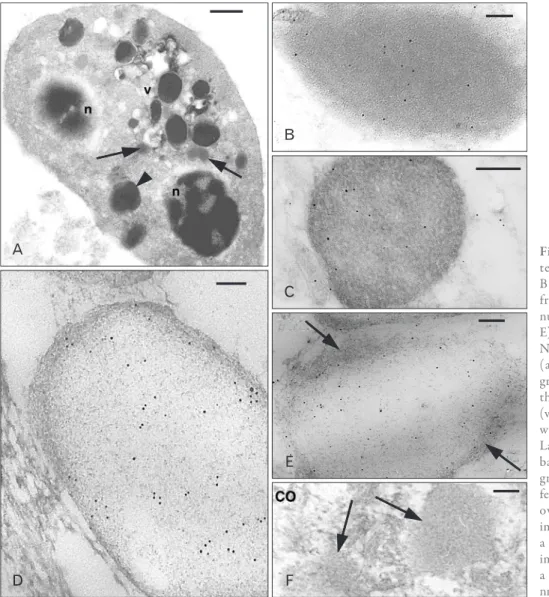
Fig. 3. Ultrastructural view of a he- te ro philic granulocyte (A) and Ac- BD-27 immunoreactive granules from tissue granulocytes (B, C), gra- nulocytes iso lated from the blood (D, E), and a control (CO) section (F). (A) Numerous lar ger and denser granules (arrowhead) and smaller and paler granules (arrows) are present between the two nuclear sections (n), vesicle (v). (B) Immunolabeled dense granule with even and dark background. (C) Labeled dense granule with a reti culate background. (D) Pale granule from a granulocyte isolated from the blood featuring a meshwork of fine filaments over a pale background. (E) Other immunolabeled pale granule showing a fibrous periphery (arrows). (F) Two immunonegative granules (arrows) in a CO section. Scale bars=1 µm (A), 50 nm (B, D, E), 100 nm (C, F).
connectives and in the apical blastema, and their cytoplasm contained immunofluorescent granules (Fig. 2C, D). Few immunolabeled cells were also seen within the wound epidermis and also in the epidermis of the regenerating tail during early stages of regeneration (Fig. 2E, F). The nature of these granular cells was better defined using the electron microscope.
Ultrastructural immunocytochemistry
The systematic observation of the entire skin and the muscle tissues located underneath the dermis in all the grids examined under the electron microscope, including the inter-muscle connective tissue with the sparse blood vessels, confirmed that few cells containing medium-dense cyto plasmic granules were immunopositive (Fig. 3A‒C).
No labeling was present in the dark and ovoidal granules of the frequent melanophores encountered in the dermis and subdermal connectives.
The cells containing variably immunolabeled granules, both present in the isolated blood sections, or within or outside blood vessels in connective tissues and among the mesenchymal blastema showed a relatively smooth surface, and were mainly identified as heterophilic granulocytes (Fig. 3A). Their heterochromatic nucleus was polymorphic and two or more nuclear sections were often seen in these cells. Granules of larger and denser type, probably cor- re s ponding to primary (azurophilic) granules of mam- malian granulocytes, and other smaller granules with less electron-density, corresponding to the specific granules of mammalian granulocytes, were observed in these cells.
The immunoreactivity for Ac-BD-27 indicated that most frequently labeled were the denser and larger granules of heterophil granulocytes while the remaining cytoplasm was immunonegative (Fig. 3B). Other sparsely labeled dense granules showed a reticulate background (Fig. 3C).
Both the cytoplasm and the variably large granules ob- served in granulocytes from the blood pellets were paler then in granulocytes present among tissues, suggesting a partial extraction of cell components during preparation of the blood pellets. The content of the immunolabeled granules varied from a fine filamentous meshwork (Fig. 3D) to broad pale areas apparently empty with no gold labeling (Fig. 3E).
Controls showed no labeling in both cells and granules of granulocytes (Fig. 3F). Small granules, likely corresponding to the specific granules of mammalian granulocytes, were generally unlabeled. Also the rare basophilic granulocytes
encountered in tissues or isolated in the blood pellet con- tained numerous large granules (over 0.5 µm), some of which appeared weakly labeled (data not shown).
Although the normal epidermis did not contain granu- locytes, during wounding and regeneration of the tail, sparse heterophilic granulocytes were seen in the basal layers or among suprabasal keratinocytes in apical areas of the wound epidermis (Figs. 2B, 4A). Among their numerous granules, some granules showed a low (few sparse gold particles) to a higher immunolabeling (more numerous and evenly distri buted gold particles) (Fig. 4B). Rare granulocytes also possessed heterogenous granules containing a dense core and a paler halo suggesting they belong to the eosinophilic type (Fig. 4C). Sparse granulocytes with labeled large and dense granules were also observed in few instances within the multilayered regenerating (wound) epidermis of the more apical region of the regenerating tail (Fig. 5A, B). Occasional cytoplasmic labeling was seen in these cells although most labeling remained inside the granules.
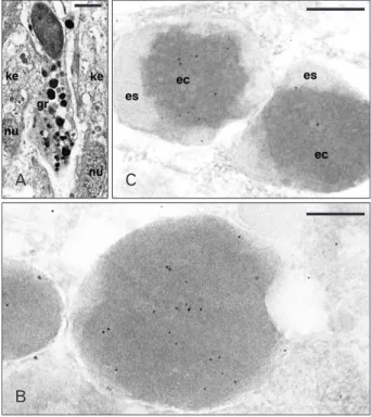
Fig. 4. Ultrastructure of a granulocytes (A) and Ac-BD-27 immu- nolabeled granules (B, C) within granulocytes of the wound epidermis.
(A) Elongated granulocyte (gr) infiltrated between two keratinocytes of the wound epithelium (ke; nu, nuclei). (B) Detail of immunolabeled dense granules. (C) Two unusual types of low labeled heterogenous granules (es, external space of the granule; ec, electron-dense core).
Scale bars=2 µm (A), 100 nm (B), 250 nm (C).
Discussion
Numerous studies in mammals, lizards, snake and turtle have shown that antimicrobial peptides are mainly stored and released from granulocytes and keratinocytes under microbial challenge [5, 17, 18-22], although other cell types can also form these peptides constitutionally or under microbial or injury challenge [9, 23].
Previous studies on early stages of healing of the skin over the stump of an amputated tail in lizards indicated a massive invasion of granulocytes underneath the scab and infiltrated among migrating keratinocytes and often engulfed with phagocytosed material [3-5, 17]. In the dermis and in normal or regenerating epidermis isolated granulocytes are difficult to be seen under the light microscope. However using the transmission electron microscopy survey these cells can be identified, particularly at the tip of the regenerating tail. The present morphological qualitative study confirms previous observations on the localization of other beta-defensins known to be highly expressed in lizards and other reptiles.
In normal conditions both beta-defensins and cathelicidins
are stored in heterophilic and basophilic granulocytes while epithelial, connective, muscle cells do not contain or express these peptides. Lizard blood monocytes and macrophages do not appear to contain beta-defensin immunoreactivity.
Also other cell types present in different organs of A.
carolinenis, such as liver hepatocytes and macrophages, intestine enterocytes, fibroblasts and cells of the seminiferous tubes in testes, alveolar cells and macrophages in the lung, and cardiomyocytes in the heart, and mucous cells in the tongue, do not usually show any immunolabeling for both beta-defensins or cathelicidins (Alibardi, unpublished ob- servations). Only granulocytes in the blood, in regenerating and normal dermis, and in the regenerating blastema of the tail have shown some immunopositive granules for Ac- BD-15 and -27, and for cathelicidins previously identified by molecular biology methods [7, 24]. In particular, a large immunolabeling of primary granules of granulocytes, intracellular localization and also immunolocalization or released beta-defensins were noticed in activated or dege- nerating heterophilic granulocytes involved in phago cytosis.
This process is very intense in the stump of an amputated tail or limb of lizards during the firts 3-10 days post-injury.
The expression data obtained from different tissues in A.
carolinensis by RT-PCR in previous studies [7, 24] probably reflects the content in blood and granulocytes in the tested organs although also other, non hematogenous cells, may become acti vated under microbial challenge and release antimicrobial peptides. In particular for BD-27 no expression was obtained in the normal skin, intestine and testis, and very weak for other organs. The intestine, liver and normal skin, were also devoid of cathelicidin expression while the regenerating skin showed up-regulation of transcripts for CATH-1 (unpublished data) [7]. However the expression of BD-27 was later observed in the wounded and regenerating skin of A. carolinensis (Dalla Valle et al., unpublished observations).
In the present study, although a variable labeling for Ac-BD-27 was observed in the large granules of lizard granulocytes, the aspect and content of the granules in cells separated from the blood by centrifugation appears as empty, likely due to the loss of some protein content due to the embedding process of the blood pellets during resin penetration. As a result, the granules from the isolated blood granulocytes contained a fine meshwork of filamentous material in a very electron-pale, almost empty matrix. Despite of this apparently poor content preservation the only labeled Fig. 5. Ac-BD-27 immunolabeled granulocytes present in the
regenerating epidermis of the tail (ke, keratinocytes). (A) Most gold particles are diffuse over the dense granules (gr) while the cytoplasm is negative. (B) Detail on a labeled dense but heterogenous granule (gr) near the nucleus (nu), possibly in a stage of discharging its content.
Scale bars=150 µm (A, B).
cell structures were these granules, although the labeling was generally low. It is unknown whether also for lizard defensins, the cleavage is intracellular or extracellular and catalized by a specific granulocyte elastase or serine-proteases like in human and avian granulocytes [16, 25].
Like for the localization of Ac-BD-15, also Ac-BD-27 immu noreactivity is present in wounded and regenerating epidermis, although it appears confined to granulocytes mixed with migrating keratinocytes and is not found within keratinocytes. This immunoreactivity parallels the immu- nolabeling observed for cathelicidins (Alibardi, personal observations).
In conclusion beta-defensins (and cathelicidins) in lizard tissues appear confined to granulocytes in normal conditions but become activated or their levels increases after wounding and tissue regeneration where also activated keratinocytes produce and likely secrete these peptides to limit microbial invasion or for other, still unknown, biological processes.
Acknowledgements
The study was mainly self-supported (Comparative Histolab, in particular the optical and electronic microscopic equipment) and with a Grant from the Italian Ministry of Education and Scientific Research (Grant 2008 AXS E-002).
References
1. Cox PG. Some aspects of tail regeneration in the lizard Anolis carolinensis. I. A description based on histology and auto- radiography. J Exp Zool 1969;171:127-49.
2. Zika JM. A histological study of the regenerative response in a lizard, Anolis carolinensis. J Exp Zool 1969;172:1-8.
3. Alibardi L. Ultrastructural features of the process of wound healing after tail and limb amputation in lizard. Acta Zool 2010;
91:306-18.
4. Alibardi L, Toni M. Wound keratins in the regenerating epi- dermis of lizard suggest that the wound reaction is similar in the tail and limb. J Exp Zool A Comp Exp Biol 2005;303:845-60.
5. Alibardi L, Celeghin A, Dalla Valle L. Wounding in lizards results in the release of beta-defensins at the wound site and formation of an antimicrobial barrier. Dev Comp Immunol 2012;36:557-65.
6. Dalla Valle L, Benato F, Maistro S, Quinzani S, Alibardi L.
Bioinformatic and molecular characterization of beta-defensins- like peptides isolated from the green lizard Anolis carolinensis.
Dev Comp Immunol 2012;36:222-9.
7. Dalla Valle L, Benato F, Paccanaro MC, Alibardi L. Bioinformatic and molecular characterization of cathelicidin-like peptides
isolated from the green lizard Anolis carolinensis (Reptilia:
Lepidosauria: Iguanidae). Ital J Zool 2013;80:177-86.
8. Sahl HG, Pag U, Bonness S, Wagner S, Antcheva N, Tossi A.
Mammalian defensins: structures and mechanism of antibiotic activity. J Leukoc Biol 2005;77:466-75.
9. Selsted ME, Ouellette AJ. Mammalian defensins in the anti- microbial immune response. Nat Immunol 2005;6:551-7.
10. Chattopadhyay S, Sinha NK, Banerjee S, Roy D, Chattopadhyay D, Roy S. Small cationic protein from a marine turtle has beta- defensin-like fold and antibacterial and antiviral activity. Pro- teins 2006;64:524-31.
11. Lakshminarayanan R, Vivekanandan S, Samy RP, Banerjee Y, Chi-Jin EO, Teo KW, Jois SD, Kini RM, Valiyaveettil S. Structure, self-assembly, and dual role of a beta-defensin-like peptide from the Chinese soft-shelled turtle eggshell matrix. J Am Chem Soc 2008;130:4660-8.
12. Stegemann C, Kolobov A Jr, Leonova YF, Knappe D, Shamova O, Ovchinnikova TV, Kokryakov VN, Hoffmann R. Isolation, purification and de novo sequencing of TBD-1, the first beta- defensin from leukocytes of reptiles. Proteomics 2009;9:1364-73.
13. Benato F, Dalla Valle L, Skobo T, Alibardi L. Biomolecular identification of beta-defensin-like peptides from the skin of the soft-shelled turtle Apalone spinifera. J Exp Zool B Mol Dev Evol 2013;320:210-7.
14. Zanetti M. Cathelicidins, multifunctional peptides of the innate immunity. J Leukoc Biol 2004;75:39-48.
15. Zelezetsky I, Pontillo A, Puzzi L, Antcheva N, Segat L, Pacor S, Crovella S, Tossi A. Evolution of the primate cathelicidin.
Correlation between structural variations and antimicrobial activity. J Biol Chem 2006;281:19861-71.
16. Panyutich A, Shi J, Boutz PL, Zhao C, Ganz T. Porcine poly- morphonuclear leukocytes generate extracellular micro bicidal activity by elastase-mediated activation of secreted proprotegrins.
Infect Immun 1997;65:978-85.
17. Alibardi L. Granulocytes of reptiles contain Beta-defensin.like peptides: a comparative ultrastructural survey. J Morphol 2013;
274:877-86.
18. Evans EW, Beach GG, Wunderlich J, Harmon BG. Isolation of antimicrobial peptides from avian heterophils. J Leukoc Biol 1994;56:661-5.
19. Stolzenberg ED, Anderson GM, Ackermann MR, Whitlock RH, Zasloff M. Epithelial antibiotic induced in states of disease. Proc Natl Acad Sci U S A 1997;94:8686-90.
20. Oren A, Ganz T, Liu L, Meerloo T. In human epidermis, beta- defensin 2 is packaged in lamellar bodies. Exp Mol Pathol 2003;74:180-2.
21. Braff MH, Di Nardo A, Gallo RL. Keratinocytes store the antimicrobial peptide cathelicidin in lamellar bodies. J Invest Dermatol 2005;124:394-400.
22. Menzies BE, Kenoyer A. Staphylococcus aureus infection of epidermal keratinocytes promotes expression of innate antimicrobial peptides. Infect Immun 2005;73:5241-4.
23. Rivas-Santiago B, Schwander SK, Sarabia C, Diamond G, Klein- Patel ME, Hernandez-Pando R, Ellner JJ, Sada E. Human {beta}-
defensin 2 is expressed and associated with Mycobacterium tuberculosis during infection of human alveolar epithelial cells.
Infect Immun 2005;73:4505-11.
24. Dalla Valle L, Benato F, Maistro S, Quinzani S, Alibardi L.
Bioinformatic and molecular characterization of beta-defensins-
like peptides isolated from the green lizard Anolis carolinensis.
Dev Comp Immunol 2011;36:222-9.
25. van Dijk A, Molhoek EM, Bikker FJ, Yu PL, Veldhuizen EJ, Haagsman HP. Avian cathelicidins: paradigms for the develop- ment of anti-infectives. Vet Microbiol 2011;153:27-36.