Association of Apolipoprotein B/Apolipoprotein A1 Ratio and Coronary Artery Stenosis and Plaques Detected by Multi-Detector Computed Tomography in Healthy Population
Despite the noninvasiveness and accuracy of multidetector computed tomography (MDCT), its use as a routine screening tool for occult coronary atherosclerosis is unclear. We investigated whether the ratio of apolipoprotein B (apoB) to apolipoprotein A1 (apoA1), an indicator of the balance between atherogenic and atheroprotective cholesterol transport could predict occult coronary atherosclerosis detected by MDCT. We collected the data of 1,401 subjects (877 men and 524 women) who participated in a routine health screening examination of Asan Medical Center. Significant coronary artery stenosis defined as
> 50% stenosis was detected in 114 subjects (8.1%). An increase in apoB/A1 quartiles was associated with increased percentages of subjects with significant coronary stenosis and noncalcified plaques (NCAP). After adjustment for confounding variables, each 0.1 increase in serum apoB/A1 was significantly associated with increased odds ratios (ORs) for coronary stenosis and NCAP of 1.23 and 1.18, respectively. The optimal apoB/A1 ratio cut off value for MDCT detection of significant coronary stenosis was 0.58, which had a sensitivity of 70.2% and a specificity of 48.2% (area under the curve, 0.61; 95% CI, 0.58- 0.63, P < 0.001). Our results indicate that apoB/A1 ratio is a good indicator of occult coronary atherosclerosis detected by coronary MDCT.
Key Words: Apolipoproteins B; Apolipoproteins A1; Coronary Disease; Multidetector Computed Tomography
Chang Hee Jung,1 Jenie Yoonoo Hwang,1 Mi Seon Shin,1 Ji Hee Yu,1 Eun Hee Kim,2 Sung Jin Bae,2 Dong Hyun Yang,3 Joon-Won Kang,3 Joong-Yeol Park,1 Hong-Kyu Kim,2 and Woo Je Lee1 Departments of 1Internal Medicine, 2Health Screening and Promotion Center, 3Radiology, Asan Medical Center, University of Ulsan College of Medicine, Seoul, Korea
Received: 16 August 2012 Accepted: 26 February 2013 Address for Correspondence:
Woo Je Lee, MD
Department of Internal Medicine, Asan Medical Center, University of Ulsan College of Medicine, 88 Olympic-ro 43-gil, Songpa-gu, Seoul 138-736, Korea
Tel:+82.2-3010-5882, Fax:+82.2-3010-6962 E-mail: [email protected]
http://dx.doi.org/10.3346/jkms.2013.28.5.709 • J Korean Med Sci 2013; 28: 709-716 Cardiovascular Disorders
INTRODUCTION
Coronary heart disease (CHD) is one of the leading causes of morbidity and mortality throughout the world. Dyslipidemias, including high low-density lipoprotein cholesterol (LDL-C) and triglyceride (TG) concentrations and low high-density lipoprotein cholesterol (HDL-C) concentration, are risk factors for CHD (1).
Efforts have been made to determine novel lipoprotein ab- normalities, other than conventional dyslipidemias, predictive of future cardiovascular diseases (CVD). The ratio of apolipo- protein B (apoB) to apolipoprotein A1 (apoA1), an indicator of the balance between atherogenic and atheroprotective choles- terol transport (2), has been found to be independently associ- ated with CVD (3). This ratio has been shown to predict cardio- vascular risk more accurately and more strongly than either apoB or apoA1 alone or any of the other cholesterol indexes (3).
To reduce the morbidity and mortality associated with CHD, efforts have been made to identify subjects at increased risk of CHD. Coronary multidetector computed tomography (MDCT) is an established quantitative and objective method for the non- invasive evaluation of coronary atherosclerosis, which has been widely used diagnostically to detect significant coronary steno-
sis and/or atherosclerotic plaques (4). Its diagnostic accuracy has been found to be similar to that of the reference standard conventional coronary angiography (5).
Despite the noninvasiveness and accuracy of MDCT, howev- er, its use as a routine screening tool for occult coronary athero- sclerosis is unclear, due to radiation exposure and cost-effective- ness. In addition, the specific patient groups who would benefit from coronary MDCT have not yet been determined (6).
To identify the specific patient groups who would benefit from coronary MDCT, we have investigated whether apoB/A1 ratio could predict occult coronary atherosclerosis detected by coronary MDCT and we determined the optimal cut-off value of apoB/A1 ratio in detecting subjects with occult coronary ath- erosclerosis.
MATERIALS AND METHODS Study participants
We retrospectively enrolled 1,844 Korean individuals who had undergone coronary CT angiography using 64-slice MDCT and measurements of serum apoB and apoA1 concentrations dur- ing general routine health evaluations at the Asan Medical Cen-
ter (AMC, Seoul, Korea) from January 2008 to December 2009.
Each subject completed a questionnaire assessing medications, history of previous medical and/or surgical diseases, and drink- ing and smoking habits. Drinking habits were categorized as never or rarely, 1-3 times/week, 4-6 times/week, or almost ev- ery day; smoking habits were categorized as never, previous or current.
History of CVD was based on each subject’s history of angi- na, myocardial infarction and/or cerebrovascular accidents.
Subjects with diabetes were defined as those with fasting plas- ma glucose (FPG) levels of ≥ 126 mg/dL and/or taking antidia- betic medications. Hypertension was defined as systolic and/or diastolic blood pressures (BP) ≥ 140/90 mmHg or administra- tion of antihypertensive medications. We excluded subjects with chest pain, a history of CVD or percutaneous coronary in- tervention, or prior coronary arterial bypass surgery (n = 91), as well as those who had taken drugs for more than 6 months or within the previous 12 months that could potentially affect lipid metabolism (n = 200). In addition, subjects with abnormal liver (aspartate aminotransferase, AST and/or alanine aminotrans- ferase, ALT ≥ 2.5 × upper limit of normal value, n = 78), kidney (serum creatinine > 1.5 in men and > 1.4 in women, n = 5) and thyroid function (TSH < 0.4 or > 5.0 mU/L and/or free T4 < 10.3 or > 24.5 pM/L, n = 69) were excluded as liver disease and thy- roid disease have been known to be associated with changes in apolipoproteins (7, 8), and decreased renal function could also affect the metabolism of apolipoproteins (9). After exclusion of ineligible subjects, 1,401 participants (877 men, mean age 53.8 ± 9.8 yr; and 524 women, mean age 53.9 ± 9.8) were deemed eli- gible and were included in this study.
Measurements
Height and weight were measured while subjects were wearing light clothing without shoes. Body mass index (BMI) was calcu- lated as weight in kilograms divided by the square of height in meters. Waist circumference (cm) was measured midway be- tween the costal margin and the iliac crest at the end of a nor- mal expiration. BP was measured on the right arm after a rest
≥ 5 min, using an automatic manometer with an appropriate cuff size.
After overnight fasting, early morning blood samples were drawn from the antecubital vein into vacuum tubes and subse- quently analyzed by a central, certified laboratory at the AMC.
Measurements included the concentrations of uric acid, fasting glucose, insulin, and high-sensitive C-reactive protein (hsCRP) and concentrations of lipid parameters, including apoB and apoA1.
Fasting total cholesterol, HDL-C, LDL-C, TG, and uric acid concentrations were measured by an enzymatic colorimetric method using a Toshiba 200FR Neo (Toshiba Medical System Co., Ltd., Tokyo, Japan). Serum apoB and apoA1 concentrations
were measured by a turbidometric method using a Cobas In- tegra C-6000 analyzer (Roche Diagnostics, Basel, Switzerland), hsCRP concentrations using an immunoturbidimetric method (Toshiba), and FPG concentrations by an enzymatic colorimet- ric method using a Toshiba 200 FR autoanalyzer (Tos hiba). He- moglobin A1c (Hb A1c) concentrations were determin ed by ion- exchange high-performance liquid chromatography (Bio-Rad Laboratories, Inc., Hercules, CA, USA) and serum insulin con- centrations by an immunoradiometric assay (TFB Co., Ltd, To- kyo, Japan). Homeostatic model assessment of insulin resis- tance (HOMA-IR) was calculated as the product of fasting se- rum insulin (micro units per milliliter) and fasting plasma glu- cose (millimoles) concentrations, divided by 22.5. All enzyme activities were mea sured at 37°C.
MDCT and data acquisition
MDCT examinations were performed using a VCT XT 64-slice MDCT scanner (GE Healthcare, Milwaukee, WI, USA). Subjects with a heart rate > 70 beats/min were given beta-blocking agents before MDCT. A standard scanning protocol was used, with 64 × 0.624 mm slice collimation, 350 ms rotation time, 100-120 kVp tube voltage, and 500-800 mA tube current, according to subject body habitus. All scans were performed using electro- cardiogram-gated dose modulation. A bolus of 80 mL iomeprol (Iomeron 400; Bracco, Milan, Italy) was intravenously injected (4 mL/s), followed by a saline flush of 50 mL.
A region of interest was defined on the ascending thoracic aorta, and image acquisition was automatically initiated once a selected threshold (120 Hounsfield units [HU]) was reached, with bolus tracking. Each subject’s electrocardiogram was si- multaneously recorded to allow for retrospective segmental data reconstruction. Images were initially reconstructed at mid- diastolic or end-systolic phase of the cardiac cycle. The average radiation dose for MDCT was 4.7 ± 1.6 mSv.
MDCT data analysis
All data were evaluated on a remote workstation (Advantage Workstation; GE Healthcare). Each lesion was identified using a multiplanar reconstruction technique and the maximal in- tensity projection of short-axis, two-chamber, and four-cham- ber views. Coronary artery stenosis and plaque characteristics were analyzed on a segmental basis according to American Heart Association criteria (10).
The contrast-enhanced portion of the coronary lumen was semi-automatically traced at the maximal stenotic site and com- pared with the mean value of the proximal and distal reference sites. Stenosis > 50% was defined as significant.
Plaques were defined as structures > 1 mm2 within and/or adjacent to the vessel lumen. Plaques consisting of calcified tis- sue occupying more than 50% of the plaque area (density > 130 HU in native scans) were classified as calcified, plaques with
< 50% calcium were classified as mixed, and plaques without any calcium were classified as noncalcified lesions (11).
Coronary artery calcium scores (CACS) were measured as described previously (12). Participants, on the basis of the CACS, were categorized in the following manner: no, 0; mild, 0.1 to 100; moderate to severe, > 100.1.
Statistical analysis
Continuous variables with normal distribution were expressed as mean ± SD, whereas continuous variables with skewed dis- tribution were expressed as median (and interquartile range).
Categorical variables were expressed as proportions (%). The apoB/A1 quartiles were Q1 ≤ 0.47, Q2 = 0.48-0.60, Q3 = 0.61- 0.74, and Q4 ≥ 0.75. Demographic and biochemical character- istics of the study population assorted according to apoB/A1 quartiles were compared using one-way analysis of variance (ANOVA) or the Kruskal-Wallis test for continuous variables and the chi-square test for categorical variables. The apoB/A1 ratios of subjects with stenosis and without significant stenosis were compared using the unpaired Student’s t-test or the Mann- Whitney U-test.
Multivariate logistic regression analysis, after adjustment for confounding variables, was used to calculate the odds ratios (ORs) of apoB/A1 ratio for significant coronary stenosis or pla- ques. To assess the utility of apoB/A1 ratio as a marker for sig- nificant coronary stenosis, we constructed receiver operating characteristics (ROC) curves and calculated the areas under the curve (AUC). The distance on the ROC curve of apoB/A1 was calculated by plotting the sensitivity against (1-specificity).
The AUC and the crucial points were determined using Med- Calc® version 11.6.1.0 for Windows (MedCalc Software, Mar- iakerke, Belgium). All other statistical analyses were performed using SPSS version 14.0 for Windows (SPSS Inc., Chicago, IL, USA). A P value < 0.05 was considered statistically significant.
Ethics statement
This study was approved by the institutional review board of the Asan Medical Center (IRB No. 2011-0728). All subjects enrolled in this study provided written informed consent.
RESULTS
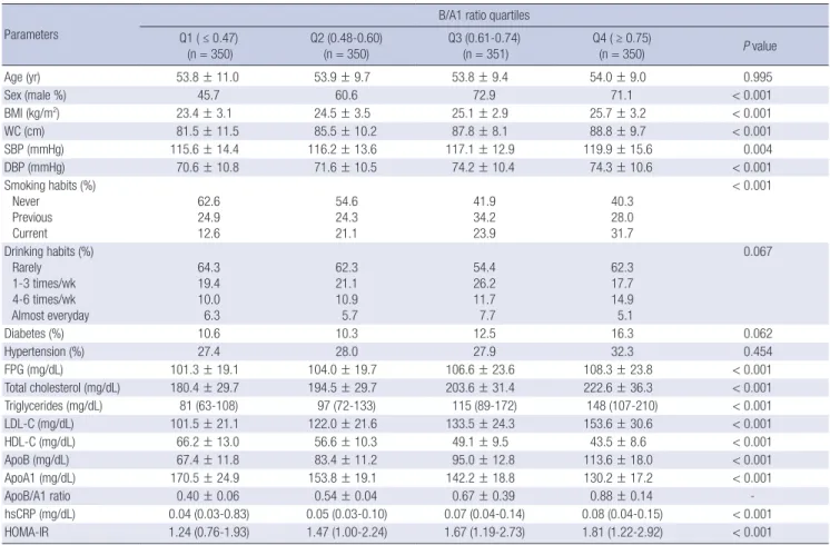
Baseline characteristics according to apoB/A1 quartiles The clinical and biochemical characteristics of the study popu- lation classified according to apoB/A1 quartiles are presented in Table 1. We observed positive relationships between apoB/
A1 quartiles and BMI, WC, SBP, DBP, FPG, uric acid, total cho- lesterol, TG, LDL-C, hsCRP, HOMA-IR, and percentages of male subjects and current smokers (P for trend < 0.001 each except for SBP), and a significant negative relationship between apoB/
A1 quartiles and HDL-C (P for trend < 0.001). We observed no
relationship between apoB/A1 quartiles and the percentages of subjects with diabetes and hypertension.
Coronary CT angiography findings relative to apoB/A1 quartiles
Significant coronary artery stenosis was detectable in 114 sub- jects (8.1%), 89 men and 25 women. An increase in apoB/A1 quartiles was associated with an increased percentage of sub- jects with significant coronary stenosis (P for trend = 0.003, Ta- ble 2). Of the 114 patients with significant stenosis, 87 (76.3%) had single-vessel disease. We also observed a positive relation- ship between apoB/A1 quartiles and increased percentage of subjects with plaques (P for trend = 0.001, Table 2). Among pla- que subtypes, only noncalcified plaques (NCAP) differed sig- nificantly across the apoB/A1 quartiles (P for trend < 0.001, Ta- ble 2). Regarding to CACS, subjects with higher CACS were ob- served in those with higher apoB/A1 quartiles (P for trend = 0.001, Table 2)
Fig. 1 shows the distribution of apoB/A1 ratios according to the presence of significant coronary stenosis. We found that apoB/A1 ratios were significantly higher in subjects with than without stenosis.
Among the 114 subjects with significant stenosis detected by MDCT, 51 (44.7%, 41 men and 10 women) underwent conven- tional coronary angiography. Angiography indicated coronary stenosis consistent with the lesions detected by MDCT in 45 subjects (88.2%), but milder coronary stenosis than indicated by MDCT in 6 subjects (11.8%). Revascularization was perform- ed in 31 subjects (68.9%), 28 (62.2%) by percutaneous coronary intervention and 3 (6.7%) by coronary artery bypass surgery.
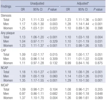
Relationship between apoB/A1 and risk of significant coronary stenosis
Table 3 shows the relationship between apoB/A1 ratio and risk of significant coronary stenosis. For each 0.1 increment in apoB/
A1 ratio, the ORs for significant stenosis, and any plaques in all subjects increased significantly, even after adjustment for con- founding variables such as age, sex, BMI, SBP, DBP, WC, smok- ing and drinking habits, hypertension, diabetes, FPG, hsCRP and HOMA-IR. Multivariate analysis showed that, among pla- que subtypes, only NCAP showed a significant association with apoB/A1 ratio. When we calculated the ORs for significant cor- onary stenosis in men and women separately, we observed sig- nificance only in men (Table 3).
The predictive value of apoB/A1 ratio in detecting significant coronary stenosis
ROC analysis revealed that the optimal apoB/A1 ratio cutoff for detecting significant coronary stenosis by MDCT was 0.58, which had a sensitivity of 70.2% and a specificity of 48.2% (Fig. 2, AUC, 0.61; 95% CI, 0.58-0.63, P < 0.001). Using this cut-off value, the
OR for significant coronary stenosis was 2.06 (95% CI, 1.31-3.24, P = 0.002) after adjusting for age, sex, BMI, SBP, DBP, WC, smo- king and drinking habits, hypertension ,diabetes, FPG, hsCRP and HOMA-IR.
DISCUSSION
We assessed the association between serum apoB/A1 ratios and significant coronary artery stenosis, as determined by MD- Table 1. Baseline patient characteristics according to apoB/A1 ratio quartiles
Parameters
B/A1 ratio quartiles Q1 ( ≤ 0.47)
(n = 350) Q2 (0.48-0.60)
(n = 350) Q3 (0.61-0.74)
(n = 351) Q4 ( ≥ 0.75)
(n = 350) P value
Age (yr) 53.8 ± 11.0 53.9 ± 9.7 53.8 ± 9.4 54.0 ± 9.0 0.995
Sex (male %) 45.7 60.6 72.9 71.1 < 0.001
BMI (kg/m2) 23.4 ± 3.1 24.5 ± 3.5 25.1 ± 2.9 25.7 ± 3.2 < 0.001
WC (cm) 81.5 ± 11.5 85.5 ± 10.2 87.8 ± 8.1 88.8 ± 9.7 < 0.001
SBP (mmHg) 115.6 ± 14.4 116.2 ± 13.6 117.1 ± 12.9 119.9 ± 15.6 0.004
DBP (mmHg) 70.6 ± 10.8 71.6 ± 10.5 74.2 ± 10.4 74.3 ± 10.6 < 0.001
Smoking habits (%) Never
Previous Current
62.6 24.9 12.6
54.6 24.3 21.1
41.9 34.2 23.9
40.3 28.0 31.7
< 0.001
Drinking habits (%) Rarely 1-3 times/wk 4-6 times/wk Almost everyday
64.3 19.4 10.0 6.3
62.3 21.1 10.9 5.7
54.4 26.2 11.7 7.7
62.3 17.7 14.9 5.1
0.067
Diabetes (%) 10.6 10.3 12.5 16.3 0.062
Hypertension (%) 27.4 28.0 27.9 32.3 0.454
FPG (mg/dL) 101.3 ± 19.1 104.0 ± 19.7 106.6 ± 23.6 108.3 ± 23.8 < 0.001
Total cholesterol (mg/dL) 180.4 ± 29.7 194.5 ± 29.7 203.6 ± 31.4 222.6 ± 36.3 < 0.001
Triglycerides (mg/dL) 81 (63-108) 97 (72-133) 115 (89-172) 148 (107-210) < 0.001
LDL-C (mg/dL) 101.5 ± 21.1 122.0 ± 21.6 133.5 ± 24.3 153.6 ± 30.6 < 0.001
HDL-C (mg/dL) 66.2 ± 13.0 56.6 ± 10.3 49.1 ± 9.5 43.5 ± 8.6 < 0.001
ApoB (mg/dL) 67.4 ± 11.8 83.4 ± 11.2 95.0 ± 12.8 113.6 ± 18.0 < 0.001
ApoA1 (mg/dL) 170.5 ± 24.9 153.8 ± 19.1 142.2 ± 18.8 130.2 ± 17.2 < 0.001
ApoB/A1 ratio 0.40 ± 0.06 0.54 ± 0.04 0.67 ± 0.39 0.88 ± 0.14 -
hsCRP (mg/dL) 0.04 (0.03-0.83) 0.05 (0.03-0.10) 0.07 (0.04-0.14) 0.08 (0.04-0.15) < 0.001
HOMA-IR 1.24 (0.76-1.93) 1.47 (1.00-2.24) 1.67 (1.19-2.73) 1.81 (1.22-2.92) < 0.001
BMI, body mass index; WC, waist circumference; SBP, systolic blood pressure; DBP, diastolic blood pressure; FPG, fasting plasma glucose; LDL-C, low density lipoprotein-cho- lesterol; HDL-C, high density lipoprotein-cholesterol; ApoB, apolipoprotein B; ApoA1, apolipoprotein A1; hsCRP, high sensitive C-reactive protein; HOMA-IR, homeostasis model of insulin resistance.
Table 2. Coronary CT angiography results in patients assorted by apoB/A1 ratio quartiles Characteristics, No. (%)
B/A1 ratio quartiles Q1 ( ≤ 0.47)
(n = 350) Q2 (0.48-0.60)
(n = 350) Q3 (0.61-0.74)
(n = 351) Q4 ( ≥ 0.75)
(n = 350) P value
Significant stenosis 19 (5.4) 20 (5.7) 33 (9.4) 42 (12.0) 0.003
Number of stenosed vessel 1
2 3
15 (4.3) 1 (0.3) 3 (0.9)
19 (5.4) 0 (0) 1 (0.3)
22 (6.3) 7 (2.0) 3 (0.9)
32 (9.1) 8 (2.3) 1 (0.3)
0.009
Plaques
Any plaque 83 (23.7) 100 (28.6) 120 (34.2) 130 (37.1) 0.001
Calcified (CAP) 47 (13.4) 52 (14.9) 57 (16.2) 72 (20.6) 0.062
Noncalcified (NCAP) 28 (8.0) 41 (11.7) 71 (20.2) 59 (16.9) < 0.001
Mixed (MCAP) 15 (4.3) 23 (6.6) 27 (7.7) 27 (7.7) 0.217
CACS 0 0.1-100 > 100.1
72.5 20.1 7.4
69.1 22.9 8.0
67.3 24.2 8.5
56.0 33.4 10.6
0.001
CAP, calcified plaque; NCAP, noncalcified plaque; MCAP, mixed calcified plaque; CACS, coronary artery calcium score.
A
Fig. 1. Box-whisker plots of apoB/A1 ratios according to the presence of significant coronary stenosis in total subjects (A), men (B), and women (C). The ends of each whisker indicate the 5th and 95th percentiles of apoB/A1 ratio.
ApoB/A1 ratio
Total
Stenosis (-) Stenosis (+)
2.0 1.5 1.0 0.5 0.0
P < 0.001
ApoB/A1 ratio
Women
Stenosis (-) Stenosis (+)
1.5
1.0
0.5
0.0
P = 0.024
C
ApoB/A1 ratio
Men
Stenosis (-) Stenosis (+)
2.0 1.5 1.0 0.5 0.0
P = 0.003
B
Table 3. Multivariate analysis of the association between apoB/A1 ratio increments of 0.1 with significant coronary stenosis and plaque subtypes
Findings Unadjusted Adjusted*
OR 95% CI P value OR 95% CI P value
Stenosis Total Men Women
1.21 1.17 1.25
1.11-1.33 1.05-1.30 1.03-1.51
< 0.001 0.003 0.023
1.23 1.28 1.10
1.11-1.36 1.14-1.44 0.89-1.36
< 0.001
< 0.001 0.398 Any plaque
Total Men Women
1.13 1.04 1.23
1.06-1.20 0.97-1.11 1.11-1.37
< 0.001 0.329
< 0.001 1.10 1.10 1.11
1.03-1.18 1.01-1.19 0.98-1.26
0.004 0.031 0.105 CAP
Total Men Women
1.09 1.05 1.11
1.02-1.17 0.96-1.14 0.97-1.28
0.015 0.309 0.132
1.08 1.11 0.99
1.00-1.17 1.01-1.22 0.84-1.16
0.051 0.028 0.875 NCAP
Total Men Women
1.18 1.09 1.35
1.10-1.27 1.00-1.19 1.17-1.55
< 0.001 0.060
< 0.001 1.18 1.14 1.29
1.08-1.28 1.03-1.26 1.10-1.50
< 0.001 0.010 0.001 MCAP
Total Men Women
1.09 0.97 1.37
0.98-1.21 0.86-1.11 1.10-1.70
0.104 0.682 0.004
1.08 1.03 1.26
0.96-1.21 0.90-1.18 0.98-1.61
0.205 0.648 0.069
*Adjusted for age, BMI, SBP, DBP, WC, smoking, drinking habits, HTN, diabetes, FPG, hsCRP and HOMA-IR (and sex in the total patients). CAP, calcified plaque; NCAP, non- calcified plaque; MCAP, mixed calcified plaque.
CT, in a large number of Koreans without typical chest pain and/
or a previous history of CVD. Furthermore, we attempted to de- termine the optimal apoB/A1 ratio to identify subjects who might benefit from coronary MDCT. To our knowledge, this is the first study to investigate the ability of serum apoB/A1 ratios to pre- dict coronary atherosclerosis detected by coronary MDCT. We found that an increase in serum apoB/A1 ratio was associated with significant coronary atherosclerosis and NCAP, indepen- dent of conventional risk factors. In addition, we found that an apoB/A1 ratio > 0.58 was optimal for identifying subjects with significant coronary atherosclerosis by MDCT.
Fig. 2. Receiver operating characteristic (ROC) curve and optimal apoB/A1 ratio cut- off value (•) for detecting significant coronary stenosis by multidetector computed to- mography (MDCT).
Sensitivity
100-Specificity
0 20 40 60 80 100
100
80
60
40
20
0 AUC = 0.61 (95% CI = 0.58-0.63, P < 0.001
Total
Sensitivity: 70.2 Specificity: 48.2 Criterion: > 0.58
Atherosclerosis is a diffuse pathological process character- ized by the deposition of lipids and other blood-borne materi- als within the arterial wall of almost all vascular territories (13).
Cholesterol accumulation within vascular walls plays a central role in atherogenesis (13). Although, lipid abnormalities have been considered major risk factors for the development of CHD, many individuals who develop CHD have normal serum lipid
concentrations (14). Apolipoproteins play pivotal roles in cho- lesterol transport and metabolism (2), and apoB/A1 ratio, a mar- ker of the balance between proatherogenic and antiatherogenic lipoproteins (2), may be a good indicator of atherosclerosis in- cluding coronary artery disease.
A high apoB/A1 ratio has been shown to be good marker of future CVD, including myocardial infarction and stroke (3, 15).
Moreover, this ratio has been shown to be a better indicator than any other lipid measurements, including conventional lipid ratios, of the presence and/or severity of coronary artery disease detected by conventional coronary angiography (16- 18). However, despite the close relationship between serum apoB/A1 ratio and CHD detected by conventional coronary an- giography, no previous study attempted to determine the opti- mal apoB/A1 cut-off value for identifying the subjects with sig- nificant coronary stenosis, especially using coronary MDCT.
Approximately 3%-5% of asymptomatic individuals have coronary artery disease (19, 20), and about 5.2% of asymptom- atic individuals have significant coronary artery stenosis, as de- tected by MDCT (21). In comparison, we found that 8.1% of our subjects had significant stenosis on MDCT. Of these subjects, 27.2% underwent revascularization via percutaneous coronary intervention or coronary artery bypass surgery. These findings indicate the importance of identifying asymptomatic individu- als who are at high risk for CHD, in preventing future morbidity and mortality.
The recent introduction of 64-slice MDCT has enabled the noninvasive characterization of atherosclerotic lesions and the determination of luminal and vessel wall alterations (22). MDCT has been shown to have a high diagnostic accuracy as compar- ed with invasive coronary angiography (23). In addition, MDCT allowed us to recognize the plaque burden in subjects with nor- mal perfusion (24, 25). However, despite these advantages and accuracy, its routine use for screening of occult CHD is contro- versial due to its radiation exposure and cost-effectiveness. De- spite this controversy, it is currently widely used as a screening tool for CHD in Korea (26). Furthermore, the prevalence of oc- cult CHD in apparently healthy individuals was not negligible (21). In this background, our study attempted to use apoB/A1 ratios to identify groups of subjects who might benefit from MDCT.
We found that serum apoB/A1 ratios were associated with the presence of plaques, especially NCAPs. Although the amount of calcified plaque (CAP) is highly related to overall plaque bur- den, CAP represents only about 20% of the total atherosclerotic plaque burden (27), and has been associated with advanced stages of atherosclerosis; in contrast, NCAPs are considered a feature of early atherosclerosis (28). Furthermore, there is in- creasing evidence suggesting that NCAPs may be associated with acute coronary syndrome (29, 30). Although no study to date has investigated the association between serum apoB/A1
ratio and the type of coronary plaques, our results indicate that serum apoB/A1 ratio may be especially useful in detecting le- sions with early atherosclerosis.
We observed positive relationships between apoB/A1 quar- tiles and BMI, WC, and smoking habits, in agreement with pre- vious results showing that BMI, waist-to-hip ratio, and cigarette smoking were positively correlated with apoB and negatively correlated with apoA1 concentrations (31). Even after adjusting for these confounding factors, however, we observed an inde- pendent association between elevated apoB/A1 ratio and sig- nificant coronary stenosis on MDCT.
There are many advantages to measuring serum apoB and apoA1 concentrations. For example, these measurements do not require samples from fasting individuals (32). In addition, the methods used to measure apoB and apoA1 have been in- ternationally standardized in reference materials from the World Health Organization and the International Federation of Clini- cal Chemistry and Laboratory Medicine (32). Therefore, mea- surement of serum apoB/A1 ratio may be easily accessible for detecting subjects at risk for occult CHD.
Our study had several limitations. First, due to its cross-sec- tional design, we could not determine whether there was a cau- sal relationship between apoB/A1 ratio and coronary artery stenosis. Second, participants were relatively healthy, and al- though large numbers were analyzed, only small percentages, especially of women, had significant stenosis on MDCT. Third, the study population consisted exclusively of Koreans, so our results may not be applicable to other ethnic groups. Forth, MD- CT only reflects the anatomic severity, not functional ischemia and it has limitation in judging the vulnerability of plaque (33).
Finally, as we did not perform conventional coronary angiogra- phy in all subjects with significant stenosis on MDCT, we may have overdiagnosed significant coronary stenosis. This possibil- ity was unlikely, however, due to the 88.2% concordance rate we observed between conventional coronary angiography and MDCT.
In conclusion, our results indicate that apoB/A1 ratio is a good indicator of occult coronary atherosclerosis detected by coro- nary MDCT and that subjects with apoB/A1 ratio greater than 0.58 are good candidates for coronary MDCT.
DISCLOSURE
The authors have no conflicts of interest to disclose.
REFERENCES
1. Expert Panel on Detection, Evaluation, and Treatment of High Blood Cholesterol in Adults. Executive Summary of the Third Report of the Na- tional Cholesterol Education Program (NCEP) Expert Panel on Detec- tion, Evaluation, and Treatment of High Blood Cholesterol in Adults
(Adult Treatment Panel III). JAMA 2001; 285: 2486-97.
2. Marcovina S, Packard CJ. Measurement and meaning of apolipoprotein AI and apolipoprotein B plasma levels. J Intern Med 2006; 259: 437-46.
3. Yusuf S, Hawken S, Ounpuu S, Dans T, Avezum A, Lanas F, McQueen M, Budaj A, Pais P, Varigos J, et al. Effect of potentially modifiable risk fac- tors associated with myocardial infarction in 52 countries (the INTER- HEART Study): case-control study. Lancet 2004; 364: 937-52.
4. Budoff MJ, Achenbach S, Blumenthal RS, Carr JJ, Goldin JG, Greenland P, Guerci AD, Lima JA, Rader DJ, Rubin GD, et al. Assessment of coro- nary artery disease by cardiac computed tomography: a scientific state- ment from the American Heart Association Committee on Cardiovascu- lar Imaging and Intervention, Council on Cardiovascular Radiology and Intervention, and Committee on Cardiac Imaging, Council on Clin- ical Cardiology. Circulation 2006; 114: 1761-91.
5. Foster G, Shah H, Sarraf G, Ahmadi N, Budoff M. Detection of noncalci- fied and mixed plaque by multirow detector computed tomography. Ex- pert Rev Cardiovasc Ther 2009; 7: 57-64.
6. Achenbach S, Daniel WG. Current role of cardiac computed tomogra- phy. Herz 2007; 32: 97-107.
7. Peppa M, Betsi G, Dimitriadis G. Lipid abnormalities and cardiometa- bolic risk in patients with overt and subclinical thyroid disease. J Lipids 2011; 2011: 575840.
8. Seidel D. Lipoproteins in liver disease. J Clin Chem Clin Biochem 1987;
25: 541-51.
9. Batista MC, Welty FK, Diffenderfer MR, Sarnak MJ, Schaefer EJ, Lamon- Fava S, Asztalos BF, Dolnikowski GG, Brousseau ME, Marsh JB. Apoli- poprotein A-I, B-100, and B-48 metabolism in subjects with chronic kid- ney disease, obesity, and the metabolic syndrome. Metabolism 2004; 53:
1255-61.
10. Austen WG, Edwards JE, Frye RL, Gensini GG, Gott VL, Griffith LS, Mc- Goon DC, Murphy ML, Roe BB. A reporting system on patients evaluat- ed for coronary artery disease: report of the Ad Hoc Committee for Grad- ing of Coronary Artery Disease, Council on Cardiovascular Surgery, Ame- rican Heart Association. Circulation 1975; 51: 5-40.
11. Leber AW, Becker A, Knez A, von Ziegler F, Sirol M, Nikolaou K, Ohne- sorge B, Fayad ZA, Becker CR, Reiser M, et al. Accuracy of 64-slice com- puted tomography to classify and quantify plaque volumes in the proxi- mal coronary system: a comparative study using intravascular ultra- sound. J Am Coll Cardiol 2006; 47: 672-7.
12. Agatston AS, Janowitz WR, Hildner FJ, Zusmer NR, Viamonte M Jr, De- trano R. Quantification of coronary artery calcium using ultrafast com- puted tomography. J Am Coll Cardiol 1990; 15: 827-32.
13. Badimon JJ, Ibanez B, Cimmino G. Genesis and dynamics of atheroscle- rotic lesions: implications for early detection. Cerebrovasc Dis 2009; 27:
38-47.
14. Holmes DR Jr, Elveback LR, Frye RL, Kottke BA, Ellefson RD. Associa- tion of risk factor variables and coronary artery disease documented with angiography. Circulation 1981; 63: 293-9.
15. Walldius G, Jungner I, Holme I, Aastveit AH, Kolar W, Steiner E. High apolipoprotein B, low apolipoprotein A-I, and improvement in the pre- diction of fatal myocardial infarction (AMORIS study): a prospective study. Lancet 2001; 358: 2026-33.
16. Noma A, Yokosuka T, Kitamura K. Plasma lipids and apolipoproteins as discriminators for presence and severity of angiographically defined cor- onary artery disease. Atherosclerosis 1983; 49: 1-7.
17. Reinhart RA, Gani K, Arndt MR, Broste SK. Apolipoproteins A-I and B as predictors of angiographically defined coronary artery disease. Arch Intern Med 1990; 150: 1629-33.
18. Enkhmaa B, Anuurad E, Zhang Z, Pearson TA, Berglund L. Usefulness of apolipoprotein B/apolipoprotein A-I ratio to predict coronary artery disease independent of the metabolic syndrome in African Americans.
Am J Cardiol 2010; 106: 1264-9.
19. Thaulow E, Erikssen J, Sandvik L, Erikssen G, Jorgensen L, Cohn PF. Ini- tial clinical presentation of cardiac disease in asymptomatic men with silent myocardial ischemia and angiographically documented coronary artery disease (the Oslo Ischemia Study). Am J Cardiol 1993; 72: 629-33.
20. Pilote L, Pashkow F, Thomas JD, Snader CE, Harvey SA, Marwick TH, Lauer MS. Clinical yield and cost of exercise treadmill testing to screen for coronary artery disease in asymptomatic adults. Am J Cardiol 1998;
81: 219-24.
21. Choi EK, Choi SI, Rivera JJ, Nasir K, Chang SA, Chun EJ, Kim HK, Choi DJ, Blumenthal RS, Chang HJ. Coronary computed tomography angiog- raphy as a screening tool for the detection of occult coronary artery dis- ease in asymptomatic individuals. J Am Coll Cardiol 2008; 52: 357-65.
22. Schuijf JD, van Werkhoven JM, Pundziute G, Jukema JW, Decramer I, Stokkel MP, Dibbets-Schneider P, Schalij MJ, Reiber JH, van der Wall EE, et al. Invasive versus noninvasive evaluation of coronary artery dis- ease. JACC Cardiovasc Imaging 2008; 1: 190-9.
23. Vanhoenacker PK, Heijenbrok-Kal MH, Van Heste R, Decramer I, Van Hoe LR, Wijns W, Hunink MG. Diagnostic performance of multidetector CT angiography for assessment of coronary artery disease: meta-analy- sis. Radiology 2007; 244: 419-28.
24. Hacker M, Jakobs T, Hack N, Nikolaou K, Becker C, von Ziegler F, Knez A, König A, Klauss V, Reiser M, et al. Sixty-four slice spiral CT angiogra- phy does not predict the functional relevance of coronary artery stenoses in patients with stable angina. Eur J Nucl Med Mol Imaging 2007; 34:
4-10.
25. Schuijf JD, Wijns W, Jukema JW, Atsma DE, de Roos A, Lamb HJ, Stok- kel MP, Dibbets-Schneider P, Decramer I, De Bondt P, et al. Relation- ship between noninvasive coronary angiography with multi-slice com- puted tomography and myocardial perfusion imaging. J Am Coll Cardi- ol 2006; 48: 2508-14.
26. Nam HJ, Jung IH, Kim J, Kim JH, Suh J, Kim HS, Kim HK, Jung YJ, Kang JW, Lee S. Association between brachial-ankle pulse wave velocity and occult coronary artery disease detected by multi-detector computed to- mography. Int J Cardiol 2012; 157: 227-32.
27. Rumberger JA, Simons DB, Fitzpatrick LA, Sheedy PF, Schwartz RS.
Coronary artery calcium area by electron-beam computed tomography and coronary atherosclerotic plaque area: a histopathologic correlative study. Circulation 1995; 92: 2157-62.
28. Stary HC, Chandler AB, Dinsmore RE, Fuster V, Glagov S, Insull W Jr, Rosenfeld ME, Schwartz CJ, Wagner WD, Wissler RW. A definition of advanced types of atherosclerotic lesions and a histological classification of atherosclerosis: a report from the Committee on Vascular Lesions of the Council on Arteriosclerosis, American Heart Association. Circulation 1995; 92: 1355-74.
29. Hoffmann U, Moselewski F, Nieman K, Jang IK, Ferencik M, Rahman AM, Cury RC, Abbara S, Joneidi-Jafari H, Achenbach S, et al. Noninva- sive assessment of plaque morphology and composition in culprit and stable lesions in acute coronary syndrome and stable lesions in stable
angina by multidetector computed tomography. J Am Coll Cardiol 2006;
47: 1655-62.
30. Fujii K, Kobayashi Y, Mintz GS, Takebayashi H, Dangas G, Moussa I, Mehran R, Lansky AJ, Kreps E, Collins M, et al. Intravascular ultrasound assessment of ulcerated ruptured plaques: a comparison of culprit and nonculprit lesions of patients with acute coronary syndromes and lesions in patients without acute coronary syndromes. Circulation 2003; 108:
2473-8.
31. Kinlay S, Dobson AJ, Heller RF, Dickeson JE, Ryan S. Lipid and apolipo-
protein levels in an Australian community. Med J Aust 1991; 154: 170-5.
32. Marcovina SM, Albers JJ, Kennedy H, Mei JV, Henderson LO, Hannon WH. International Federation of Clinical Chemistry standardization project for measurements of apolipoproteins A-I and B: IV. comparabili- ty of apolipoprotein B values by use of International Reference Material.
Clin Chem 1994; 40: 586-92.
33. Waxman S, Ishibashi F, Muller JE. Detection and treatment of vulnerable plaques and vulnerable patients: novel approaches to prevention of cor- onary events. Circulation 2006; 114: 2390-411.