Received June 22, 2013, Revised August 19, 2013, Accepted for publication September 20, 2013
Corresponding author: Hae Jun Song, Department of Dermatology, Korea University Guro Hospital, 148 Gurodong-ro, Guro-gu, Seoul 152-703, Korea. Tel: 82-2-2626-1308, Fax: 82-2-838-2359, E-mail:
This is an Open Access article distributed under the terms of the Creative Commons Attribution Non-Commercial License (http://
creativecommons.org/licenses/by-nc/3.0) which permits unrestricted non-commercial use, distribution, and reproduction in any medium, provided the original work is properly cited.
ORIGINAL ARTICLE
The Antimicrobial Activity of (-)-Epigallocatehin-3-Gallate and Green Tea Extracts against Pseudomonas aeruginosa and Escherichia coli Isolated from Skin Wounds
Jiehyun Jeon, Joo Ha Kim, Chang Kyu Lee1, Chil Hwan Oh, Hae Jun Song
Departments of Dermatology and 1Laboratory Medicine, Korea University College of Medicine, Seoul, Korea
Background: Skin infections with Gram-negative bacteria are sometimes challenging to treat, because these bacteria show multidrug resistance against commonly used antibiotics and patients with Gram-negative bacterial infection overall have deteriorated in conditions in many cases. Studies have shown that epigallocatechin gallate (EGCG) and green tea extracts (GTE) inhibit the growth of several Gram-positive bacteria species. Objective: The purpose of this study was to investigate the minimum inhibitory concentrations (MICs) of EGCG and GTE in Pseudomonas aeruginosa and Escherichia coli, and assess the use of these chemicals as an alternative or adjunct topical antimicrobial agent against P. aeruginosa and E. coli with multidrug resistance. Methods: The MICs of EGCG, GTE, and other tested antibiotics were measured and compared to determine the antibacterial efficacy and the differences in pattern of resistance. Results: The P. aeru- ginosa and E. coli strains used in this study showed multidrug resistance. EGCG inhibited the growth of P. aeruginosa at a MIC level of 200∼400 μg/ml. The MIC of GTE was a 1 : 16 dilution for P. aeruginosa. EGCG showed antimicrobial activity against E. coli at a MIC of 400 μg/ml. In the case of GTE, the MIC was a dilution between 1:8 and 1:4 for E.
coli. Conclusion: EGCG and GTE showed potential as alternative or adjunct topical antimicrobial agents for
infections that are resistant to traditional antibiotic therapy.
(Ann Dermatol 26(5) 564∼569, 2014) -Keywords-
Escherichia coli, Epigallocatechin gallate, Gram-negative bacterial infection, Microbial sensitivity tests, Pseudomo- nas aeruginosa
INTRODUCTION
Skin infections with Pseudomonas aeruginosa and Esch- erichia coli occur more frequently in patients who are immunologically compromised, elderly, or who are in intensive care units (ICU). These patients may have other comorbidities, including deteriorated renal and hepatic function, administration of multiple other drugs, and complications from chronic diseases. Thus, the admini- stration of systemic antibiotics to these patients has a higher risk of side effects. In addition, there is a narrow range of antibiotics that can be used to treat infection, because many strains of P. aeruginosa and E. coli show resistance to certain antibiotics.
Useful topical treatment for Gram-negative bacterial infection is limited. The use of topical antibiotics and antiseptics are also limited due to bacterial resistance and the inherent nature of treatment agents. For instance, some antibiotics/antiseptics contain superoxide-generating hydrogen peroxide. Further, potassium permanganate and chlorhexidine are cytotoxic and can inhibit the regener- ation of cells for wound repair1,2.
Epigallocatechin gallate (EGCG) is a major catechin found in green tea extracts (GTE), and has remarkable anti- bacterial activity as well as anti-oxidant, anti-inflammatory, and anti-cancer effects. Further, EGCG can induce epithelial
proliferation and differentiation3-5. EGCG is known to be active against Gram-positive bacteria4,6. It is also known that the minimum inhibitory concentration (MIC) of EGCG in Gram-negative bacteria appears to be 8- to 16-fold higher than that in Gram-positive bacteria6. The relatively lower antimicrobial activity of catechins against Gram- negative bacteria is due to the protection of the Gram- negative bacteria by the outer membrane and lipopoly- saccharides.
This study was conducted to assess the possible use of EGCG and GTE as antibacterial agents in Gram-negative P. aeruginosa and E. coli, regardless of antibiotic resistance status.
MATERIALS AND METHODS
Bacterial strains
The studied bacterial strains were isolated from the skin wounds of patients admitted to the ICU at our University Hospital. Twenty-two strains in total were studied, and consisted of 10 P. aeruginosa strains, 10 E. coli strains, and 2 reference strains (P. aeruginosa ATCC 27853, E. coli ATCC 25922) for quality control. Cultured strains were kept at a turbidity matching a 0.5 McFarland standard and placed in each well with a final concentration of 2.5×10⁵ colony-forming unit (CFU) /ml.
Antibiotic susceptibility tests of the bacterial strains Specimens from the skin wounds of the ICU patients were inoculated and cultured on blood agar medium and MacConkey agar medium for 24 to 48 hours at 35oC. The identification of bacterial isolates was performed manually and processed simultaneously using the ATB ID system (BioMériuex, Marcy l’Etoile, France). Antibiotic suscepti- bility was assessed using the disk diffusion method following the guidelines published by the Clinical and Laboratory Standards Institute (CLSI)7 for ampicillin, ami- kacin, aztreonam, cefoxitin, cefepime, cephalothin, cefo- taxime, ciprofloxacin, ceftazidime, gentamicin, imipenem, piperacillin, piperacillin/tazobactam, and sulperazone. The results of the antibiotic susceptibility tests of isolated strains were compared with the results of EGCG and GTE susceptibility tests.
EGCG and GTE preparation
EGCG (Sigma, St. Louis, MO, USA) in powder form with 95% purity was dissolved in normal saline and serially diluted with cation-adjusted Mueller-Hinton broth (MHB) starting from a maximum concentration of 800 μg/ml to 0.4 μg/ml with 12 levels of dilution. Two grams of dried green tea leaves (product name: Halla; Amorepacific
Corporation, Seoul, Korea) were brewed with 100 ml of boiling water for 10 min. After removing the tea leaves, the infusion was left for another 10 min and then cooled down to below 40oC. The green tea infusion was filtered twice using Whatman filter paper (Whatman International Ltd., Maidstone, UK). Final filtration by a filter with a pore size of 0.22 μm was performed to remove possible microbial contaminants. Crude GTE was diluted from un- diluted extract in 12 dilution stages (1:1∼1:2,048) by successively adding two-fold MHB.
Measuring minimal inhibitory concentration
Gentamicin (Sigma) and ciprofloxacin (Fluka, Saint Gallen, Switzerland) were used as reference antibiotics to assess the antimicrobial activity of EGCG and GTE against P.
aeruginosa8. Ampicillin (Sigma) and ciprofloxacin were tested against E. coli. Ciprofloxacin was dissolved in 0.1 N HCl, and other antibiotics and EGCG were dissolved in normal saline. The antibiotics were serially diluted with cation-adjusted Muller-Hinton broth from a concentration of 256 μg/ml to 0.125 μg/ml, and 200 μl of the serially diluted antibiotics, EGCG or GTE was placed in a 96-well plate (Corning, Acton, MA, USA). E. coli and P. aeruginosa were inoculated in every well at a concentration of 2.5×10⁵ CFU/ml. The MIC was visually determined after 16∼20 hours of incubation at 37oC according to the broth microdilution methods suggested by the CLSI8.
RESULTS
Antibiotic susceptibility of the studied strains of P.
aeruginosa and E. coli
1) P. aeruginosa
Five of the ten collected strains and the reference strain were susceptible to all of the tested antibiotics, while the other five strains showed multidrug resistance. These multidrug-resistant strains were commonly resistant to ciprofloxacin, gentamicin, piperacillin, and piperacillin/
tazobactam. All ten of the studied strains were susceptible to imipenem and ceftazidime (Table 1).
2) E. coli
Only one of the collected E. coli strains and the reference strain were susceptible to all of the tested antibiotics. Nine of the ten strains were resistant to at least one antibiotic (up to nine antibiotics), although they did not show any similarities in their patterns of resistance. All of the E. coli strains were susceptible to cefoxitin, amikacin, imipenem, piperacillin/tazobactam, and cefoperazone/sulbactam (Table 1).
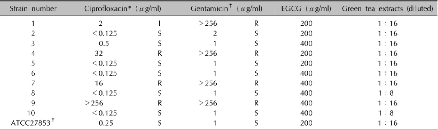
Table 2. MICs of tested antibiotic agents against Pseudomonas aeruginosa
Strain number Ciprofloxacin* (μg/ml) Gentamicin† (μg/ml) EGCG (μg/ml) Green tea extracts (diluted)
1 2 I >256 R 200 1:16
2 <0.125 S 2 S 200 1:16
3 0.5 S 1 S 400 1:16
4 32 R >256 R 200 1:16
5 <0.125 S 1 S 200 1:16
6 <0.125 S 1 S 400 1:16
7 16 R >256 R 400 1:16
8 <0.125 S 1 S 400 1:8
9 >256 R >256 R 400 1:16
10 <0.125 S 1 S 400 1:8
ATCC27853‡ 0.25 S 1 S 200 1:16
MIC: minimum inhibitory concentration, EGCG: epigallocatechin gallate, I: intermediate, R: resistant, S: sensitive. *MIC breakpoints of gentamicin: S, ≤4 μg/ml; I, 8 μg/ml; R, ≥16 μg/ml. †MIC breakpoints of ciprofloxacin: S, ≤1 μg/ml; I, 2 μg/ml; R, ≥4 μg/ml. ‡Acceptable limits for quality control to monitor the accuracy of the MICs of ATCC27853: gentamicin, 0.5∼2 μg/ml;
ciprofloxacin, 0.12∼1 μg/ml.
Table 1. Antibiotic susceptibility test results of 20 clinical isolates used in this study
Strain AM CP CTX CE CI CX GM AN AZ CZ IM PI PT SP
1 Pseudomonas aeruginosa R R R R I S S R R R
2 P. aeruginosa S S S S S S S S S S
3 P. aeruginosa S S S S I S S S S S
4 P. aeruginosa I R R R I I S R R I
5 P. aeruginosa S S S S S S S S S S
6 P. aeruginosa S S S S I S S S S S
7 P. aeruginosa R R R R I S S R R R
8 P. aeruginosa S S S S S S S S S S
9 P. aeruginosa R R R S R S S R R I
10 P. aeruginosa S S S S S S S S S S
ATCC27853
P. aeruginosa S S S S S S S S S S
1 Escherichia coli R R S S S R R S S S S R S S
2 E. coli R S S S S R S S S S S S S S
3 E. coli S S S S S R S S S S S S S S
4 E. coli R R R S R S S S R R S R S S
5 E. coli R R R S R R R S R R S R S S
6 E. coli S S S S S S S S S S S S S S
7 E. coli R R R S R R R S R R S R S S
8 E. coli R R R S R R R S R R S R S S
9 E. coli R S S S S R S S S S S R S S
10 E. coli R S S S S S R S S S S R S S
ATCC25922 E. coli
S S S S S S S S S S S S S S
AM: ampicillin, CP: cephalothin, CTX: cefotaxime, CE: cefoxitin, CI: cefepime, CX: ciprofloxacin, GM: gentamicin, AN: amikacin, AZ: aztreonam, CZ: ceftazidime, IM: imipenem, PI: piperacillin, PT: piperacillin/tazobactam, SP: sulperazone, R: resistant, I: intermediate, S: sensitive.
MICs of ciprofloxacin, gentamicin, EGCG, and GTE against P. aeruginosa
1) Three P. aeruginosa strains were resistant to ciproflo- xacin. However, the growth of one strain was not suppre-
ssed, even at a concentration of 256 μg/ml. The reference strain and six of the studied P. aeruginosa strains were susceptible to gentamicin, with MICs around 1 μg/ ml.
The other four P. aeruginosa strains showed a high degree of resistance to gentamicin, even up to 256 μg/ml (Table 2).
Table 3. MICs of tested antibiotic agents against Escherichia coli
Strain number Ciprofloxacin* (μg/ml) Ampicillin† (μg/ml) EGCG (μg/ml) Green tea extracts (diluted)
1 16 R >256 R 400 1:8
2 64 R 256 R 400 1:8
3 32 R 4 S 400 1:4
4 0.25 S >256 R 400 1:4
5 128 R >256 R 400 1:4
6 <0.125 S 4 S 400 1:8
7 32 R >256 R 400 1:4
8 256 R >256 R 400 1:8
9 16 R >256 R 400 1:4
10 0.25 S >256 R 400 1:4
ATCC25922‡ <0.125 S 2 S 400 1:16
MIC: minimum inhibitory concentration, R: resistant, S: sensitive. *MIC breakpoints of ciprofloxacin: S, ≤8 μg/ml; I, 16 μg/ml;
R, ≥32 μg/ml. †MIC breakpoints of ampicillin: S, ≤1 μg/ml; I, 2 μg/ml; R, ≥4 μg/ml. ‡Acceptable limits for quality control to monitor the accuracy of the MICs of ATCC25922: ampicillin, 2∼8 μg/ml; ciprofloxacin, 0.004∼0.016 μg/ml.
2) The MIC of EGCG against six of the P. aeruginosa strains was 400 μg/ml, while the MIC of the other four strains and reference strain was 200 μg/ml. Two of the ten P. aeruginosa strains were susceptible an 8-fold dilution of crude GTE, whereas the other eight strains and the reference strain were susceptible at a 16-fold dilution (Table 2).
3) The MIC of EGCG and the dilution levels of GTE were not related to the degree of resistance of P. aeruginosa to ciprofloxacin and gentamicin.
MICs of ciprofloxacin, ampicillin, EGCG, and GTE against E. coli
1) Three of the ten strains of E. coli and the reference strain were susceptible to ciprofloxacin. The E. coli studied had a higher MIC and a higher incidence of resistance to antibiotics than P. aeruginosa. Eight of the E.
coli strains were resistant to ampicillin, with uninhibited growth at a concentration of 256 μg/ml (Table 3).
2) The MIC of EGCG against all E. coli strains, including reference strain, was 400 μg/ml. In the case of GTE, the growth of the reference strain was inhibited at a 1:16 dilution. The MIC of GTE for four E. coli strains was an 8-fold dilution, whereas the MIC of the other six E. coli strains was a 4-fold dilution (Table 3).
3) The degree of resistance of the E. coli strains to cipro- floxacin and ampicillin was not related to the MIC of EGCG and GTE.
DISCUSSION
Skin and soft tissue infections with P. aeruginosa and E.
coli have been an issue since both incidence and drug resistance are increasing. In healthy people, P. aeruginosa rarely causes infection; however, it poses a serious health risk in hospitals, where it is responsible for about 10% of in-hospital infections in immunocompromised patients, including those with cancer, diabetes, and hematological disorders, as well as patients undergoing transplant, receiving implants, and being treated with corticosteroids and antibiotics9. In Taiwan, it was reported that E. coli was responsible for almost one-fifth of skin and soft tissue infections10. In Europe, E. coli accounted for 10.8% of infections, and the corresponding rate was 7.2% for North America11. With the observed increase in the rate of bacterial resistance to an extended spectrum of antibiotics, clinical cures and the selection of active antibiotics for empirical treatment may be more difficult to achieve12. A major component of green tea is a flavonol known as catechin13. (−)-Epicatechin (EC), (−)-epigallocatechin (EGC), (−)-epicatechin gallate (ECG), and EGCG are the four main catechins. In particular, EGCG is found only in green tea, comprising 40%∼50% of green tea catechins and is thought to be primarily responsible for the antibacterial and bactericidal properties of green tea4-6,14,15. Galloca- techin and gallate are necessary moieties for antibacterial activity, with gallate-conjugated (−)-ECG and (−)-EGCG demonstrating more powerful antibacterial activity than non-gallate-conjugated (−)-EGC and (−)-EC14.
It has been reported that the bactericidal effect of EGCG is stronger in Gram-positive bacteria than in Gram-negative bacteria, owing to the different amounts of EGCG absor-
bed by the bacterial cells6,15,16. While the MIC of EGCG against Staphylococcus aureus, Staphylococcus epidermi- dis, Staphylococcus hominis, and Staphylococcus hae- molyticus has been reported to be 50∼100 μg/ml, the MIC of EGCG against Klebsiella pneumoniae, Salmonella typhi, and Proteus mirabilis is much higher (800 μg/ml)6. GTE has also shown various degrees of antibacterial activity, and a wide range of susceptibility against different strains of the same species6,17,18. One proposed mechanism for the bactericidal action of catechin is that the negatively charged EGCG combines with the positively-charged bacterial lipid polysaccharide membrane generating hydrogen peroxide (H2O2), which damages the bacterial membrane3,15. Gram-negative bacteria are generally more resistant to catechins than Gram-positive bacteria, due to the presence of strong negative charge of lipopolysacc- harides on the exterior outer member of Gram-negative bacteria16. EGCG is known to have unique dual actions, and it protects human keratinocytes and fibroblasts against H2O2 by reversing the H2O2-induced decrease of supero- xide dismutase (SOD) and glutathione peroxidase19,20. The amount of EGCG or catechins in green tea differs depending on the product and extraction method.
However, when green tea is infused in hot water for 3 min in a proportion of 1 g of leaves to 100 ml of water, the tea usually contains 250∼280 mg of solids, of which 30%∼
42% are catechins3. When green tea or EGCG capsules are orally administered, only 0.2%∼2.0% of the ingested EGCG is intestinally absorbed and appears in the blood17,18. Considering its low plasma concentrations and the reported MIC of EGCG against Gram-negative and Gram-positive bacteria, topical application of EGCG on infected lesions is more desirable than systemic admini- stration, since the concentrations required to treat bacterial infections of the skin cannot be reached through drinking green tea.
Based on the previously reported proportion of catechins in green tea infusions, the estimated concentration of EGCG in the crude infusion of GTE prepared in this study was roughly 400∼800 μg/ml21,22. At a GTE dilution level of 1:16, the EGCG concentration was about 25∼50 μg/
ml. Despite the low concentration of EGCG in the tested GTE, GTE showed antibacterial activity equivalent to 400 μg/ml EGCG. This can be explained through the syner- gistic antibacterial action of other polyphenols in GTE, such as ECG. Considering that even a 16-fold dilution of crude GTE showed effective antibacterial activity, GTE may be practical and economically feasible for use as an alternative for topical antibiotics or as dressing agents in clinical practices. Furthermore, the clinical application of catechin is plausible, since catechin is very stable under
physical manipulations, such as freezing and heating, and can be refrigerated in an aqueous solution for over a month with good stability23,24.
The bacterial strains in this study were isolated from the ulcers and sores of long-stay patients admitted to the ICU.
Thus, the incidence of multidrug resistant strains was higher than community acquired strains. However, bacterial strains showing multidrug resistance did not have the same pattern of resistance against EGCG or GTE, and showed susceptibility to EGCG or GTE independent of the antibiotic resistance status. Though the antibacterial effects of EGCG and GTE varied with the individual strains of bacteria, consistent levels of effectiveness were seen regard- less of the susceptibility of bacteria to the reference antibiotics. Since EGCG exerts antibacterial effects through diverse mechanisms in vivo, the effective MICs of EGCG against P. aeruginosa and E. coli on skin lesions will be lower than the MICs seen in this study.
Better antimicrobial effects can be expected from GTE than EGCG, since GTE contains various types of catechins with antibacterial activity other than the four major catechins: EGCG, ECG, EGC, and EC. Furthermore, GTE is easily available and would be more cost-effective. Although more studies on their mechanisms of action are needed, EGCG and GTE have great potential for use as topical antimicrobial agents with systemic antibiotics to manage skin infections. Several experiments that tested the ability of EGCG to synergistically inhibit methicillin-resistant S.
aureus with concomitant use of oxytetracycline, carbapenem, and ampicillin/sulbactam in vitro demonstrated that EGCG synergistically inhibited bacterial growth25-27. An in vivo study of chronic E. coli bacterial prostatitis rat model showed synergistic effects between an oral gavage of 300 mg/kg body weight of catechin concentrate and cipro- floxacin28.
The clinical application of EGCG and GTE is worth considering as a therapeutic in pursuit of overcoming the increasing antibiotic resistance of bacteria and further studies are needed29.
REFERENCES
1. Sagripanti JL, Bonifacino A. Cytotoxicity of liquid disinfectants.
Surg Infect (Larchmt) 2000;1:3-14.
2. Hidalgo E, Dominguez C. Mechanisms underlying chlor- hexidine-induced cytotoxicity. Toxicol In Vitro 2001;15:
271-276.
3. Balentine DA, Wiseman SA, Bouwens LC. The chemistry of tea flavonoids. Crit Rev Food Sci Nutr 1997;37:693-704.
4. Taylor PW, Hamilton-Miller JM, Stapleton PD. Antimicrobial properties of green tea catechins. Food Sci Technol Bull 2005;2:71-81.
5. Hamilton-Miller JM. Antimicrobial properties of tea (Camellia sinensis L.). Antimicrob Agents Chemother 1995;39:2375- 2377.
6. Yoda Y, Hu ZQ, Zhao WH, Shimamura T. Different suscep- tibilities of Staphylococcus and Gram-negative rods to epigallocatechin gallate. J Infect Chemother 2004;10:55-58.
7. Clinical and Laboratory Standards Institute. Performance standards for antimicrobial disk susceptibility tests. Appro- ved standard M2-A7. 7th ed. Wayne, PA, USA: NCCLS, 2000.
8. Clinical and Laboratory Standards Institute. Methods for dilution antimicrobial susceptibility tests for bacteria that grow aerobically. Approved Standard M7-A6. 6th ed. Way- ne, PA, USA: NCCLS, 2003.
9. Andonova M, Urumova V. Immune surveillance mechanisms of the skin against the stealth infection strategy of Pseu- domonas aeruginosa-review. Comp Immunol Microbiol Infect Dis 2013;36:433-448.
10. Chang CM, Lee HC, Lee NY, Lee IW, Wu CJ, Chen PL, et al.
Community-acquired Klebsiella pneumoniae complicated skin and soft-tissue infections of extremities: emphasis on cirrhotic patients and gas formation. Infection 2008;36:
328-334.
11. Moet GJ, Jones RN, Biedenbach DJ, Stilwell MG, Fritsche TR. Contemporary causes of skin and soft tissue infections in North America, Latin America, and Europe: report from the SENTRY Antimicrobial Surveillance Program (1998-2004).
Diagn Microbiol Infect Dis 2007;57:7-13.
12. Ruef C. Complicated skin and soft-tissue infections--consider gram-negative pathogens. Infection 2008;36:295.
13. Graham HN. Green tea composition, consumption, and polyphenol chemistry. Prev Med 1992;21:334-350.
14. Toda M, Okubo S, Ikigai H, Shimamura T. Antibacterial and anti-hemolysin activities of tea catechins and their structural relatives. Nihon Saikingaku Zasshi 1990;45:561-566.
15. Arakawa H, Maeda M, Okubo S, Shimamura T. Role of hydrogen peroxide in bactericidal action of catechin. Biol Pharm Bull 2004;27:277-281.
16. Ikigai H, Nakae T, Hara Y, Shimamura T. Bactericidal catechins damage the lipid bilayer. Biochim Biophys Acta 1993;1147:132-136.
17. Nakagawa K, Miyazawa T. Absorption and distribution of tea catechin, (-)-epigallocatechin-3-gallate, in the rat. J Nutr Sci Vitaminol (Tokyo) 1997;43:679-684.
18. Nakagawa K, Okuda S, Miyazawa T. Dose-dependent incor- poration of tea catechins, (-)-epigallocatechin-3-gallate and
(-)-epigallocatechin, into human plasma. Biosci Biotechnol Biochem 1997;61:1981-1985.
19. Feng B, Fang Y, Wei SM. Effect and mechanism of epigall- ocatechin-3-gallate (EGCG). against the hydrogen peroxide- induced oxidative damage in human dermal fibroblasts. J Cosmet Sci 2013;64:35-44.
20. Elbling L, Herbacek I, Weiss RM, Jantschitsch C, Micksche M, Gerner C, et al. Hydrogen peroxide mediates EGCG- induced antioxidant protection in human keratinocytes. Free Radic Biol Med 2010;49:1444-1452.
21. Khokhar S, Magnusdottir SG. Total phenol, catechin, and caffeine contents of teas commonly consumed in the United kingdom. J Agric Food Chem 2002;50:565-570.
22. El-Shahawi MS, Hamza A, Bahaffi SO, Al-Sibaai AA, Abduljabbar TN. Analysis of some selected catechins and caffeine in green tea by high performance liquid chrom- atography. Food Chem 2012;134:2268-2275.
23. Chow HH, Cai Y, Hakim IA, Crowell JA, Shahi F, Brooks CA, et al. Pharmacokinetics and safety of green tea poly- phenols after multiple-dose administration of epigallo- catechin gallate and polyphenon E in healthy individuals.
Clin Cancer Res 2003;9:3312-3319.
24. Morita O, Kirkpatrick JB, Tamaki Y, Chengelis CP, Beck MJ, Bruner RH. Safety assessment of heat-sterilized green tea catechin preparation: a 6-month repeat-dose study in rats.
Food Chem Toxicol 2009;47:1760-1770.
25. Novy P, Rondevaldova J, Kourimska L, Kokoska L. Synergistic interactions of epigallocatechin gallate and oxytetracycline against various drug resistant Staphylococcus aureus strains in vitro. Phytomedicine 2013;20:432-435.
26. Hu ZQ, Zhao WH, Asano N, Yoda Y, Hara Y, Shimamura T.
Epigallocatechin gallate synergistically enhances the activity of carbapenems against methicillin-resistant Staphylococcus aureus. Antimicrob Agents Chemother 2002;46:558-560.
27. Hu ZQ, Zhao WH, Hara Y, Shimamura T. Epigallocatechin gallate synergy with ampicillin/sulbactam against 28 clinical isolates of methicillin-resistant Staphylococcus aureus. J An- timicrob Chemother 2001;48:361-364.
28. Lee YS, Han CH, Kang SH, Lee SJ, Kim SW, Shin OR, et al.
Synergistic effect between catechin and ciprofloxacin on chronic bacterial prostatitis rat model. Int J Urol 2005;12:
383-389.
29. Navon-Venezia S, Ben-Ami R, Carmeli Y. Update on Pseu- domonas aeruginosa and Acinetobacter baumannii infec- tions in the healthcare setting. Curr Opin Infect Dis 2005;
18:306-313.