Local Recurrence at the Bronchial Stump Site of Post-Operative Non-Small Cell Lung Cancer Patients:
Comparison of CT Findings and Bronchoscopy
1Sanghyeok Lim, M.D., Woocheol Kwon, M.D., Seung-Whan Cha, M.D.,
Myung Soon Kim, M.D., Daekeon Lim, M.D., Yunjoo Park, M.D., Won-Yeon Lee, M.D.2, Sang-Ha Kim, M.D.2, Tae Hoon Kim, M.D.3
1Department of Radiology, Wonju Christian Hospital, Yonsei University Wonju College of Medicine
2Department of Internal Medicine, Wonju Christian Hospital, Yonsei University Wonju College of Medicine
3Department of Radiology, Yongdong Severance Hospital, Yonsei University College of Medicine 이 논문은 2006학년도 연세대학교 학술연구비의 지원에 의하여 이루어진 것임.
Received August 25, 2008 ; Accepted October 14, 2008
Address reprint requests to : Woocheol Kwon, M.D., Department of Radiology, Yonsei University Wonju College of Medicine, Wonju Christian Hospital, 162 Ilsan-dong, Wonju, Gangwondo 220-701, Republic of Korea.
Tel. 82-33-741-1467 Fax. 82-33-732-8281 E-mail: [email protected]
Purpose: To compare computed tomography (CT) images and bronchoscopic findings of local tumor recurrence at the bronchial stump site in post-operative non-small cell lung cancer (NSCLC) patients.
Materials and Methods: A retrospective study was conducted to review the CT images of 9 lung cancer cases that recurred at the bronchial stump site on 576 resected prima- ry non-small cell lung cancers over a 9-year period. The CT images of the bronchial stump site recurrence were classified as: bronchial wall thickening, nodular or endo- bronchial polypoid lesion, multiplicity, and enhancement patterns. We classified the bronchoscopic findings based on the revised classification by the Japan Lung Cancer Society.
Results: The histologic types of the 9 cases of lung cancer that recurred, included 7 squamous cell carcinomas, 1 adenocarcinoma, and 1 adenoid cystic carcinoma. The CT findings included bronchial wall thickening with nodules (n = 6) and endobronchial polypoid nodules (n = 3) with heterogeneous enhancement. The CT findings were fur- ther classified as nodular infiltrating type (n = 5), polypoid type (n = 3) and superficial infiltrating type (n = 1) on bronchoscopy.
Conclusion: Both a bronchoscopy and CT can be used as a complementary or alterna- tive tool in evaluating bronchial stump site recurrences.
Index words :Lung, neoplasms
Neoplasm recurrence, local Tomography, X-ray computed Bronchoscopy
Carcinoma, non-small cell lung
The recurrence of non-small cell lung cancer (NSCLC) is frequent after such treatments as a curative resection, chemotherapy, radiation therapy or multimodal thera- py. In patients with stage I disease confirmed at surgery, the overall reported recurrence rates range from 27 to 39% (1-3). Among them, 10 to 20% of cases show lo- coregional recurrences, while 60 to 70% of cases show distant recurrences (1, 4, 5). Locoregional recurrence means recurrent disease in the ipsilateral hilar or medi- astinal lymph nodes as well as in the surgical margin, in- cluding the bronchial stump, pleura, and chest wall (6, 7).
In the past days, most cases of the recurring lung can- cers were found in advanced stages because they had been diagnosed using a chest X-ray and bronchoscopy.
Nowadays, technical advances in both chest computed tomography (CT) and bronchoscopy have been helpful in diagnosing the locoregionally recurring lung cancers in their earlier stages (7-10). Previous studies have sug- gested a relationship between bronchoscopic findings and chest X-ray findings of bronchial stump recurrence (8). To our knowledge, however, there has been no study that has suggested a relationship between the CT findings and bronchoscopic findings of bronchial stump recurrence.
Thus, the aim of our study was to present the CT im- ages individually with the corresponding bronchoscopic findings of local tumor recurrence at the bronchial stump site, based on recently the reported bronchoscop- ic classification.
Materials and Methods
No specific approval was deemed necessary by our in- stitutional review board for this retrospective study.
Informed consent was obtained from all patients before performing follow-up chest CT scans.
Study Design
From January 1995 to December 2004, 3872 patients were diagnosed with lung cancer at our institution. Of these, 576 patients with under stage IIIA NSCLC under- went radical surgery. The stages of all patients were re- ported in accordance with the New International Staging System for Lung Cancer. Staging definitions for the T (primary tumor), N (regional lymph nodes), and M (distant metastasis) components were in accordance with the International Staging System for Lung Cancer (11, 12). In this study, bronchial stump cancer recur-
rence was defined as recurrent disease occurring in the surgical margin of bronchus only. We did not include re- gional lymph node, chest wall, or pleural disease recur- rence. A total of 12 cases (2.08%) had recurrent disease at the bronchial stump sites. In all cases, the resection margin was microscopically negative for tumor cells at the time of surgery. Three of the twelve cases were ex- cluded from the study because their CT images were unavailable on our image data base. Therefore, our study included 9 patients (8 men, 1 woman, mean age:
66 years, age range: 56 to 74 years). During the follow- up periods, the patients underwent a CT or bron- choscopy as scheduled at the three month, six month and one year mark after surgery. If any abnormal find- ings were identified on the chest X-rays or symptoms, a CT or bronchoscopy was performed. Any identified re- current disease was confirmed histopathologically by a bronchoscopic biopsy.
We retrospectively acquired individual information about the histopathologic types of lung cancers, opera- tion types, postoperative pathologic staging, time inter- val between operation and recurrence, and broncho- scopic findings from medical records.
CT scans were obtained using a single-detector helical CT scanner (Tomoscan SR-7000, Philips Medical Systems, Best, Netherlands), a 4-slice CT scanner (LightSpeed, GE Medical Systems, Milwaukee, Wis.), or a 16-slice CT scanner (LightSpeed Pro 16, GE Medical Systems, Milwaukee, Wis.). The CT protocol was con- sisted of a 5.0- (single-detector helical CT scanner), 2.5- (multi-slice CT scanners) mm collimation, pitch of 1.0 (Tomoscan SR-7000, LightSpeed Pro 16), 1.5 (LightSpeed) at 120 kVp, 180-200 mAs, 40 seconds after the administration of 100-120 mL (3 mL/sec via the in- travenous route) of contrast medium (Ultravist, Schering, Seoul, Korea), and covering the lung apices to both adrenal glands.
Chest CT Image Evaluation
All CT images were reviewed as a consensus interpre- tation by two radiologists who are specialists in chest imaging. The images were photographed at the medi- astinal (width, 350 H; level, 50 H) and lung (width, 1600 H; level, -600 H) window settings. The CT scans were assessed for the presence and distribution of the recog- nized features of bronchial stump site recurrence, in- cluding bronchial wall thickening, nodular and endo- bronchial polypoid lesion, multiplicity, and enhance- ment patterns. Bronchial wall thickening was defined as
the difference between the pathologic site and the nor- mal site, which shows smooth tapering. A nodular infil- trating type was defined as the round protrusion, from the bronchial stump to the lumen. The polypoid type was defined as the lobular protrusion with a single stalk, from the bronchus to the lumen.
Bronchoscopy Evaluation
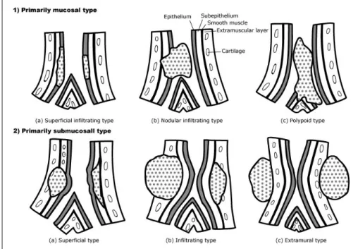
Two physicians and pulmonary specialists reviewed all the lesions on the bronchoscopy as a consensus inter- pretation, and classified them based on the broncho- scopic findings of the revised classification by the Japan Lung Cancer Society (Fig. 1) (13). The bronchoscopic findings were divided into mucosal invasion type and submucosal invasion type. The mucosal invasion type means tumor growth mainly in the mucosal epithelium, and is subclassified as follows: superficial infiltrating type (thickened type), nodular infiltrating type, and polypoid type. The superficial infiltrating types of recur- rences is characterized as mucosal changes, such as red- ness or a whitish patch at the mucosa, without a contour abnormality. The nodular infiltrating type is character- ized as mucosal change with the round mucosal protru- sion from the bronchus to the lumen. The polypoid type is characterized as the lobular mucosal protrusion with a single stalk, from the bronchus to the lumen.
Submucosal types show tumor growth in layers deeper than the lamina propria and are classified into the super- ficial type, infiltrating type, and extramural type.
Results
Out of 576 patients, 12 (2.08%) had bronchial stump recurrences. The histologic types of 9 cases of recurred cancers for which the CT images were available, includ- ed 7 squamous cell carcinomas, 1 adenocarcinoma, and 1 adenoid cystic carcinoma. The surgical resections in- volved lobectomy (n = 2), bilobectomy (n = 1), and pneumonectomy (n = 6). The post-operative pathologic stages of the tumors included stage IB (n = 3), IIB (n = 4), and IIIA (n = 2) at the time of surgery. The interval between the time of operation and recurrence ranged from 17 to 72 months (mean 32 ± 9 months). The rela- tionships between age, sex, histopathologic type, opera- tion type, TNM staging, the interval between operation and recurrence, bronchoscopic findings, and CT find- ings for individual patients are described in Table 1.
The CT scans revealed that all recurrences were in the form of soft tissues occupying the space around stump site. Further, the CT scans demonstrated nodular lesions with heterogeneous enhancement in all cases. Three of the cases had multiple recurrence sites at the time of re- currence, while 2 cases had nodular infiltrating types and 1 case had a polypoid type.
The bronchoscopic findings found that all of the le- sions were classified as mucosal invasion types (no sub- mucosal invasion type were observed). The broncho- scopic findings showed 5 nodular infiltrating types, 3 polypoid types, and 1 superficial infiltrating type of re-
Fig. 1. Classification of Bronchoscopic Findings (Revised classification by the Japan Lung Cancer Society) (11).
currences.
The 6 cases of superficial infiltrating and nodular type recurrences demonstrated nodular infiltrations around the bronchial stumps and into the bronchial wall. The 3 remaining polypoid type recurrences demonstrated lob- ular infiltration to the lumen on the CT scan. Grossly, these bronchoscopic and CT image findings correspond- ed well with each other.
Discussion
Radical resection is considered as an adequate treat- ment for providing survival benefits for early stages of NSCLC, especially for the patients without metastasis into the lymph nodes. However, even after radical re- section, recurrences of lung cancer are not unusual events, occurring in 27 to 39% of patients (1-3). There may be tumor recurrence at the bronchial stump, even when the margin of the resected specimen is negative for cancer and the resected margin is sufficiently distant from the tumor. One of the reason for this is even a visu- al examination of the stump site following a curative re- section does not reveal any tumor, but pathologists re- port the presence of microscopic, residual tumor tissue at the bronchial resection margin in approximately 4- 5% of all lung resections (14).
In this study, the recurrences at the bronchial stump
were seen in 2.08% (12 cases) of all cases of resected lung cancers, and this result is similar to the stump re- currence rates of previous studies (7, 8). However, the cases described by Jang et al. (7) were mostly submucos- al type (7), whereas all of the recurrences in the current study were of mucosal type on bronchoscopic findings (5 nodular infiltrating types, 3 polypoid types, and 1 su- perficial infiltrating type). This difference may be due to the fact that we targeted patients who had recurrences at the bronchial stump sites and excluded those who had recurrences at the lymph nodes. Another possibility for the discrepancy is that we used the revised classifica- tion by the Japan Lung Cancer Society for the classifica- tion of bronchoscopic findings.
All the nodular infiltrative type recurrences were from squamous cell carcinomas, which was consistent with the results of other studies and demonstrated heteroge- neous enhancing nodules on the CT scan (8). Three out of the 9 cases had endobronchial polypoid nodules. One of them was from a squamous cell carcinoma, and the other polypoid type recurrences were from adenocarci- noma and adenoid cystic carcinoma. Jang et al. (7) re- ported the bronchial stump recurrences from squamous cell carcinomas only (7). However, the study of Miura et al. that preceded the aforementioned study of Jang et al.
reported recurrences from various types of lung cancer (8). It is clear that lung cancer recurrences at bronchial Table 1. Individual Information of the Patients
Age / Histopathologic Recur Bronchoscopic CT Findings
No. Sex Type Operation Stage
Period Finding Features Enhancement
Pattern
1 66 /M SqCC P IB 48 months Superficial Nodule Heterogeneous
infiltrating enhancement
2 69 /M SqCC P IIIA 17 months Nodular Multiple nodules Heterogeneous
infiltrating enhancement
3 56 /M SqCC P IIB 22 months Nodular Nodule Heterogeneous
infiltrating enhancement
4 65 /M SqCC B IIB 24 months Nodular Multiple nodules Heterogeneous
infiltrating enhancement
5 73 /M SqCC P IB 24 months Nodular Nodule Heterogeneous
infiltrating enhancement
6 63 /F SqCC P IIIA 31 months Nodular Nodule Heterogeneous
infiltrating enhancement
7 70 /M AdenoCA L IB 26 months Polypoid Endobronchial Heterogeneous
polypoid nodule enhancement
8 64 /M SqCC P IIB 27 months Polypoid Endobronchial Heterogeneous
polypoid nodule enhancement
9 67 /M ACC L IIB 72 months Polypoid Multiple endobronchial Heterogeneous
polypoid nodules enhancement SqCC = Squamous cell carcinoma, AdenoCA = Adenocarcinoma, ACC = Adenoid cystic carcinoma
P = Pneumonectomy, L=lobectomy, B=bilobectomy
stump are not limited to squamous cell carcinomas.
A bronchoscopy is a sufficient tool to evaluating mu- cosal changes and contour changes in the bronchial lu- men. However, there is a tendency to underestimate the infiltrating degree and perilesional conditions. On bron- choscopy, there was one superficial infiltrating type re- currence detected in this study. But in same case, on the CT findings, there was a heterogeneous enhancing nodular soft tissue lesion at the bronchial stump site. On the other hand, the bronchoscopy is thought to be more sensitive than a chest CT when evaluating early local re- currences that do not show bronchial wall thickening or nodular infiltration. In summary, when a bronchoscopy reveals the possibility of local infiltration, a cross-sec- tional examination, such as a CT, should be performed in order to know how much the cancer has advanced.
However, when testing for early recurrent cancer that is not easily visible on a chest CT, the bronchoscopy is more advantageous. In that sense, the two examinations complement each other.
Five cases of the nodular infiltrative type showed nodular lesions originating from a post-operative bronchial stump site on CT findings (Fig. 2). Both the su- perficial infiltrative type and nodular infiltrative type of recurrences appeared as heterogeneous enhancing nod- ules that showed an imprint and direct invasion of the adjacent bronchus or trachea. In contrast, the three polypoid type recurrences were found to be endo- bronchial polypoid nodules. The recurrences of the polypoid type seem to originate from the superficial lay- er, while some invade into the bronchial lumen (case 7), and others invade into both the bronchial lumen and in-
A B
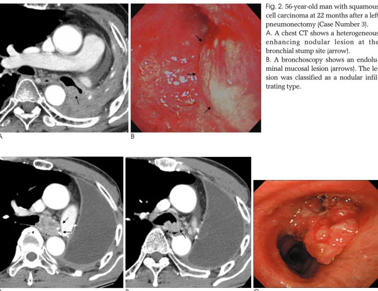
Fig. 2. 56-year-old man with squamous cell carcinoma at 22 months after a left pneumonectomy (Case Number 3).
A. A chest CT shows a heterogeneous enhancing nodular lesion at the bronchial stump site (arrow).
B. A bronchoscopy shows an endolu- minal mucosal lesion (arrows). The le- sion was classified as a nodular infil- trating type.
A B C
Fig. 3. 64-year-old man with squamous cell carcinoma at 27 months after a left pneumonectomy (Case Number 8).
A. A CT image shows an endobronchial mass and heterogeneous enhancement at the adjacent bronchial stump (arrows).
B. A heterogeneous enhancing mass involves carina (arrows).
C. On bronchoscopy, there is a polypoid mass filling left main bronchus involving carina and classified as a polypoid type.
NOTE: The bronchoscopic images were flipped horizontally to show the same point of view for both the CT and bronchoscopy.
to the bronchial wall (case 8, 9) (Fig. 3). Considering this result, we propose that the recurrence of the polypoid type may originate at a more superficial layer of the bronchus, compared to the other types, such as superfi- cial or nodular infiltrative types. According to the previ- ous study, residual tumor cells can infiltrate the entire bronchial wall or can be confined to a specific part of the bronchial wall and the involved part can be classi- fied as mucosal, such as an in situ or invasive carcinoma (14).
There are several limitations to our study: first, sam- ple size was small because of the low incidence of bronchial stump recurrence after primary radical surgery. Accordingly, the ability of our data to represent all the recurrences at the bronchial stump site is limited, and we did not perform any statistical analysis to deter- mine any correlations between bronchoscopic findings, CT findings, and other factors. Secondly, the study was unable to identify the origin of the recurrent tumors pathologically, because additional operations were not performed in some patients. So, we could not pinpoint the bronchial layer that gave rise to the tumor. We ex- pect it to be proven that the polypoid type recurrences originate from the mucosal layers, while the superficial infiltrative type and nodular infiltrative type recur- rences originate from extra-mucosal layer. Another con- cern is that we used 3 different CT scanners with differ- ent protocols, and, as a result, we may not have ob- tained consistent result.
In conclusion, the correlation with bronchoscopic and CT findings is possible in verifying the presence of a re- currence at the bronchial stump site. In other words, both a bronchoscopy and CT can be used as comple- mentary or alternative tools in evaluating bronchial stump site recurrences. Also, when referring to chest CT findings for polypoid type and nodular infiltrative type recurrences, the origin of the tumor may lead to differ- ent types of recurrence. In effect, the CT findings for re- current lung cancer at the bronchial stump are nodules
with heterogeneous enhancement.
References
1. Pairolero PC, Williams DE, Bergstralh EJ, Piehler JM, Bernatz PE, Payne WS. Postsurgical stage I bronchogenic carcinoma: morbid implications of recurrent disease. Ann Thorac Surg 1984;38:331- 338
2. Martini N, Bains MS, Burt ME, Zakowski MF, McCormack P, Rusch VW, et al. Incidence of local recurrence and second prima- ry tumors in resected stage I lung cancer. J Thorac Cardiovasc Surg 1995;109:120-129
3. Harpole DH Jr, Herndon JE 2nd, Wolfe WG, Iglehart JD, Marks JR. A prognostic model of recurrence and death in stage I non- small cell lung cancer utilizing presentation, histopathology, and oncoprotein expression. Cancer Res 1995;55:51-56
4. Matthews MJ, Kanhouwa S, Pickren J, Robinette D. Frequency of residual and metastatic tumor in patients undergoing curative re- section for lung cancer. Cancer Chemother Rep 3. 1973;4:63-67 5. Feld R, Rubinstein LV, Weisenberger TH. Sites of recurrence stage
I non-small-cell lung cancer: a guide for future studies. J Clin Oncol 1984;2:1352-1358
6. Yano T, Yokoyama H, Inoue T, Asoh H, Tayama K, Takai E, et al.
The first site of recurrence after complete resection in non-small- cell carcinoma of the lung. Comparison between pN0 disease and pN2 disease. J Thorac Cardiovasc Sug 1994;108:680-683
7. Jang KM, Lee KS, Shim YM, Han D, Kim H, Kwon OJ, et al. The rates and CT patterns of locoregional recurrence after resection surgery of lung cancer: correlation with histopathology and tumor staging. J Thorac Imaging 2003;18:225-230
8. Miura H, Konaka C, Kato H, Kawate N, Taira O. Recurrence at the bronchial stump after resection of lung cancer. Ann Surg 1994;219:
306-309
9. Peters JC, Desai KK. CT demonstration of postpneumonectomy tu- mor recurrence. AJR Am J Roentgenol 1983;141:259-262
10. Glazer HS, Aronberg DJ, Sagel SS, Emami B. Utility of CT in de- tecting postpneumonectomy carcinoma recurrence. AJR Am J Roentgenol 1984;142:487-494
11. Mountain CF. Revisions in the international system for staging lung cancer. Chest 1997;111:1710-1717
12. Mountain CF. A new international staging system for lung cancer.
Chest 1986;89(4 Suppl):225S-233S
13. The Japan Lung Cancer Society. Classification of lung cancer Tokyo:
Kanehara, 2000:32-34
14. Wind J, Smit EJ, Senan S, Eerenberg JP. Residual disease at the bronchial stump after curative resection for lung cancer. Eur J Cardiothorac Sur 2007;32:29-34
대한영상의학회지 2009;60:325-331
비소세포 폐암 수술 후 기관지 잘린 끝의 국소재발:
전산화 단층촬영과 기관지 내시경 소견의 비교1
1연세대학교 원주의과대학 원주기독병원 영상의학과
2연세대학교 원주의과대학 원주기독병원 호흡기내과
3연세대학교 의과대학 영동세브란스병원 영상의학과
임상혁∙권우철∙차승환∙김명순∙임대건∙박연주∙리원연2∙김상하2∙김태훈3
목적: 수술 후 비소세포 폐암 환자에 있어서, 기관지 잘린 끝에 국소 재발암의 전산화 단층촬영 소견과 기관지 내시 경 소견을 비교하고자 하였다.
대상과 방법: 근치적 수술을 받은 총 576명의 원발성 비소세포 폐암 환자 중, 기관지 잘린 끝에 재발을 보인 9명의 환자를 대상으로 후향적 연구를 진행하였다. 9명의 환자를 기관지 내시경 소견을 the bronchoscopic findings of the revised classification by the Japan Lung Cancer Society에 따라 분류하였으며, 이후 전산화 단층촬영 소견과 비교, 분석하였다.
결과: 환자들의 원발암은 조직학적으로 7명의 편평세포암종, 1명의 샘암종, 1명의 샘낭암종이었다. 기관지 내시경 소견으로는 결절 침윤형이 5예, 용종형이 3예, 얕은 침윤형이 1예였다. 전산화 단층촬영 소견에서 결절을 보인 예는 6개, 기관지 내강에 소엽상을 보인 예가 3개였으며 9예 모두 불균일한 조영증강을 보였다.
결론: 기관지 잘린 끝의 국소 재발의 진단에서, 기관지 내시경 소견과 흉부 CT 소견은 상관관계를 보이며, 각각의 결점을 충족시켜주는 상호보완적인 도구로 사용할 수 있다.