서 론
Clostridium difficile은 그람 양성, 혐기성, 아포생성막대 CLINICAL ARTICLE
J Korean Neurotraumatol Soc 2011;7:92-98 ISSN 1738-8708
균으로서 입원 환자에 발생하는 설사의 주요한 원인 균주 이며, 1978년 항생제와 연관된 Clostridium difficile 관련 설사(Clostridium difficile-associated diarrhea: CDAD)의 원인으로 처음 기술되었다.14) CDAD는 항생제와 연관된 설 사의 약 15~20%를 차지하며 경한 설사부터 생명을 위협하 는 장염까지 다양한 증상으로 발현된다. 전격성 장염(Ful- minant colitis)도 1~3%에서 발생하며 독성 거대결장(Toxic megacolon)이 동반되는 경우 사망률이 25~40%에 달한
다.15,23,24) CDAD가 심각하게 진행된 상태인 위막성 대장염
Received: August 22, 2011 / Revised: September 15, 2011 Accepted: September 15, 2011
Address for correspondence: Byung Moon Cho, MD, Department of Neurosurgery, Kangdong Sacred Heart Hospital, Hallym University College of Medicine, 150 Seongnae-gil, Gang- dong-gu, Seoul 134-701, Korea
Tel: +82-2-2224-2236, Fax: +82-2-473-7387 E-mail: [email protected]
Clostridium Difficile 관련 설사가 발생한 신경외과 환자의 임상적 특징과 위험인자들에 대한 분석
한림대학교 의과대학 강동성심병원 신경외과학교실
김용상 .전홍준 .남수아 .조병문 .박세혁 .오세문
Analysis of Clinical Characteristics and Risk Factors to Neurosurgical Patients with Clostridium Difficile -Associated Diarrhea
Yong-Sang Kim, MD, Hong-Jun Jeon, MD, Su-A Nam, SN, Byung Moon Cho, MD, Se-Hyuck Park MD and Se Moon Oh, MD
Department of Neurosurgery, Kangdong Sacred Heart Hospital, Hallym University College of Medicine, Seoul, Korea
Objective: The risk factors of Clostridium difficile-associated diarrhea (CDAD) are well known in medical part. However, there have been a few studies of CDAD about neurosurgical patients. The aim of this study was to investigate clinical char- acteristics and risk factors of neurosurgical patients with CDAD. Methods: We retrospectively reviewed the record of eighty-five patients with CDAD between January 2007 and December 2010. They made a diagnosis that used a toxin assay, stool culture and sigmoidoscopy or colonoscopy. We analyzed the association factor such as age, gender, operation, enteral feeding, length of Intensive Care Unit stay, prophylactic antibiotics, duration and number of antibiotics, types of antibiot- ics, diarrhea onset time, toxin assay, serum albumin, C-reactive protein, pseudomembranous colitis, recurrence rates be- tween the neurologically impaired group and well-active group. Results: Of 15 parameters, 6 parameters were significant- ly associated with neurologically impaired group in univariate analysis. The enteral feeding and bed ridden state (p<0.001) frequently had practiced and the intensive care unit stay had longer (p=0.001) and the diarrhea onset time (p=0.034) from the last antibiotics use administered prior to development of the CDAD was lesser. The pseudomembranous colitis and CDAD recurrence had more appeared in impaired group (p<0.001). On multivariate analysis, longer intensive care unit stay (p=0.024) and increasing cumulative days of antibiotic administration (p=0.007) correlated with pseudomembra- nous colitis. Conclusion: The data suggest that it may make a vulnerable influence to development of the CDAD in neu- rologically impaired patients. To prevent the CDAD, we need to practice stricter guidelines about antibiotic usage and minimize length of stay in ICU. (J Korean Neurotraumatol Soc 2011;7:92-98)
KEY WORDS: Clostridium difficile-associated diarrhea ㆍAntibiotics ㆍPseudomembranous colitis ㆍNeurosurgery.
(Pseudo-Membranous Colitis: PMC)은 Clostridium difficile 에 의해 대장점막에 위막을 형성하는 경우를 말한다. 현재 광범위 항생제 사용이 널리 보급된 이후 CDAD는 병원 내 감염에서 4번째로 흔한 부분을 차지하고 있고,17) CDAD의 발생과 관련된 많은 위험 인자가 보고되었다.1,6,10,20) 특히 신 경외과 영역에서는 수술을 시행한 환자들은 대부분 수술 후 항생제를 사용하고 뇌수막염 및 뇌농양 등의 합병증이 있을 경우는 장기간 항생제를 사용하게 된다. 또한 신경학 적 손상으로 의식기능이 저하된 경우 급성기간 중환자실 치료가 필요하며 다른 요로 감염이나 폐렴 및 카테타 관련 감염 등의 발생 가능성이 많아지고, CDAD의 위험 요인으 로 여겨지는 금식이 수일 동안 이뤄지기도 하며 위장관 식 이 등의 처치도 더불어 시행될 수 있다.3,5) 이 중 일반적인 다른 환자들과 차이는 신경학적 손상으로 의식기능의 저하 가 발생할 수 있다는 점이다. 이러한 이유로 저자들은 신경 외과영역에서 CDAD로 확진 된 환자와 특히 CDAD가 심 각하게 진행된 상태인 위막성 대장염이 발생한 환자들의 임상양상 및 검사실 결과 분석을 통해 CDAD 발생의 특징 과 이와 관련된 위험 인자를 비교 분석하고자 하였다.
대상 및 방법
대 상
2007년 1월부터 2010년 12월까지 본원 신경외과에서 수 술 전후 혹은 호흡기 및 요로 감염 등, 감염치료의 목적으 로 항생제 사용 후 설사를 일으킨 총 550명의 성인 환자들 중 CDAD로 진단된 85명 (15.5%)의 환자를 대상으로 하였 다. 85명의 환자는 두부외상 32명, 뇌혈관질환 26명, 뇌종양 17명이었고 그 외 질환 10명이 포함되었다. 그리고 대상환자 중 73명이 신경외과 수술을 받은 환자였다.
CDAD의 진단기준은 정맥주사용 항생제 사용 후 하루 3회 이상의 수양성 설사가 2일 이상 지속되는 경우에 Clos- tridium difficile 독소 A와 대변 배양검사를 시행하여 양성 이거나, Clostridium difficile 독소가 음성일지라도 내시경
소견과 내시경 조직검사에서 CDAD가 확인된 경우로 진단 하였다. 그러나 전신상태가 좋지 않은 환자의 경우, 내시경이 힘들므로 복부 컴퓨터단층촬영을 하여 위막성 대장염으로 판독될 경우에도 본 연구에 포함시켰다.7)
방 법
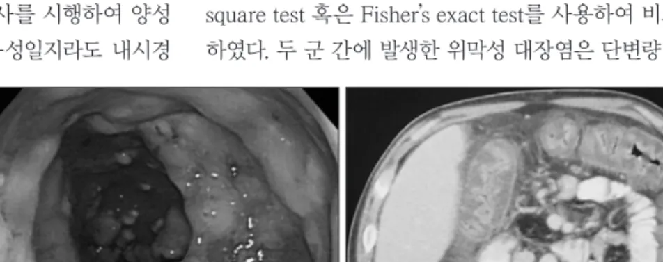
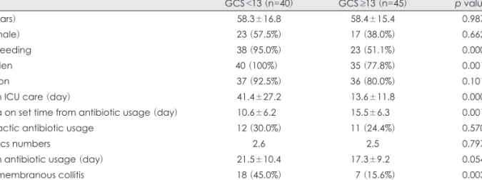
CDAD로 진단된 총 85명의 환자에 대하여 후향적 방법 으로 의무기록을 검토하였다. Glasgow coma scale상 13점 을 기준으로 의식이 저하된 40명의 환자군과 의식이 정상 에 가까운 45명의 환자군으로 나누었다. 각 군에서 임상적 인자로 연령, 성별, 수술 여부, 위장관 식이(Enteral feeding), 독립적인 보행 불가상태(Bed ridden state), 수술 전 예방적 항생제 사용 유무, 수술 후 사용한 항생제 종류와 수, 항생 제 사용기간, 중환자실 재원기간, 항생제 사용 후 설사 발생 까지의 기간, CDAD 재발 및 위막성 대장염 발생 여부에 대 해 비교 조사하였다. 치료 종결은 치료 항생제를 7~10일 사 용 후 설사가 발생하지 않는 경우이며, 재발은 CDAD로 치 료 종결 후 다시 진단받을 경우로 정의하였다. 위막성 대장 염은 내시경에서 2~10 mm의 특징적인 “adherent yellow plaque” (Figure 1A)를 보이거나 복부 컴퓨터단층촬영상
“thumb-printing”과 동반하여 현저히 두터워진 대장벽, 점 막 및 점막 하 부종이 있을 시에 진단하였다 (Figure 1B). 혈 액 및 면역학적 인자로는 진단 시일의 혈청 알부민 (정상치:
3.8~5.3 g/dL), C-reactive protein (CRP, 정상치: 0~3 mg/
L) 수치 및 Clostridium difficile 독소 A 유무를 비교 조사 하였다. 또한 CDAD의 치료를 위해 사용된 경구 항생제들 의 사용 효과를 알아보았다.
통계적 분석
통계적 분석은 SPSS 통계 프로그램(Windows ver. 12.0) 을 이용하였으며 연속 변수에 대해서는 Independent- Sample t-test를 사용하였고 범주형 변수에 대해서는 Chi- square test 혹은 Fisher’s exact test를 사용하여 비교 분석 하였다. 두 군 간에 발생한 위막성 대장염은 단변량 분석으
FIGURE 1. Descending colon covered with yellowish pseudo-membrane on sigmoidoscopy (A) and CT scan of the abdomen (B) showing gross thickening of the large bowel wall and obliteration
of the lumen.
A B
로 위막성 대장염 유무에 따른 유의한 차이를 보인 위험인 자들에 대해 다변량 분석인 Logistic regressions analysis 를 시행하였고 통계 결과는 p 값이 0.05 이하인 경우를 유 의한 것으로 간주하였다.
결 과
임상 양상 비교
임상 지표 중 위장관 식이와 침상 안정상태는 의식저하군 에서 빈도가 높았고, 중환자실 재원기간은 의식저하 환자 군이 평균 41.4±27.2일로 의식정상군의 13.6±11.8일보다
재원기간이 길었다 (p<0.001, t-test)(Table 1). 항생제 사용 후 설사 발생까지의 기간은 의식저하군은 평균 10.6±6.2 일로 의식정상군 보다 평균 4.9일이 빠르게 나타났고 위막 성 대장염의 빈도 (n=18, 45%)가 유의적으로 높게 나타났 다 (p<0.005, t-test)(Table 1). 혈액 및 면역학적 지표는 의 식저하군에서 CRP가 평균 61.3±57.1 mg/L이었으며 14명 (35%) 만이 독소A에 양성을 보였지만 의식정상군 (n=21, 47%)과의 통계적인 유의성을 보이지 않았다. 그러나 알부 민은 의식저하군에서 평균 2.9±0.3 g/dL로 상대적으로 저 하된 소견을 보였다 (p=0.008, t-test)(Table 2).
TABLE 1. Comparisons of clinical characteristics between neurologically impaired and well active group
GCS<13 (n=40) GCS≥13 (n=45) p value*
Age (years) 58.3±16.8 58.4±15.4 0.987
Sex (female) 23 (57.5%) 17 (38.0%) 0.662
Enteral feeding 38 (95.0%) 23 (51.1%) 0.000
Bed ridden 40 (100%)0 35 (77.8%) 0.001
Operation 37 (92.5%) 36 (80.0%) 0.101
Duration ICU care (day) 41.4±27.2 13.6±11.8 0.000
Diarrhea on set time from antibiotic usage (day) 10.6±6.20 15.5±6.30 0.001
Prophylactic antibiotic usage 12 (30.0%) 11 (24.4%) 0.570
Antibiotics numbers 2.6 2.5 0.797
Duration antibiotic usage (day) 21.5±10.4 17.3±9.20 0.054
Pseudomembranous collitis 18 (45.0%) 07 (15.6%) 0.003
CDAD recurrence 14 (35.0%) 02 (04.4%) 0.001
*continuous variable calculated by using Student-t test and categorical variable by using chi-square test. GCS: Glasgow Coma Scale, ICU: intensive care unit, CDAD: Clostridium difficile-associated diarrhea
TABLE 2. Comparisons of laboratory data between neurologically impaired and well active group
GCS<13 (n=40) GCS≥13 (n=45) p value*
Serum albumin (g/dL) 02.9±0.30 03.2±0.40 0.008
Serum CRP (mg/L) 61.3±57.1 51.7±75.4 0.518
Toxin assay (positive) 14 (35%) 21 (47%) 0.281
*p value: continuous variable calculated by using Student-t test and categorical variable by using chi-square test. GCS:
Glasgow Coma Scale, CRP: C-reactive protein
TABLE 3. Use of antibiotics between neurologically impaired and well active group
Number of course (%) GCS<13 (n=40)(%) GCS≥13 (n=45)(%) p value*
Penicillins and beta lactamase 33 (38.8) 17 (51.5) 16 (48.5) ns
Quinolone 28 (32.9) 10 (35.7) 18 (64.3) ns
Cephalosporin
1st 13 (15.3) 06 (46.2) 07 (53.8) ns
2nd 03 (3.5) 01 (33.3) 02 (66.7) ns
3rd 58 (68.2) 29 (50.0) 29 (50.0) ns
4th 09 (10.6) 02 (22.2) 07 (77.8) ns
Aminoglycoside 39 (45.9) 19 (48.7) 20 (51.3) ns
Carbapenem 14 (16.5) 07 (50.0) 07 (50.0) ns
Glycopeptide 37 (43.5) 19 (51.4) 18 (48.6) ns
*chi-square test. ns: non-specific, GCS: Glasgow Coma Scale, 1st: 1st cephalosporin, 2nd: 2nd cephalosporin, 3rd: 3rd ceph- alosporin, 4th: 4th cephalosporin
항생제 사용
모든 환자에서 항생제를 정맥 투여되었고, 대부분 2개 이 상의 항생제 병합요법을 시행하여 CDAD의 원인 항생제를 정확히 규명하기는 어려웠지만, 의식저하군과 의식정상군 간에 사용 항생제 종류에 따른 분석상 두 군에 유효한 차 이는 없었다 (p>0.05, chi-square test)(Table 3). 두 군 모 두 3세대 cephalosporin과 aminoglycoside, glycopeptides, penicillins and beta lactamase, quinolone계열 등을 주로 사용하였다 (Table 3). 의식저하군에서 평균적으로 사용한 항생제 수와 항생제 사용기간은 각각 2.6개 및 21.6±10.4 일로 의식정상군 (2.5개, 17.3±9.2일) 과의 통계적 유의성은 보이지 않았다 (p>0.05, t-test)(Table 1).
위막성 대장염(Pseudomembranous colitis)의 발생에 관 한 분석
전체 환자 85명 중 위막성 대장염 환자는 25명 (29.4%)이 었으며, 위험 요인의 단변량 분석을 시행한 결과, 위막성 대 장염이 발생한 경우는 의식저하군에서 발생률 (n=18, 45%) 이 높았으며 (p=0.003, chi-square test)(Table 1), 위장관 식 이를 시행하는 경우 (p=0.007, chi-square test)와 중환자 실 재원기간이 긴 경우 (p<0.001, t-test)에 위막성 대장염이 발생하지 않은 군과 유의한 차이를 보였다 (Table 4). 또한 항생제 사용과 관련하여 항생제 수 (p=0.206, t-test)는 유
의적 차이를 보이지 않았으나 수술 전 예방적 항생제를 사 용한 경우 (p=0.038, chi-square test)와 항생제 사용이 긴 경우 (p<0.001, t-test)에 위막성 대장염이 발생하지 않은 군과 유의한 차이가 나타났다 (Table 4). 단변량 분석에서 유의적 결과를 보인 요인에 대해 다변량 로지스틱 회귀 분 석을 시행한 결과 의식저하군과 위장관 식이 및 예방적 항 생제 사용은 위막성 대장염 발생과 연관성을 보이지 않았 다 (Table 5). 그러나 28일 이상의 중환자실 치료기간(odds ratio (OR), 7.5; 95 percent confidence interval (CI), 1.3- 43.3; p=0.024)와 21일 이상의 항생제 사용(OR, 7.1; 95 percent CI, 1.7-29.8; p=0.007)은 의미 있게 나타났다 (Ta- ble 5).
치료반응
CDAD의 일차 치료 경구용 항생제는 metronidazole 74 명 (87.1%), vancomycin 11명 (12.9%)이었다. 일차 항생제의 반응률은 87.0% (n=74)였다 (Figure 2). Metronidazole을 일차로 복용한 74명 중 64명 (86.5%)은 호전되었으며 그 중 12명 (16.2%)은 재발되었다. 재발한 12명은 vancomycin 으로 7~8일 정도 교체 복용하여 호전되었다. Metronidazole 에 호전이 없는 10명 (13.5%) 중 8명은 vancomycin으로 변 경하였고 2명은 vancomycin을 7~10일 정도 추가 복용하 여 호전되었다. Vancomycin을 일차 복용한 11명 중 10명 TABLE 4. Univariate analysis of risk factors for pseudomembranous colitis among the Clostridium difficile-associated diarrhea
PMC group (n=25) Non-PMC group (n=60) p value*
Age (years) 57.4±13.2 58.7±17.1 0.743
Sex (female) 09 (36.0%) 25 (41.7%) 0.632
Enteral feeding 23 (92.0%) 38 (63.3%) 0.007
Bed ridden 24 (96.0%) 51 (85.0%) 0.155
Operation 22 (88.0%) 51 (85.0%) 0.721
Duration ICU care (day) 45.2±27.0 19.0±19.1 0.000
Prophylactic antibiotic usage 12 (48.0%) 11 (18.3%) 0.038
Antibiotic numbers 2.8 2.5 0.206
Duration antibiotic usage (day) 26.6±10.6 16.5±8.10 0.000
Toxin assay (positive) 10 (40.0%) 25 (41.7%) 0.889
*continuous variable calculated by using Student-t test and categorical variable by using chi-square test. ICU: intensive care unit, PMC: pseudomembranous colitis
TABLE 5. Multivariate regression analysis of risk factors for pseudomembranous colitis among the Clostridium difficile-associated diarrhea
Adjusted odds ratio 95% confidence interval p value*
Neurologically impaired state 0.7 0.1-4.1 0.682
Enteral feeding 2.1 0.2-17.3 0.505
Duration ICU care (>26 days) 7.5 1.3-43.3 0.024
Prophylactic antibiotic usage 3.7 0.9-15.2 0.068
Duration antibiotic usage (>19 days) 7.1 1.7-29.8 0.007
*multivariate analyses were performed by using a binary logistic regression. ICU: intensive care unit
(90.9%)은 호전되었지만, 그 중 4명 (36.4%)은 재발되었고 1 명은 호전이 없었다. 재발한 4명은 metronidazole을 추가 하여 약 10일 정도의 재치료로 호전되었고 vancomycin의 일차 복용에 호전을 보이지 않은 1명 (9.1%)은 metronida- zole을 추가하여 약 10일 정도의 연장 치료로 호전되었다 (Figure 2). CDAD로 인한 사망 환자는 없었으며 기존 질환 악화로 5명, 패혈증으로 3명이 사망하였다. 치료반응 기간 은 의식저하 군과 의식정상군 간에 유의한 차이가 없었으나 재발여부는 의식저하군에서 많은 빈도를 보였다 (p<0.001, chi-square test)(Table 1).
고 찰
Clostridium difficile은 포자를 형성하며 물, 토양, 자연계 에 흔히 존재하는데 약 100여 종이 알려져 있고, 이 중 일부 만이 인간에게 병원체로 존재한다. 정상 성인의 약 3~5%
에서는 무증상 보균상태로 존재하며, 원내 입원 환자의 경 우는 약 25~30%가 무증상 보균상태로 나타난다.16,26) 감염 의 전파는 경구감염(fecal-oral route)으로 이뤄져 원내에 선 주변 환경과 사람의 손이나 의료기구의 접촉을 통해서 이뤄진다. CDAD의 발현은 항생제를 사용 후 정상 장내 세 균총이 줄어든 경우에 Clostridium difficile의 증식으로 일 어나며 위막성 대장염으로 진행된 경우에는 90~100% 균이 검출된다.8)
CDAD의 진단 방법으로는 대변에서 독소를 검출하는 방법과 대변 배양검사로 균주를 분리한 후 독소를 검출하 는 방법이 있다. 하지만, 임상적으로 대변 배양검사의 경우 검체 채취에 주의를 요하며 배양에 장시간이 소요되어 일
반적으로 독소의 검출과 내시경을 통해 진단이 이루어진다.
독소의 검출 방법으로는 세포배양 세포독성법(cell culture cytotoxic assay), 효소면역법(immunoenzyme assay), latex 법(latex test) 등이 있으며 각각의 검사는 70~100%의 민감 도와 특이도를 보인다.8) 모든 균주가 독소 A와 B를 분비하 는 것은 아니며 Clostridium difficile의 1~2%는 독소 B만 을 분비하고 약 5~25%는 독소를 생산하지 않음으로 장염 이나 설사를 유발하지 않는다.12)
CDAD 발생 위험 요인은 항생제 사용 외에도 고령, 신부전, 위장관 시술, 복부 수술, 재원기간, 면역 기능의 저하, 영양 결핍 등이 알려져 있다.1,7,20) 항생제 사용 후 CDAD의 임상 증상은 1일에서 6주 정도로 폭넓게 나타날 수 있지만 전형 적으로 1~2주 이내에 나타나고 상당한 수양성 혹은 점액 성 설사 및 고열을 동반할 수 있다. 또한 심한 경우에는 탈 수와 전해질 불균형 및 저단백혈증 등이 동반될 수 있
다.7,11) 일반적으로 CDAD와 관련 사망률은 2~5%로 높지
않지만 고령의 환자나 전신상태가 위중한 환자에게서는 10~20%로 높게 나타날 수 있다.28)
McFarland 등18)은 전향적 연구를 통해 65세 이상 연령을 위험인자로, Crabtree 등5)은 성별 중에 여성을 위험인자로 보고하였으나, 본 연구에선 의식 기능 상태에 따른 분류상 연령이나 성별은 차이를 보이진 않았다 (Table 1). 환자 상태 와 관련하여 의식저하군에서 CDAD의 위험인자인 위장관 식이 (p<0.001, chi-square test)와 침상 생활 (p=0.001, chi-square test)의 빈도가 높았으며, 중환자실 재원기간이 오랜 환자에서 의료인 및 의료기구에 의한 접촉 등의 위험 인자에 대해 노출이 잦으므로 CDAD 발생이 높다고 추정 된다.3,5) 연구에 포함되진 않았으나 의식정상 환자에서 급성 FIGURE 2. Clinical response of Clostridium difficile-associated colitis (CDAD) to use antibiotics.
CDAD total patient=85
Cure: 10 (90.9%)
Cure: 64 (86.5%) Fail: 10 (13.5%) Fail: 1 (9.1%)
1st treatment
2nd treatment
Recurrence Metronidazole+Vancomycin n=4 (36.4%)
Metronidazole+Vancomycin n=1 Vancomycin n=8
Metronidazole+Vancomycin n=2
Vancomycin n=12 (16.2%)
Metronidazole n=74 (87.1%) Vancomycin n=11 (12.9%)
기에 중환자실 치료를 받았으나 항생제를 사용한 병력 없 이 위막성 대장염이 발생한 경우가 있었는데 이는 전파 감 염 가능성이 높은 환경에 노출되는 것만으로도 CDAD가 발생한다는 기존 보고와 일치하는 결과이다.8,28)
Warny 등27)은 독소에 대한 항체 반응 정도와 임상 양상 과의 관련성이 있다고 보고하였고, Price 등25)은 재발이 Clostridium difficile의 독소에 대한 면역글로불린 G 항체 가 충분하지 못해 생기는 것으로 발표하였으며 Hashimoto 등9)은 알부민 저하를 위막성 대장염의 불량한 예후인자로 언급하였다. 본 연구에서도 의식정상군보다 CDAD의 임상 증상이 의식저하군에서 평균적으로 4.9일 빠르게 보였고 알부민 수치도 2.9 g/dL로 낮게 나타났으며 CDAD의 재발 도 14예에서 보여 의식저하군 환자에서 면역기능저하를 추 정해 볼 수 있겠다.
Bartlett2)는 CDAD를 유발하는 흔한 항생제로 cepha- losporin계열과 clindamycin을 보고하였다. 본 연구에서 clindamycin의 경우 항생제 사용 빈도가 매우 적어 원인 항 생제로 영향을 미친 점은 미미하였다. 본 연구에서 CDAD 를 유발한 주요 항생제들은 3세대 cephalosporin, amino- glycoside, glycopeptide, penicillins and beta lactamase 계열 및 quinolone제제였다. Cephalosporin과 aminogly- coside는 수술 전후 감염 방지를 위해 투여되는 경우가 흔 하였고 glycopeptide는 수술 전 예방적 사용인 경우나 meth- icillin-resistant Staphylococcus aureus (MRSA)와 관련 한 폐렴과 수술부위 감염에 있어 사용이 흔하였다. Penicil- lins and beta lactamase계열 및 quinolone제는 폐렴이나 요로 감염과 같은 다른 질환 감염 등이 있을 시에 사용이 많았다. 항생제 종류에 따른 분석에서 의식저하군과 의식 정상군 사이에 CDAD 발생 비율은 차이를 보이지 않았다.
Owen 등21)은 광범위 항생제 사용으로 인한 장내 세균이 억제되었을 시에 Clostridium difficile은 포자를 통해 광범 위 항생제 사용에도 증식할 수 있다고 하였고 일반적인 항 생제 사용기간과 항생제 복합사용이 CDAD의 발생과 합 병증을 높인다고 보았다. 본 연구에서 두 군 모두 항생제 복합 사용이 주로 이루어져 이에 의한 영향력을 알기는 어 렵지만 의식저하군의 항생제 사용기간이 약 3주 이상 길게 사용한 후 재발과 위막성 대장염 발생이 높다는 것은 앞선 연구결과에 어느 정도 부합된다고 생각된다.
위막성 대장염은 Clostridium difficile에 의해 발생하는 염증성 장질환으로 대장 점막에 위막을 형성하는 경우를 말하며, CDAD가 심각하게 진행된 상태로 독소 검사의 유 무 없이 하부 위장관 내시경이나 복부 컴퓨터단층촬영상에 서 특징적인 소견이 나타나면 진단할 수 있다.7,8) Morris 등19)
에 따르면 사망률은 6∼30%에 달하며, 항생제 사용에 의해 감염된 경우가 오염된 환경에 노출되어 감염된 경우를 포 함한 수평적 감염보다 예후가 나쁘다고 보고하였다. 위험 인자에 대한 여러 보고가 있는데 Lee 등15)은 70세 이상의 고령과 20일 이상의 병원 재원 일수가 위험인자로 영향을 줄 수 있다는 것과 위험인자 유무에 따른 위막성 대장염의 예측률을 보고하였다. Park 등22)은 위장관계 수술 환자에 서 aminoglycoside계열 항생제의 장기간 사용과 혈액수혈 및 중환자실 재원기간이 위막성 대장염 발생의 독립적 위 험인자임을 보고하였다. 본 연구에서도 위막성 대장염 여부 에 따른 위험인자에 대해 다변량 분석 결과, 의식저하나 위 장관 식이가 위험인자로 영향을 끼치진 않았지만 28일 이상 의 중환자실 재원기간과 21일 이상의 항생제 사용기간이 독 립적인 위험인자가 될 수 있음을 확인하였다 (Table 5).
Cohen 등4)에 의해 발표된 최근의 지침은 CDAD의 원인 항생제를 가능하면 중단하여 대장 안의 정상 세균총이 생 성되도록 한다고 하였다. 권장하는 치료 경구 항생제로는 metronidazole과 vancomycin이 있는데 metronidazole은 장내 흡수가 잘 이루어져 설사가 호전될 때 약물 효과가 상 대적으로 감소할 수 있지만 vancomycin 과의 효험(effi- cacy)이 동등함이 확인되어 경미한 경우나 중등도인 경우 에도 사용하도록 하고 있다. Vancomycin 경우는 장내 흡 수가 거의 이루어지지 않아 상대적으로 효과가 좋으며 빠른 시간 안에 증상의 호전을 보일 수 있어 중증인 경우에 사용 하도록 하지만 vancomycin-resistant enterococci의 증가 위험도와 비용 문제를 고려해야 한다. 또한 CDAD와 관련 된 장마비나 독성 거대결장이 동반된 경우는 경구 항생제로 vancomycin과 정맥주사로 metronidazole을 사용하도록 하며, 적극적인 대장절제술까지도 고려하도록 하고 있다.
본 연구에선 CDAD에 대한 경구용 항생제는 주로 met- ronidazole이 이용되었고 경우에 따라 vancomycin이 사용 되었다. 사용효과는 metronidazole의 치료반응이 86.5%로 vancomycin의 90.9%보다 낮았지만 재발률에 있어선 vancomycin의 36.4%보다 낮은 16.2%를 보였다 (Fig. 3).
치료 실패나 재발과 관련하여 metronidazole의 경우 van- comycin으로 전환하거나 추가하였고 vancomycin의 경우 는 모두 metronidazole을 추가하여 치료되었다. CDAD와 관련하여 중한 임상경과는 보이지 않았다. 그러므로 임상 적으로 심각한 경우가 아니라면 metronidazole 사용을 고 려하고 치료실패나 재발시에 vancomycin을 추가하거나 변 경하여 치료하는 것이 좋겠다.
본 연구의 제한점은 첫째, 신경외과 전체 환자를 대상으 로 하지 않고 CDAD가 발생한 환자를 대상으로 하여서 신
경학적 결손에 따른 의식저하와 함께 CDAD 발생 여부를 고려하지 않았다는 점이다. 결과적으로 의식저하 환자에서 CDAD 발생의 독립적 위험인자를 알아볼 수는 없었다. 둘 째, 위막성 대장염 발생 여부에 수술 전 예방적 항생제 사 용의 독립적인 영향을 알기 위해선 예방적 항생제 사용 여부 만을 고려해야 했다는 점이다. 결과적으로 수술 후 일반적 인 항생제 사용에 따른 위막성 대장염 발생에 간섭(bias)을 줄 수 있다. 그러나 의식기능이 저하된 환자들에서 CDAD 가 더 많이 발생할 수 있다는 점과 항생제 사용기간 및 중 환자실 재실기간이 위막성 대장염의 위험인자로 영향을 줄 수 있다는 것을 확인할 수 있었으며 앞으로 더 많은 연구 를 하는 것이 도움이 되겠다.
결 론
본 연구에선 의식저하군에서 위장관식이, 중환자실 재원 기간, 알부민 수치저하, 항생제 사용 후 설사 발생 시점이 통 계적으로 의미가 있는 위험인자로 분석되었으며 재발과 위 막성 대장염 발생의 빈도가 많았다. 또한 위막성 대장염 발 생에 있어 항생제 사용기간과 중환자실 재실기간이 가능 성 있는 위험인자로 나타났다. 따라서 의식 기능이 정상인 군보다 CDAD 감염에 취약하여 가능한 중환자실 재원기간 을 줄이고 항생제 사용을 적게 하는 것이 도움이 될 것이다.
중심 단어: Clostridium difficile 관련설사・항생제・위막 성 대장염・신경외과.
■ The authors have no financial conflicts of interest.
REFERENCES
1) Barbut F, Petit JC. [Epidemiology, risk factors and prevention of Clostridium difficile nosocomial infections]. Pathol Biol (Paris) 48:745-755, 2000
2) Bartlett JG. Antibiotic-associated diarrhea. Clin Infect Dis 15:
573-581, 1992
3) Bulstrode NW, Bradbury AW, Barrett S, Stansby G, Mansfield AO, Nicolaides AN, et al. Clostridium difficile colitis after aortic sur- gery. Eur J Vasc Endovasc Surg 14:217-220, 1997
4) Cohen SH, Gerding DN, Johnson S, Kelly CP, Loo VG, McDonald LC, et al. Clinical practice guidelines for Clostridium difficile in- fection in adults: 2010 update by the society for healthcare epide- miology of America (SHEA) and the infectious diseases society of America (IDSA). Infect Control Hosp Epidemiol 31:431-455, 5) Crabtree T, Aitchison D, Meyers BF, Tymkew H, Smith JR, Guth-2010 rie TJ, et al. Clostridium difficile in cardiac surgery: risk factors and impact on postoperative outcome. Ann Thorac Surg 83:
1396-1402, 2007
6) Frost F, Hurley JS, Petersen HV, Casciano RN. Estimated inci- dence of Clostridium difficile infection. Emerg Infect Dis 5:
303-304, 1999
7) Gerding DN, Johnson S. Harrison’s principles of internal medi-
cine, ed 16: McGraw-Hill, pp760-762, 2005
8) Gerding DN, Johnson S, Peterson LR, Mulligan ME, Silva J Jr.
Clostridium difficile-associated diarrhea and colitis. Infect Con- trol Hosp Epidemiol 16:459-477, 1995
9) Hashimoto M, Sugawara Y, Tamura S, Kaneko J, Matsui Y, To- gashi J, et al. Clostridium difficile-associated diarrhea after liv- ing donor liver transplantation. World J Gastroenterol 13:2072- 2076, 2007
10) Hirschhorn LR, Trnka Y, Onderdonk A, Lee ML, Platt R. Epide- miology of community-acquired Clostridium difficile-associated diarrhea. J Infect Dis 169:127-133, 1994
11) Hookman P, Barkin JS. Clostridium difficile associated infection, diarrhea and colitis. World J Gastroenterol 15:1554-1580, 2009 12) Johnson S, Kent SA, O’Leary KJ, Merrigan MM, Sambol SP,
Peterson LR, et al. Fatal pseudomembranous colitis associated with a variant clostridium difficile strain not detected by toxin A immunoassay. Ann Intern Med 135:434-438, 2001
13) Kyne L, Warny M, Qamar A, Kelly CP. Association between an- tibody response to toxin A and protection against recurrent Clos- tridium difficile diarrhoea. Lancet 357:189-193, 2001
14) Larson HE, Price AB, Honour P, Borriello SP. Clostridium difficile and the aetiology of pseudomembranous colitis. Lancet 1:1063- 1066, 1978
15) Lee KS, Shin WG, Jang MK, Kim HS, Kim HS, Park CJ, et al.
Who are susceptible to pseudomembranous colitis among pa- tients with presumed antibiotic-associated diarrhea? Dis Colon Rectum 49:1552-1558, 2006
16) Loo VG, Poirier L, Miller MA, Oughton M, Libman MD, Mi- chaud S, et al. A predominantly clonal multi-institutional out- break of Clostridium difficile-associated diarrhea with high mor- bidity and mortality. N Engl J Med 353:2442-2449, 2005 17) Lyerly DM, Krivan HC, Wilkins TD. Clostridium difficile: its
disease and toxins. Clin Microbiol Rev 1:1-18, 1988
18) McFarland LV, Mulligan ME, Kwok RY, Stamm WE. Nosocomi- al acquisition of Clostridium difficile infection. N Engl J Med 320:204-210, 1989
19) Morris AM, Jobe BA, Stoney M, Sheppard BC, Deveney CW, De- veney KE. Clostridium difficile colitis: an increasingly aggres- sive iatrogenic disease? Arch Surg 137:1096-1100, 2002 20) Oldfield EC 3rd. Clostridium difficile-associated diarrhea: risk
factors, diagnostic methods, and treatment. Rev Gastroenterol Disord 4:186-195, 2004
21) Owens RC Jr, Donskey CJ, Gaynes RP, Loo VG, Muto CA. Anti- microbial-associated risk factors for Clostridium difficile infec- tion. Clin Infect Dis 46 Suppl 1:S19-S31, 2008
22) Park BS, Kim JH, Seo HI, Kim HS, Kim DH, Cho HJ, et al. Pseu- domembranous colitis after gastrointestinal operation. J Korean Surg Soc 77:106-112, 2009
23) Pépin J, Valiquette L, Alary ME, Villemure P, Pelletier A, Forget K, et al. Clostridium difficile-associated diarrhea in a region of Quebec from 1991 to 2003: a changing pattern of disease severity.
CMAJ 171:466-472, 2004
24) Pépin J, Valiquette L, Cossette B. Mortality attributable to noso- comial Clostridium difficile-associated disease during an epi- demic caused by a hypervirulent strain in Quebec. CMAJ 173:
1037-1042, 2005
25) Price AB, Davies DR. Pseudomembranous colitis. J Clin Pathol 30:1-12, 1977
26) Shim JK, Johnson S, Samore MH, Bliss DZ, Gerding DN. Prima- ry symptomless colonisation by Clostridium difficile and de- creased risk of subsequent diarrhoea. Lancet 351:633-636, 1998 27) Warny M, Vaerman JP, Avesani V, Delmée M. Human antibody
response to Clostridium difficile toxin A in relation to clinical course of infection. Infect Immun 62:384-389, 1994
28) Yassin SF, Young-Fadok TM, Zein NN, Pardi DS. Clostridium difficile-associated diarrhea and colitis. Mayo Clin Proc 76:725- 730, 2001