최근 중재적 시술의 증가와 더불어 시술과 관련된 이물, 즉 혈관 내에서 제거되지 않은 카테타, 스텐트 및 코일 등의 제거 에 올가미(snare), 집게겸자(grasping forceps) 등을 이용한 경 피적 제거가 시도되고 있다. 혈관내의 이물질 제거에 이용된 방법이 비뇨기계, 소화기계, 호흡기계로 광범위하게 사용되고 있다 (1-3).
상부 요로의 중재적 시술과 관련된 이물 및 체외충격파 쇄 석술의 적응증이 되지 않거나 비뇨기과에서 시행한 결석 제거 술 후 남아있는 작은 크기의 요로 결석들은 경피적으로 제거 하기가 쉽지 않다. 저자는 여러 중재적 시술에 사용된 이물 제 거술을 이용하여 상부 요로 이물 및 결석의 경피적 제거술에 대한 임상적 경험과 시술의 유용성 및 안정성에 대하여 알아 보고자 하였다.
대상과 방법
1996년 1월부터 2001년 5월까지 본원에서 상부 요로 이물 혹은 결석으로 경피적 제거술을 시도한 20명을 대상으로 하였
다. 남자가 13명, 여자가 7명이었으며, 나이는 15세에서 68세 (평균 54.4세)였다. 중재적 시술과 관련된 이물이 11예로, 요 관 스텐트 8예, 부러진 신루 설치술(percutaneous nephros- tomy, 이하 PCN) 카테타 2예, 요관스텐트 삽입술시 분리된 비 투과성 금속성 표식자(radiopaque metallic marker) 1예 등이 었다. 이 환자들의 기저 질환(underlying disease)은 신결석 4 예, 위암의 요관 및 방광 침범 2예, 자궁경부암의 방광 침범 1 예, 방광암 1예, 신우 요관 이행부의 협착 1예, 신결핵 1예, 척 수 손상에 의한 신경인성 방광 1예였다. 요관 스텐트를 경피적 으로 제거하게 된 이유는 요관 스텐트의 끝이 방광 내에 있지 않고 요관으로 이동한 예가 5예, 종양의 침윤 2예, 방광절제술 후 회장 우회로술(ileal conduit)을 시행하여 해부학적 접근이 불가능한 경우가 1예로, 방광경을 이용한 역행적 제거 또는 교 체가 용이하지 않거나 실패하였기 때문이었다. 2예의 PCN 카 테타는 부러진 끝이 트랙 혹은 신배 내에 위치하여 겸자 등으 로 제거가 어려웠으며, 비투과성 금속성 표식자는 요관 스텐트 삽입술시 스텐트를 삽입하는 밀대(pusher) 끝에 있는 표식자 가 떨어져 나온 것을 시술 중 발견한 경우였다.
신결석은 주로 경피적 신쇄석술 및 요관 내시경을 이용한 쇄 석술을 시행한 후의 잔여 결석이었다. 기존의 PCN 트랙을 이
상부 요로에 있는 이물 및 결석의 경피적 제거술
1신 태 범・성 창 규・김 용 주
목적: 상부 요로에 위치한 이물 및 체외충격파 쇄석술을 이용하여 제거하기 어렵거나 결석 제 거술 후 남아있는 결석에 대하여 경피적 제거술의 유용성 및 시술의 안정성에 대하여 알아보 고자 하였다.
대상과 방법: 1996년 1월에서 2001년 5월까지 본원에서 상부 요로 이물 및 결석에 대하여 경
피적 제거술을 시도한 20예를 대상으로 하였다. 이 중에서 이물은 11예로, 부러진 PCN 카테 타 2예, 스텐트의 이동때문에 방광경을 이용한 역행적 제거가 어려운 요관 스텐트 8예, 요관 스텐트의 밀대에서 분리된 비투과성 금속성 표식자 1예였다. 결석은 9예로 방사선학적으로 측 정된 결석의 크기는 장경 4-8 mm였으며, 신우 혹은 신배에 위치한 경우가 5예, 요관에 위치 한 경우가 4예였다. 모든 예에서 경피적 신루 설치술을 시행한 후 내경 7 F에서 14 F의 피포 를 삽입하여 방사선 투시 하에서 시술을 시행하였다. 이물 혹은 결석을 제거하기 위하여 결석 제거용 바스켓, 올가미, 집게 겸자, 풍선 카테타 등을 이용하였다.
결과: 1예를 제외한 전 예에서 상부 요로의 이물 혹은 결석을 제거 할 수 있었고 수술적 제거 술은 필요하지 않았다. 시술 중 혹은 시술 후에 치료를 요하는 합병증은 발생하지 않았다.
결론: 상부 요로에 위치한 이물 및 작은 크기의 결석에 대한 경피적 제거술은 안전하며 유용한 치료법으로 생각된다.
1경북대학교 의과대학 진단방사선과학교실
이 논문은 2001년 12월 5일 접수하여 2002년 4월 1일에 채택되었음.
용하여 제거가 가능한 경우가 6예, 결석 하방 부위의 협착을 동반하여 체외충격파 쇄석술의 적응증이 되지 않는 경우가 2 예, 방광 절제술 후 회장 우회로술로 인한 해부학적 변화가 1 예 이었다. 결석의 자연 배출이 어려울 것으로 보이는 환자를 대상으로 하였으며, 이물의 크기는 장경 4-8 mm 였다.
이물 제거에 앞서 PCN을 시행하였다. PCN은 환자를 복와 위 상태에서 신장의 위치와 주변 장기와의 관계를 초음파로 확 인한 후, 방사선 투시 유도 하에 21-게이지 세침으로 12번째 늑골 하방 그리고 후방 액와선과 척추 근방 근육사이에서 천 자하였다. 신우 혹은 요관 병변에 대하여 순차적 시술이 필요 한 경우는 후방 중간 소신배(posterior middle calyx)를 천자 하여 시술을 용이하게 하였다. 소신배 천자 후 유도 철사를 삽 입한 후 8-10 F 확장기로 트랙(tract)을 확장시킨 후 PCN 카 테타를 삽입하였다. 요관 스텐트 등의 이물 제거 환자 중 PCN 시술과 관련된 출혈 및 요로 폐색에 의한 감염이 없는 10예의 환자에서는 PCN과 이물 또는 결석 제거를 동시에 시행하였으 며, 6예에서는 PCN 후 일차적으로 비뇨기과적 결석 제거술 후
에 잔여 결석에 대하여 제거술을 시행하였다. 4예에서는 감염 및 신우내의 혈종으로 PCN만 시행하고 소변이 맑아질 때까지 며칠 기다린 후 경피적 제거술을 시행하였다.
8예의 요관 스텐트 중 6예에서는 7-10 F 피포(sheath)를 통하여, 루프 지름 25-35 mm gooseneck 올가미(Amplatz gooseneck snare, Microvena, MN, U.S.A.)나 0.038 인치 유 도 철사(Wire guide, Cook group company, Blooming-ton, U.S.A.)로 20-30 mm의 올가미를 만들어 스텐트를 제거하였 다 (Fig. 1). 스텐트의 상단부가 상부의 신배까지 이동하여 스 네어 루프로 스텐트를 잡을 수 없는 2예에서는 다음과 같은 기 법을 사용하였다. 집게 겸자(grasping forceps)로 스텐트를 잡 아서 제거하거나, 코브라 모양의 카테타로 스텐트 주위를 유도 철사로 감은 후 유도철사 끝을 집게겸자로 잡아서 피포 밖으 로 나오게 하여 루프를 형성한 후에 스텐트 제거를 시도하였 다 (Fig. 2).
부러진 PCN 카테타 2예 중 1예는 gooseneck 올가미를 이 용하여 제거하려 하였다. 그러나 올가미가 목표물에 도달하지
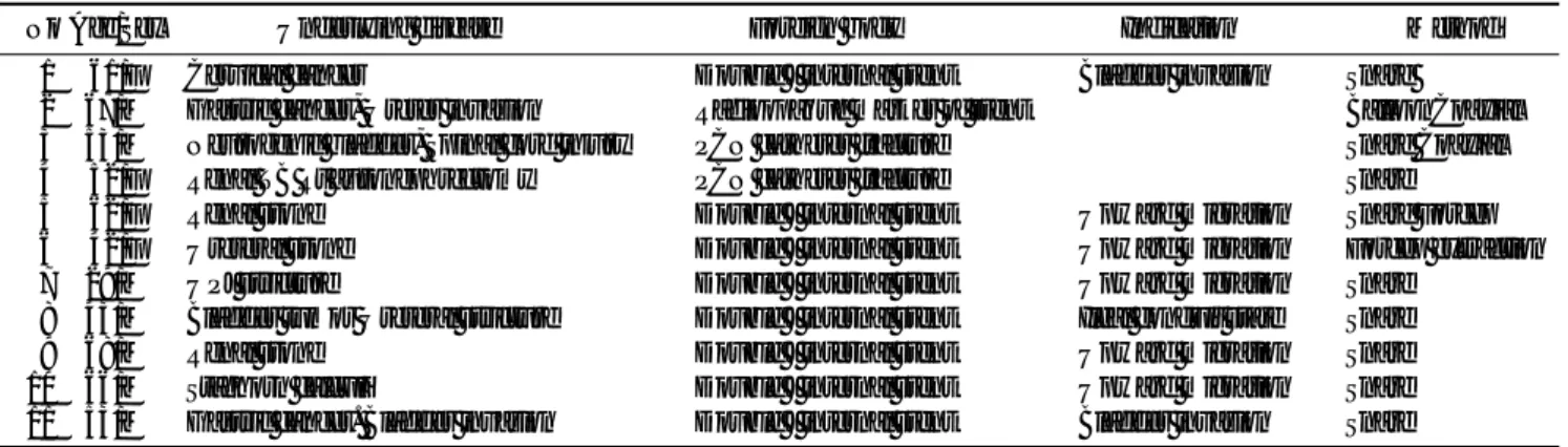
A B
Fig. 1. Gastric cancer with ureters and bladder invasion in a 53-year-old man (Patient 11, Table 1)
A. The tip of a internal stent is encir- cled by a gooseneck snare and then ex- tracted from the kidney.
B. Double J internal ureteral stent was inserted and PCN was done.
Table 1. Summary of Patients with Percutaneous Management of Upper Tract Foreign Bodies
No Age/Sex Underlying disease Foreign body Indication Method
01 61/F Cervical cancer Double J internal stent Bladder invasion Snare
02 67/M Gastric cancer, Ureter invasion Radioopaque marker of stent BalloonCoaxial 03 53/M Neurogenic bladder, Spinal cord injury PCN catheter fracture Snare Coaxial
04 52/F Renal TB Rt autonephrectomy PCN catheter fracture Snare
05 62/F Renal stone Double J internal stent Upward migration Snare Forcep
06 42/F Ureteral stone Double J internal stent Upward migration Forcep extraction
07 29/M UPJ stricture Double J internal stent Upward migration Snare
08 43/M Bladder tumor Ureteral stricture Double J internal stent Ileal conduit state Snare
09 68/M Renal stone Double J internal stent Upward migration Snare
10 66/M Staghorn calculi Double J internal stent Upward migration Snare
11 53/M Gastric cancer. Bladder invasion Double J internal stent Bladder invasion Snare Note.-UPJ = Ureteropelvic junction, TB = Tuberculosis
않아 유도철사를 부러진 카테타 내강을 통과시킨 후 스네어의 접근을 쉽게 하는 동축기법(coaxial technique), 즉 유도 철사 를 따라 스네어를 넣어 부러진 카테타를 쉽게 잡을 수 있도록 하여 제거에 성공하였다. 다른 1예에서는 기존의 트랙을 통해 부러진 카테타로의 접근이 어려웠다. 이물이 있는 신배를 직접 천자하여 제거하려 하였으나 신배의 공간이 협소하여 올가미 의 접근이 어려워 유도철사를 기존의 트랙에서 신우로 통과시 키고 새로운 트랙에서 gooseneck 올가미로 유도철사의 자유 로운 끝부분을 잡아서 C자 모양의 루프를 만들어 하방의 신배 에서 이물로의 접근을 쉽게 하였다. 그러한 후에 하방의 신배 로 10 F 피포를 삽입하여 gooseneck 올가미를 이용하여 부러 진 카테타를 제거하였다 (Fig. 3). 요관의 금속성 표식자의 제
거 시에도, 동축기법을 이용하여 목표물 내로 유도철사를 통과 시킨 후 3 mm 풍선 카테타를 표식자내에서 팽창시킨 후 제거 하였다 (Fig. 4).
경피적으로 제거를 시도한 상부 요로 결석은 크기에 따라 10-14 F의 피포를 이용하였다. 결석의 크기가 커서 피포내로 결석이 나오지 않은 경우에는 유도철사를 깊이 삽입한 후 결 석을 피포와 바스켓 사이에 끼워 피포와 동시에 제거하였다.
요관-방광 접합부위 협착 및 결석에 의한 폐색 증상을 동반한 2예의 환자에서는 PCN 카테타를 설치하여 감압시켰다. 그런 후에 유도철사를 방광으로 통과시킨 후 10 F Nelaton 카테타 를 요도를 통하여 방광으로 삽입하였다. 카테타를 통하여 gooseneck 올가미를 삽입하여 유도철사를 요도를 통하여 빼
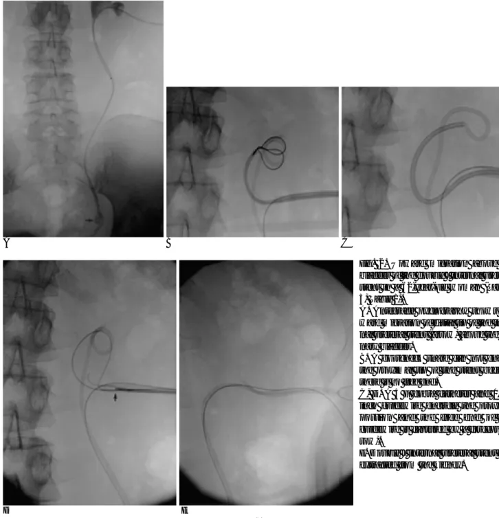
D E
Fig. 2. Upward migration above the bladder of the double J internal ureteral stent in a 62-year-old woman (Patient 5, Table 1).
A. Antegrade pyelography shows up- ward migration of distal tip of the inter- nal ureteral stent (arrow) above the uri- nary bladder.
B. A gooseneck snare can not engage the proximal tip of the stent because there is no free end.
C, D. A 5 F cobra catheter and 0.035 inch guidewire encircle the proximal portion and the free end of the guidewire is captured by a forceps (ar- row).
E. Double J internal ureteral stent was extracted from the kidney.
A B C
낸 후, 10 F 피포를 유도철사의 유도 하에 요관으로 삽입한 후 결석제거용 바스켓으로 결석을 제거하였다 (Fig. 5). 경피적으 로 요관 스텐트를 제거한 환자에서는 새로운 요관 스텐트를 재 삽입하였다. 소변의 배출과 트랙의 지혈을 위하여 8-12 F PCN 카테타를 다시 삽입하였다. 2-3일 후에 추적 검사를 시행하여 잔여물의 유무, 요로폐색, 조영제의 유출 등의 이상소견을 확 인하여 카테타 제거 여부를 결정하였다. 대상과 시술의 적응증 및 방법은 Table 1과 2에서 요약하였다.
결 과
경피적 제거술을 시도한 20예 중 1예를 제외한 모든 예에서 성공적으로 이물 혹은 결석을 제거하였다. 1예에서는 결석이 피포 내에 들어오지 않아 바스켓에 포획된 결석을 피포와 동 시에 제거하도록 시도하여 보았다. 그러나 시술 중에 결석을 신장 바깥의 트랙에서 놓쳤으며 결석 주위의 공간이 없어 결 석을 제거하는데 실패하였다. 초음파로 결석이 신장 바깥의 연
부조직에 위치하고 있는 것을 확인한 후 시술을 마쳤으며 1개 월 후 추적검사에서 변화가 없음을 확인하였다. 전 예에서 시 술과 관련된 주위 장기의 손상, 치료를 요하는 출혈, 조영제의 누출 등의 합병증은 없었다.
고 찰
최근 혈관계의 유치 카테타(indwelling catheter)의 사용 및 중재적 시술의 증가로 시술과 관련된 체내의 이물, 즉 유도 철 사, 부러진 카테타, 스텐트, 코일 등에 대한 경피적 제거술이 빈번하게 이루어지고 있다. 이러한 중재적 시술의 범위가 혈관 계뿐만 아니라 간 담도계 및 비뇨기계 분야의 시술에도 널리 이용되고 있다 (1-3). 요로계에서는 요관 스텐트 혹은 PCN 카테타 삽입술과 관련된 이물이 대부분을 차지한다. 요관 스텐 트는 합병증 즉 스텐트 폐쇄, 스텐트의 이동, 부러짐, 가피 (encrustation) 혹은 석회화가 발생할 경우에 그 스텐트를 제 거해야 한다 (4). 스텐트의 제거는 일차적으로 방광경을 이용 한 역행적 제거 및 교체를 주로 이용한다. 스텐트의 하단부가 요관으로 상방 이동한 경우에도 요관 내시경을 이용하여 제거 하기도 한다 (4-6). 그러나 스텐트로의 접근이 어렵거나 역행 적 제거가 기술적으로 어려운 경우는 경피적 제거술을 시도한 다. 경피적 제거술에는 방사선 투시 하에 올가미, 집게 겸자, 결석 제거용 바스켓을 이용하는 방법과 신장내시경을 이용하 는 방법이 있다. 이 중 방사선 투시 하 제거술이 트랙의 크기 가 작고, 덜 침습적이며 국소 마취로 시술이 가능하고 스텐트 제거 후에 8-10 F 카테타를 삽입할 수 있는 장점을 가지고 있다 (7). 이물질을 제거하는 기구로써 Troy 등(8)은 집게 겸 자를 이용한 경험을 보고하였고 Yeung 등(7)은 집게 겸자 및 올가미를 이용하여 이물 제거를 시도한 경험을 보고하였다. 저 자들은 주로 gooseneck 올가미를 이용하거나 0.038 인치 유 도철사를 굽혀 자가형 올가미를 이용하였다. 올가미는 이물의
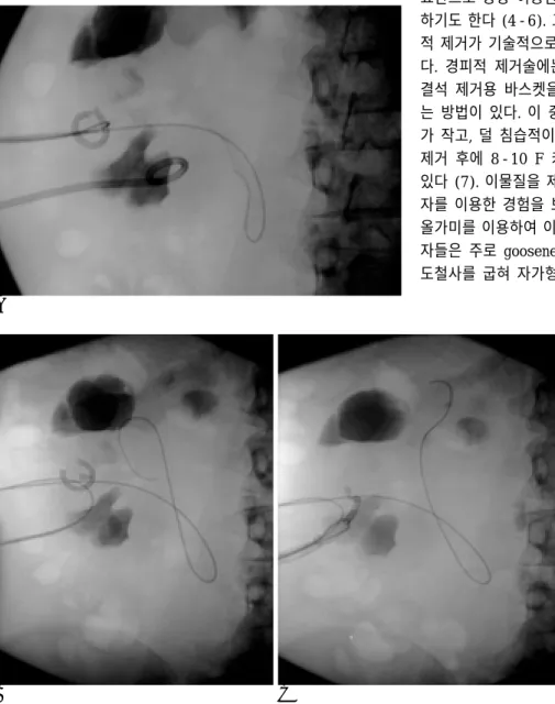
B A
C
Fig. 3. A fragmented PCN catheter in a 52-year-old woman with renal tubercu- losis (Patient 4, Table 1).
A. A foreign body containing calyx was punctured. Trial to extract the frag- ment by gooseneck snare was failed, because there was no free space.
B. A 0.035 inch guide wire was inserted to the renal pelvis from the previous PCN tract. The free end of the guidewire was captured by the goose- neck snare through the new PCN tract.
C. A 10 F sheath was inserted through previous PCN tract, under the guid- ance of C-looped guidewire. After then, the free end of the fragmented catheter was captured by a gooseneck snare through the sheath.
일부분을 걸 수 있어야 제거가 가능하며 이물의 일부를 걸 수 없는 경우는 집게 겸자를 이용할 수 있다. Savader 등(9)은 담 도 폐쇄 환자에서 내시경을 이용하여 삽입한 스텐트의 경피적 제거에 대해 보고하였다. 그들은 스텐트의 유리된 끝부분이 없
어 올가미로 이물을 잡을 수 없는 경우에 유도철사로 스텐트 를 감아 십이지장으로 통과시키고 유도철사의 끝부분을 gooseneck 올가미로 잡아서 루프를 만들어 스텐트를 제거하 였다. 저자들의 경우에도 스텐트의 끝이 상부의 신배까지 이동
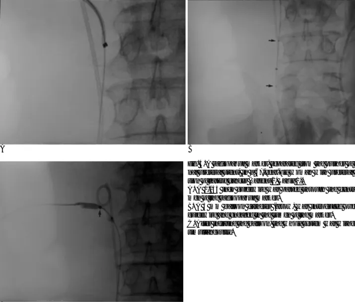
A
C
B
Fig. 4. A radiopaque marker, separated from the pusher of inter- nal ureteral stent, in a 67-year-old woman with ureteral inva- sion of gastric cancer (Patient 2, Table 1).
A. A 0.035 inch guidewire was passed through the central lu- men of the radioopaque marker.
B. A 3 mm balloon catheter (arrow) was introduced over the guidewire and engaged in the lumen of the marker.
C. After inflating the balloon, the whole system was withdrawn simultaneously.
Table 2. Summary of Patients with Percutaneous Stone Removal No Age/
Underlying Disease Previous
Location Size (mm) Method
Sex Intervention
1 62/M Renal & ureteral stone URS Renal pelvis 3×4 Stone basket
2 15/F Ureteral stone UVJ stricture UVJ 2×4 Transurethral approach Stone basket
3 53/F Ureteral stones UVJ stricture URS UVJ 2×4 Transurethral approach Stone basket 4 68/F Renal TB Renal & ureteral stone URS Renal pelvis 6×8 Stone basket
5 59/M Renal & Ureteral stone URS Renal pelvis 5×6 Stone basket
6 52/M Staghorn calculi PCNL Mid ureter 8×6 Stone basket
7 56/M Neurogenic bladder Ileal conduit Proximal ureter 8×8 Stone basket Ureteral stone
8 66/M Single kidney Renal stone calyx 5×6 Stone basket
9 61/M Staghorn calculi PCNL calyx 4×5 Stone basket
Note.-URS = Ureteroscopic removal of stone, UVJ = ureterovesicular junction, PCNL = Percutaneous nephrolithotomy
하거나 스텐트와 신우 사이의 공간이 부족하여 스네어를 스텐 트에 직접 걸 수 없는 경우에는, 트랙으로 후크 모양의 카테타 를 삽입하여 유도철사로 스텐트 주위를 감아, 유도 철사의 끝 을 집게 겸자 혹은 올가미로 잡아서 빼낸 후 루프를 만들어 스 텐트 제거를 시도하였다. 부러진 PCN 카테타 제거시에 기존 의 트랙에서 제거가 어려워 이중으로 트랙을 형성하여 이물로 의 접근을 쉽게 하여 이물 제거에 성공하였다. Lang 등(10)은 녹각석이나 다발성 신결석의 제거를 위하여 다수의 경피적 접 근경로를 이용하였으며, 위의 방법이 결석제거율을 향상시키며 신기능 손상등의 부작용의 증가와는 관련이 없는 것으로 보고 하였다.
상부 요로 결석의 치료는 체외충격파쇄석술을 이용하는 방 법과 경피적 신쇄석술이 있다. 경피적 신쇄석술은 방사선 투시 하 혹은 신내시경 유도 하에 집게겸자, 바스켓을 이용한 제거 법, 초음파 쇄석기 혹은 전기 수압 쇄석기를 이용한 분쇄법, 작 은 결석이나 침전물의 제거에 이용되는 세척법을 결합하여 사 용한다 (8). 경피적 신쇄석술의 적응증은 신결석 제거를 위한 수술의 과거력이 있는 환자에서 재발한 경우, 수술을 받을 수 없는 고위험군 환자, 수술 후 잔여결석이 있는 경우이다. 경피 적 신쇄석술은 기존의 관혈적인 수술법에 비하여 짧은 시술 시 간과 적은 합병증 및 낮은 이환율 등의 장점을 가지고 있다 (11, 12).
저자들은 결석의 크기가 작거나 요관 폐색 혹은 수술 후 변 화 등에 의하여 결석의 자연 배출이 어려운 환자에서 기존의 경피적 신쇄석술 트랙 혹은 신루 설치술 트랙을 이용하여 국
소마취 하에서 결석 제거를 시도하였다. 주로 결석 제거용 바 스켓을 사용하였다. 그러나 신우, 신배 혹은 요관의 공간이 협 소하여 바스켓을 이용한 제거가 어려울 경우는, 조영제가 희석 된 생리식염수을 주입하여 공간을 확보한 후에 결석 제거를 시 도하였다.
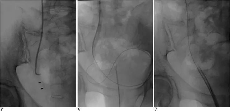
저자들은 요관-방광 접합부위 협착 및 결석에 의한 폐색 증 상을 동반한 2예의 환자에서 요도를 통한 역행적인 방법으로 결석제거를 시도하였다. Baere 등(13)이 요관 스텐트의 교체 시에 방사선 투시 하에서 0.018 인치 유도철사로 올가미를 만 들어 스텐트를 교체한 방법 및 Yedlicka 등(14)이 gooseneck 올가미를 이용하여 요도를 통한 요관 스텐트의 제거 및 교체 한 방법을 응용하였다. 2예 모두에서 성공적으로 결석을 제거 할 수 있었다. 저자들의 경험으로 하부 요관의 협착을 동반한 결석 환자에서 요관 내시경 혹은 체외충격파 쇄석술을 이용한 결석 제거가 어려울 경우에 바스켓을 이용한 결석 제거술이 유 용한 방법으로 생각된다.
결론적으로 저자들은 14 F 이하의 작은 지름의 피포를 통 하여 경피적으로 또는 요도를 통한 역행적인 경로를 통하여 이 물로의 접근을 시도하였다. 이물로의 접근이 어려운 경우에서 는 접근이 용이한 신배를 천자 하거나 동축기법, 후크 모양의 카테타 등으로 접근을 용이하게 하였다. 그런 후에 자가형 혹 은 goose neck 올가미, 집게겸자, 결석제거용 바스켓 등을 이 용하여 이물 제거에 성공하였다. 이러한 시술은 비뇨기계 중재 적 시술과 관련된 이물 제거 및 경피적 신쇄석술 후 잔여결석 등에 안전하게 사용될 수 있는 시술법으로 생각한다.
A B C
Fig. 5. Distal ureter stones with UVJ stricture in a 53-year-old woman ( Patient 3, Table 2).
A. Plain radiograph shows elliptical stones (arrows) in the distal ureter.
B. After failure of antegrade stone removal, a guidewire was captured by a gooseneck snare through the transurethral approach.
C. After retrograde insertion of a 10 F sheath, the stone was captured by a stone basket.
참 고 문 헌
1. Rubinstein ZJ, Morag B, Itzchak Y. Percutaneous removal of in- travascular foreign bodies. Radiology 1990;176:535-538
2. Uflacker R, Lima S, Melichar AC. Intravascular foreign bodies:
Percutaneous retrieval. Radiology 1986;160:731-735
3. Selby JB, Tegtmeyer CJ, Bittner GM. Experience with new re- trieval foreceps for foreign body removal in the vascular, urinary and biliary systems. Radiology 1990;176:535-538
4. Leroy AJ, Williams HJ, Segura JW, Patterson DE, Benson RC.
Indwelling ureteral stents: Percutaneous management of complica- tions. Radiology 1986;158:219-222
5. Niendorf DC. Kamhi B. Retrieval of indwelling ureteral stent uti- lizing Fogarty catheter. Urology 1975;6:622-623
6. Pocock RD, Stower MJ, Ferro MA, Smith PJ, Gingell JC. Double J stents: A review of 100 patients. BR J Urol 1986;58;629-633 7. Yeung EY, Carmody E, Thurston W, Ho CS. Percutaneous fluoro-
scopically guided removal of dysfunctioning ureteral stents. Radi- ology 1994;190:145-148
8. Troy RB, Streem SB, Zelch MG. Percutaneous management of up- per tract foreign bodies. J Endourol 1994;8:43-47
9. Savader SJ, Brodkin J, Osterman FA Jr. In situ formation of a loop snare for retrieval of a foreign body without free end. Cardiovasc Intervent Radiol 1996;19:298-301
10. Lang EK, Glorioso LW. Multiple percutaneous access routes to multiple calculi, calculi in caliceal diverticula, and staghorn calculi.
Radiology 1986;158:211-214
11. Castaneda-Zuniga WR, Clayman R, Smith A, Rusnak B, Herrera M, Amplatz K. Nephrostolithotomy: Percutaneous techniques for urinary calculus removal. AJR Am J Roentgenol 1982;139:721-726 12. Snyder JA, Smith AD. Staghorn calculi: Percutaneous extraction
versus anatrophic nephrolithotomy. J Urol 1986;136:351-354 13. Baere TD, Denys A, Pappas P, Chalier E, Roche A. Ureteral stents:
Exchange under fluoroscopic control as an effective alternative to cystoscopy. Radiology 1994;190:887-889
14. Yedlicka JW Jr, Carlson JE, Hunter DW, Castaneda-Zuniga WR, Amplatz K. Nitinol goose neck snare for removal of foreign bodies:
Experimental study and clinical evaluation. Radiology 1991;178:
691-693
J Korean Radiol Soc 2002;47:69-76
Address reprint requests to : Chang Kyu Seong, M.D., Department of Diagnostic Radiology, Kyungpook National University School of Medicine, 52 Sam Duk-dong 2Ga, Chung-gu, Daegu 700-721, Korea.
Tel. 82-53-420-5390 Fax. 82-53-422-2677 E-mail: sck@knu.ac.kr
Percutaneous Retrieval of Upper Urinary Tract Foreign Bodies and Calculi
1Tae Beom Shin, M.D., Chang Kyu Seong, M.D., Yong Joo Kim, M.D.
1Department of Diagnostic Radiology, Kyungpook National University School of Medicine
Purpose:To determine, when extracorporeal shockwave lithotripsy is contraindicated, the usefulness and safety of percutaneous management in the removal from the upper urinary tract of foreign bodies and calculi, or small remnants of these, retained affer percutaneous nephrolithotomy.
Materials and Methods:Between January 1996 and May 2001, we attempted to retrieve foreign bodies or cal- culi from the upper urinary tract of 20 patients, using various percutaneous techniques. There were eleven for- eign bodies, namely fragmented nephrostomy catheters (n=2), migrated ureteric stents inaccessible to retro- grade ureteroscopic management (n=8), and one metallic radiopaque marker which was separated from the pusher of the internal ureteral stent. Nine urinary tract calculi were present. These ranged in radiographically measured size from 4 to 8 mm in their largest diameter, and were found in the renal pelvis or calyx (n=5) and ureter (n=4). After percutaneous nephrostomy, all procedures involved the use of a 7-F to 14-F sheath, insert- ed under fluoroscopic guidance. Devices used for the retrieval of these objects include a stone basket retriever, loop snare, grasping forceps, and balloon catheter.
Results:In all cases except one, it was possible to retrieve calculi or other items from the upper urinary tract.
No surgical procedure was required and no significant complications were encountered in any of the cases during or after the procedures.
Conclusion:The percutaneous technique can be useful and safe in the management of foreign bodies or cal- culi present in the upper urinary tract.
Index words :Kidney, interventional procedure Kidney, percutaneous procedure Ureter, interventional procedure Ureter, calculi
Foreign bodies