www.krspine.org
Ogilvie’s Syndrome after Lumbar Spinal Surgery
Su-Keon Lee, M.D., Seung-Hwan Lee, M.D., Byeong-Mun Park, M.D., Bong-Seok Yang, M.D., Ji-Hyeon Kim, M.D., Hwan-Mo Lee, M.D.
J Korean Soc Spine Surg 2019 Jun;26(2):63-67.
Originally published online June 30, 2019;
https://doi.org/10.4184/jkss.2019.26.2.63
Korean Society of Spine Surgery
SMG-SNU Boramae Medical Center, 20, Boramae-ro 5-gil, Dongjak-gu, Seoul 07061, Korea Tel: +82-2-831-3413 Fax: +82-2-831-3414
©Copyright 2017 Korean Society of Spine Surgery pISSN 2093-4378 eISSN 2093-4386
The online version of this article, along with updated information and services, is located on the World Wide Web at:
http://www.krspine.org/DOIx.php?id=10.4184/jkss.2019.26.2.63
This is an Open Access article distributed under the terms of the Creative Commons Attribution Non-Commercial License (http://
creativecommons.org/licenses/by-nc/4.0) which permits unrestricted non-commercial use, distribution, and reproduction in any medium, provided the original work is properly cited.
Journal of Korean Society of
Spine Surgery
Ogilvie’s Syndrome after Lumbar Spinal Surgery
Su-Keon Lee, M.D., Seung-Hwan Lee, M.D., Byeong-Mun Park, M.D., Bong-Seok Yang, M.D., Ji-Hyeon Kim, M.D., Hwan-Mo Lee, M.D.
* Department of Orthopaedic Surgery, Gwangmyeong Sungae Hospital*Department of Orthopaedic Surgery, Yonsei University College of Medicine
Study Design: Case report.
Objectives: We report a case of Ogilvie’s syndrome following posterior decompression surgery in a spinal stenosis patient who presented with acute abdominal distension, nausea, and vomiting.
Summary of Literature Review: Ogilvie’s syndrome is a rare and potentially fatal disease that can easily be mistaken for postoperative ileus, and is also known as acute colonic pseudo-obstruction. Early recognition and diagnosis enable treatment prior to bowel perforation and requisite abdominal surgery.
Materials and Methods: An 82-year-old woman presented with 6 months of worsening back pain with walking intolerance due to weakness in both legs. She had hypertension, asthma, and Cushing syndrome without bowel or bladder symptoms. Further workup demonstrated the presence of central spinal stenosis on magnetic resonance imaging. The patient underwent an L2-3 laminectomy and posterior decompression. Surgery was uneventful.
Results: The patient presented with acute abdominal distension, nausea, and vomiting on postoperative day 1. The patient was initially diagnosed with adynamic ileus and treated conservatively with bowel rest, reduction in narcotic dosage, and a regimen of stool softeners, laxatives, and enemas. Despite this treatment, her clinical course failed to improve, and she demonstrated significant colonic distension radiographically. Intravenous neostigmine was administered as a bolus with a rapid and dramatic response.
Conclusion: Ogilvie’s syndrome should be included in the differential diagnosis of postoperative ileus in patients developing prolonged unexplained abdominal distension and pain after lumbar spinal surgery. Early diagnosis and initiation of conservative management can prevent major morbidity and mortality due to bowel ischemia and perforation.
Key Words: Ogilvie’s syndrome, Ileus, Lumbar surgery
Received: March 24, 2019 Revised: April 8, 2019 Accepted: June 14, 2019 Published Online: June 30, 2019
Corresponding author: Seung-Hwan Lee, M,D.
ORCID ID: Seung-Hwan Lee: https://orcid.org/0000-0002-0432-3857 Byeong-Mun Park: https://orcid.org/0000-0003-2637-4257 Department of Orthopaedic Surgery, Gwangmyeong Sungae Hospital, 36 Digital-ro, Gwangmyeong, 14241, Korea
TEL: +82-2-2680-7699, FAX: +82-2-2680-7755 E-mail: java5885@gmail.com
Ogilvie’s syndrome was first described by Sir William Heneage Ogilvie in 1948. Ogilvie’s syndrome presents with symptoms, sign and radiographic appearance of acute large bowel obstruction of non-mechanical etiology. The clinical features include abdominal distension and pain (80%), as well as nausea with or without associated vomiting (60%).
Tympanic abdomen, exists in almost 90% of patients although bowel sounds are preserved. Plain abdominal X-ray and computed tomography show varying degrees of colonic dilatation. The reason, it develops in patients of posterior spinal decompression is unknown. Interruption of the parasympathetic fibers from S2 to S4 level after spinal trauma or corrective spinal surgery, spinal anesthesia, and pharmacological agents leads to impairment of the autonomic nervous system. A kind of imbalance between sympathetic and parasympathetic stimulation, an atonic distal colon, and
a functional proximal obstruction, if left untreated can result in bowel ischemia and perforation with an estimated mortality rate of 40%.1-4)
Su-Keon Lee et al Volume 26 • Number 2 • June 30 2019
www.krspine.org 64
Case Report
This study received an exemption by the Institutional Review Board of our institute.
A 82-year-old women presented with 6 month of worsening back pain with walking intolerance due to both leg weakness . She has hypertesion, asthma, Cushing syndrome and has no bowel or bladder symptoms. Further workup demonstrated the presence of central spinal stenosis in MRI finding. MRI shows central spinal stenosis in L2-3 level with
hypertrophic ligamentum flavum. The patient underwent an L2-3 laminectomy and posterior decompression. Surgery was uneventful. Total operative time was approximately 1 hours (Fig. 1).
The patient was intact on postoperative neurological examination. Tomorrow evening, approximately 24 hours after surgery was completed, the patient developed worsening abdominal pain and nausea. Examination at this time revealed a distended abdomen with diminished bowel sounds. The immediately postoperative spine radiographs and subsequent abdominal films demonstrated marked colonic dilation and a gaseous pattern Postoperative paralytic ileus was initially diagnosed, and treatment included bowel rest; a combination of stool softeners, laxatives, and enemas; and limitation of narcotic dosage. While the patient remained clinically stable, disease progression was noted radiographically (Fig. 2).
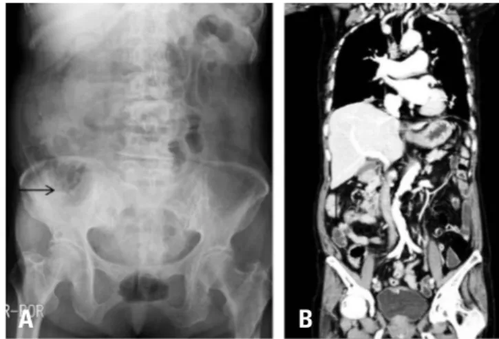
On postoperative Day 4, her symptoms of abdominal distension and nausea progressively worsened, and plain abdominal radiographs demonstrated an increase in colon diameter from 11 to 15 cm (Fig. 3). A nasogastric tube was placed, and a general surgery consultation was obtained.
The patient was transferred to the intensive care unit for treatment with neostigmine. Within 6 hours of neostigmine
Fig. 1. (A, B) Preoperative anteroposterior and lateral radiographs of an 82-year-old woman show sclerotic changes and disc space narrowing at L2-3. (C, D) Postoperative anteroposterior and lateral radiographs show L2-3 decompression.
A
C
B
D
Fig. 2. An abdominal radiograph on postoperative day 1 shows abdominal distension and fecal impaction with gas retention.
administration, the patient noted a dramatic improvement in abdominal pain and distension, which was followed by flatus and bowel movement shortly thereafter. Abdominal radiography 12 hours after neostigmine administration revealed dramatically reduced colonic distension (Fig. 4).
The patient was observed on bowel rest until the return of function. She was discharged home in good condition, and she reported continued normal bowel function at her follow-up appointment 1 week later.
Discusssion
Sir William Ogilvie first described the syndrome of colonic pseudo-obstruction in 1948 in a series of two patients with tumor invasion of the celiac plexus. At that time he theorized the cause to be sympathetic denervation of the colon.2) There have since been numerous theories regarding the etiology of the syndrome, including loss of sympathetic stimulation, prostaglandin abnormalities, transient ischemia, mechanical air-fluid lock of the bowel, diminished parasympathetic stimulation, and/or autonomic imbalance.5-9) While the underlying pathophysiology remains unclear, the syndrome is characterized by decreased gastrointestinal motility with massive colonic dilation in the absence of mechanical obstruction and with limited small bowel involvement. This distinguishes the syndrome from severe postoperative adynamic ileus, which affects the small bowel and does not respond well to neostigmine.9,10) Treatment of Ogilvie’s syndrome must be undertaken to prevent bowel ischemia and perforation, which carries a high mortality rate as noted.
Various intestinal diseases has to be ruled out before making the diagnosis of Ogilvie’s syndrome such as acute megacolon, acute mesenteric ischemia, chronic constipation, diverticulitis, hirschsprung disease, intestinal perforation, mechanical colonic obstruction, toxic megacolon, fecal impaction, and tumors.
Patients with mechanical obstruction will present with crampy abdominal pain; but the absence of pain with opiates treatment Fig. 3. (A-C) An abdominal radiograph and computed tomography scan
taken on postoperative day 4. The patient remained clinically stable, but an increased amount of air was noted in the colon, most notably in the cecum, which measured 11 cm.
Fig. 4. (A, B) An abdominal radiograph and computed tomography scan taken on postoperative day 6, at 12 hours after neostigmine administra- tion, demonstrating a dramatic reduction in colonic air and fecal impac- tion.
A
C
B
A B
Su-Keon Lee et al Volume 26 • Number 2 • June 30 2019
www.krspine.org 66
does not rule out the mechanical obstruction. Toxic megacolon patients appear very ill with fever tachycardia and abdominal tenderness with a history of bloody diarrhea. Electrolyte monitoring is a necessity. Computer tomography scan was done for both of our patients keeping in mind to rule out tumors, cecal and sigmoid volvulus, perforation, peritonism, ischemia, obstruction, and toxic megacolon.
Neostigmine, a parasympathomimetic, is an anticholinesterase, and acts to increase the acetylcholine concentration at the synapses. In this case, neostigmine’s action is directed at the imbalance in sympathetic-parasympathetic activity. Maloney and Vargas in their trial, first used guanethidine, followed by neostigmine. The improvement was noted following administration of neostigmine, proving that the pseudo- obstruction arises from parasympathetic under activity rather than sympathetic overactivity.1)
Our patient has hypertesion, asthma, cushing syndrome.
And she was accompanied with hypokalemia. It seems to be associated with ogilvie syndrome. And fortunately she had dramatical response to neostigmine. So surgical exploration was not required for our patient, and she recovered successfully in 6 days. We recommend that Ogilvie’s syndrome should be considered as a differential diagnosis in patients with postoperative significant abdominal distension who had undergone spine surgery. Early recognition and appropriate conservative treatment would be necessary to prevent complications such as bowel ischemia and perforation.
REFERENCES
1. Maloney N, Vargas HD. Acute intestinal pseudo-obstruc- tion (Ogilvie’s syndrome). Clin Colon Rectal Surg 2005 May;18(2):96-101. DOI: 10.1055/s-2005-870890.
2. Ogilvie WH. William Heneage Ogilvie 1887-1971. Large- intestine colic due to sympathetic deprivation. A new clinical syndrome. Dis Colon Rectum. 1987 Dec;30(12):984-7.
DOI: 10.1007/bf02554291.
3. Tsirikos AI, Sud A. Ogilvie’s syndrome following poste- rior spinal arthrodesis for scoliosis. Indian J Orthop. 2013 Jul;47(4):408-12. DOI: 10.4103/0019-5413.114934.
4. Vanek VW, Al-Salti M. Acute pseudo-obstruction of the colon (Ogilvie’s syndrome). An analysis of 400 cases. Dis Colon Rectum. 1986 Mar;29(3):203-10. DOI: 10.1007/
bf02555027.
5. Anuras S, Baker CR, Jr. The colon in the pseudoobstructive syndrome. Clin Gastroenterol. 1986 Oct;15(4):745-62.
6. Bachulis BL, Smith PE. Pseudoobstruction of the colon.
Am J Surg. 1978 Jul;136(1):66-72. DOI: 10.1016/0002- 9610(78)90202-7.
7. Feldman RA, Karl RC. Diagnosis and treatment of Ogilvie’s syndrome after lumbar spinal surgery. Report of three cases.
J Neurosurg. 1992 Jun;76(6):1012-6. DOI: 10.3171/
jns.1992.76.6.1012.
8. Ozkurt H, Yilmaz F, Bas N, et al. Acute colonic pseudo- obstruction (Ogilvie’s syndrome): radiologic diagnosis and medical treatment with neostigmine. Report of 4 cases. Am J Emerg Med. 2009 Jul;27(6):757.e1-4. DOI: 10.1016/
j.ajem.2008.10.020.
9. Singleton AO, Jr., Wilson M. Air-fluid obstruction of the colon. South Med J South Med J. 1967 Sep;60(9):909-13.
DOI: 10.1097/00007611-196709000-00001.
10. Livingston EH, Passaro EP, Jr. Postoperative ileus. Dig Dis Sci. 1990 Jan;35(1):121-32. DOI: 10.1007/bf01537233.
요추 수술 후에 발생한 Ogilvie’s Syndrome
이수건 • 이승환 • 박병문 • 양봉석 • 김지현 • 이환모*
광명성애병원 정형외과, *연세대학교 의과대학 정형외과학교실
연구계획: 증례 보고
목적: 요추부 후방 수술 후에 발생한 Ogilvie’s syndrome 환자를 보고하고자 한다.
선행 연구문헌의 요약: Ogilvie’s syndrome은 수술 후 장폐색으로 오인할 수 있는 드물지만 치명적인 질환이다. 급성 대장 가성폐색이라고도 알려져 있으 며, 조기 진단 및 치료을 통해 장천공 및 복부 수술을 예방할 수 있다.
대상 및 방법: 82세 여자 환자가 양하지 근력 약화를 동반한 보행 장애 및 요통으로 내원하였다. 환자는 고혈압, 천식, 쿠싱 증후군 병력이 있었다. 중심부 척추 협착증으로 진단하여, 제 2-3요추간 후궁 절제술 및 후방 감압술을 시행하였다.
결과: 환자는 수술 다음 날 복부 팽만, 오심, 구토 증상을 보였다. 장운동 감소에 의한 장폐색으로 진단하여 변완화제 투여 및 관장을 시행하고, 마약성 약 물 투여를 감소시켰으나 증세는 악화되었다. 정맥 내 네오스티그민 투여 후에 빠른 증상 완화를 보였다.
결론: Olivie’s syndrome은 척추 수술 후 장폐색을 보일 때 감별 진단으로 고려되어야 한다. 조기 진단 및 약물 투여가 장허혈과 천공으로 인한 합병증을 예방할 수 있다.
색인 단어: Ogilvie’s symrome, 장폐색, 요추 수술 약칭 제목: 수술 후 Ogilvie’s symdrome
접수일: 2019년 3월 24일 수정일: 2019년 4월 8일 게재확정일: 2019년 6월 14일 교신저자: 이승환
경기도 광명시 디지털로 36 광명성애병원 정형외과
TEL: 02-2680-7699 FAX: 02-2680-7755 E-mail: java5885@gmail.com