ABSTRACT
Background: Estrogen controls the pubertal growth spurt, growth plate closure, and accretion of bone mineral density (BMD) of long bones after biding estrogen receptor (ER).
There are two subtypes of ER, ERα and ERβ. If each ER subtype has different effects, we may control those actions by manipulating the estrogen binding intensity to each ER subtype and increase the final adult height without markedly reducing BMD or impairing reproductive functions. The purpose of our study was to compare these effects of ERα and ERβ on long bones in ovariectomized rats.
Methods: Thirty female rats were ovariectomized and randomly divided into 3 groups. The control, propylpyrazole triol (PPT), and 2,3-bis (4-hydroxyphenyl) propionitrile (DPN) groups were subcutaneously injected for 5 weeks with sesame oil, PPT as an ERα agonist, and DPN as an ERβ agonist, respectively. The crown-lump length and body weight were measured weekly.
BMD, serum levels of growth hormone (GH) and estradiol were checked before and after 5 weeks of injections. Pituitary GH1 expression levels were determined with quantitative real- time polymerase chain reaction, the proximal tibias were dissected, decalcified and stained with hematoxylin-eosin, and the thicknesses of epiphyseal plates including proliferative and hypertrophic zones were measured in 20-evenly divided sites after 5 weeks of injections.
Comparisons for auxological data, serum hormone and pituitary GH1 expression levels, BMD, and epiphyseal plate thicknesses among 3 groups before and after injections were conducted.
Results: There was no significant difference in body lengths among 3 groups. The body weights were significantly lower, but, serum GH, pituitary GH1 expression levels, and BMDs were higher in PPT group than the other 2 groups after 5 weeks of injections. There was no significant difference in the thicknesses of the total epiphyseal plate, proliferative, and hypertrophic zone among 3 groups.
Conclusion: ERα is more involved in pituitary GH secretion and bone mineral deposition than ERβ. Weight gain might be prevented with the ERα agonist.
Keywords: Growth; Bone Density; Estrogen Receptor Alpha; Estrogen Receptor Beta
Original Article
Received: May 4, 2020 Accepted: Sep 8, 2020 Address for Correspondence:
Kye Shik Shim, MD, PhD
Department of Pediatrics, Kyung Hee University Hospital at Gangdong, Kyung Hee University School of Medicine, 892 Dongnam- ro, Gangdong-gu, Seoul 05278, Korea.
E-mail: [email protected]
*Byung Ho Kang and Ja Hyang Cho contributed equally to this work.
© 2020 The Korean Academy of Medical Sciences.
This is an Open Access article distributed under the terms of the Creative Commons Attribution Non-Commercial License (https://
creativecommons.org/licenses/by-nc/4.0/) which permits unrestricted non-commercial use, distribution, and reproduction in any medium, provided the original work is properly cited.
ORCID iDs Byung Ho Kang
https://orcid.org/0000-0002-1690-7753 Ja Hyang Cho
https://orcid.org/0000-0002-5562-588X So Youn Kim
https://orcid.org/0000-0001-8309-4927 Kyoung A Jeong
https://orcid.org/0000-0002-0612-4392 Shin-Hee Kim
https://orcid.org/0000-0002-8405-5417 Chanwoo Kim
https://orcid.org/0000-0001-9914-313X Sung-Jig Lim
https://orcid.org/0000-0003-2549-8434 Kye Shik Shim
https://orcid.org/0000-0002-8004-151X
Byung Ho Kang ,1* Ja Hyang Cho ,1* So Youn Kim ,1 Kyoung A Jeong ,1 Shin-Hee Kim ,1 Chanwoo Kim ,2 Sung-Jig Lim ,3 and Kye Shik Shim 1
1 Department of Pediatrics, Kyung Hee University Hospital at Gangdong, Kyung Hee University School of Medicine, Seoul, Korea
2 Department of Nuclear Medicine, Kyung Hee University Hospital at Gangdong, Kyung Hee University School of Medicine, Seoul, Korea
3 Department of Pathology, Kyung Hee University Hospital at Gangdong, Kyung Hee University School of Medicine, Seoul, Korea
Growth and Bone Mineral Density Changes in Ovariectomized Rats
Treated with Estrogen Receptor Alpha or Beta Agonists
Endocrinology, Nutrition &
Metabolism
Funding
This research was supported by the Korean Society of Pediatric Endocrinology Grant (grant No. 2011-01).
Disclosure
The authors have no potential conflicts of interest to disclose.
Author Contributions
Conceptualization: Shim KS. Data curation:
Cho JH. Formal analysis: Kang BH.
Methodology: Kang BH, Cho JH, Kim SY, Jeong KA, Kim SH. Investigation: Kang BH.
Software: Kim C, Lim SJ. Validation: Kim C, Lim SJ. Writing - original draft: Kang BH, Cho JH, Kim SY, Jeong KA, Kim SH. Writing - review &
editing: Kim C, Lim SJ, Shim KS.
INTRODUCTION
Estrogen is known to have two opposite effects on the growth of longitudinal bones. It increases growth by increasing growth hormone (GH) and insulin like growth factor-I (IGF-I) secretions during the pubertal growth spurt, but it stops growth by promoting the closure of the growth plates with differentiating chondrocytes. It also promotes the accretion of bone mineral density (BMD) through the differentiation of osteoblasts and osteoclasts.1
In the classical pathway, the effects of estrogen occur after binding to estrogen receptors (ER, ESR). Two subtypes of ER have been identified, estrogen receptor α (ERα, ESR1) and β (ERβ, ESR 2).2
If each ER subtype has different effects on regulating pubertal onset, growth spurts, growth plate closure, acquisition of bone mineral content, and reproductive functions, we may control these actions by manipulating the estrogen binding intensity to each ER subtype.
We might be able to increase the final height in adults without markedly reducing BMD or impairing reproductive functions, even in humans.3-6
Nowadays, the incidence of precocious puberty in children is increasing in developed countries. It causes short stature due to earlier pubertal growth spurt and closure of epiphyseal plates in long bones.7 If we can elucidate the action of each ER subtype on pubertal growth spurt and epiphyseal plate fusion, we may find out the method to increase human height more efficiently.
The aim of our study was to understand the actions of each ER subtype on the pubertal growth spurt, growth plate closure, and acquisition of bone mineral content.
METHODS
Animals
Thirty female Sprague Dawley rats were housed in an approved animal facility with a 12- hour light cycle and given ad libitum access to food and water. They were ovariectomized at 4 weeks of age under intramuscular and intraperitoneal anesthesia with 30 mg/kg of tiletamine hydrochloride (HCl) and zolazepam HCl (Zoletil® 10%; Virbac, Carros Cedex, France) and 5 mg/kg of xylazine HCl (Rompun® 2%; Bayer, Leverkusen, Germany).
Drug injections
The rats were randomly divided into 3 groups (n = 10/group). Ten rats were injected with sesame oil (control group); another 10 were injected with 10 mg/kg of propylpyrazole triol (PPT®; Cayman Chemical, Ann Arbor, MI, USA) as the ERα agonist treated group (PPT group); the other 10 were injected with 10 mg/kg of 2,3-bis (4-hydroxyphenyl) propionitrile (DPN®; Cayman Chemical) as the ERβ agonist treated group (DPN group). The rats were subcutaneously injected with the same volume starting at the age of 6 weeks, 5 days per week for 5 weeks.
Body length and weight measurements
The body length (crown-rump length) and weight of each rat were measured weekly from the age of 1 through 10 weeks.
BMD analysis
Analyses of total body and lumbar vertebral BMDs of the rats were performed at the age of 6 and 10 weeks through dual energy X-ray absorptiometry (DXA) using the Lunar PIXImus mouse densitometer (Wipro GE Healthcare, Madison, WI, USA), the Norland Medical systems pDEXA Sabre (Norland Medical Systems, Fort Atkinson, WI, USA), and the Sabre Research Software (version 3.6; Norland Medical System).
Measurement of serum hormone levels
Blood samples were obtained via the tail vein of 6- and 10-week old rats, and serum levels of GH and estradiol (E2) were determined with the ELISA kits for GH (Millipore, Darmstadt, Germany) and E2 (Calbiotech, Spring Valley, CA, USA).
Quantitative real-time polymerase chain reaction (RT-PCR) analysis
After each rat was euthanized, Gh1 mRNA was extracted from the pituitary gland with RNeasy mini kit (Qiagen, Duesseldorf, Germany) according to the manufacturers' instructions. The quantitative RT-PCR analysis was performed using the ABI Power SYBR green PCR master mix (Thermo Fisher Scientific, Waltham, MA, USA) and the Step One Plus RT-PCR system (ABI). The sequences of the primer sets used for Gh1 and 18S were as follows; sense 5′-GCTGCAGACTCTCAGACTCCCTGG-3′, antisense 5′-CTGAGAAGCAGAACGCAGCCTG-3′, sense 5′-TGGTTGATCCTGCCAGTAG-3′, and antisense 5′-CGACCAAAGGAACCATAACT-3′.Quantitative histology of growth plates
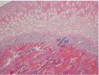
The proximal tibias of the rats were dissected, decalcified, and stained with hematoxylin- eosin. The thicknesses of their epiphyseal plates (EP) including the proliferative (PZ) and hypertrophic zones (HZ) were determined on 20-evenly divided sites in the central three- fourths of the growth plate sections using a Nikon Eclipse E800 light microscope (Nikon, Tokyo, Japan) with ImageJ software (version 1.5, NIH, USA) (Fig. 1).8
Statistics
The differences in auxological data, BMD, serum GH and E2 levels before and after injections were analyzed with the one-way analysis of variance with multiple comparisons. The Kruskall- Wallis test was used to compare the pituitary Gh1mRNA levels and histologic data among the
EP HZPZ
Fig. 1. Thickness measurement of the EP including the PZ and HZ of tibia in a rat (×100, H&E staining).
EP = epiphyseal plate, PZ = proliferative zone, HZ = hypertrophic zone, H&E = hematoxylin-eosin.
groups with the SPSS ver. 20.0. All the data were expressed as mean ± standard deviation. P <
0.05 was considered as statistically significant.
Ethics statement
The procedures used and the care of animals were approved by the Institutional Animal Care and Use Committee in the Kyung Hee University Hospital at Gangdong (approval No.
KHNMC AP 2013-011).
RESULTS
Change of auxological data
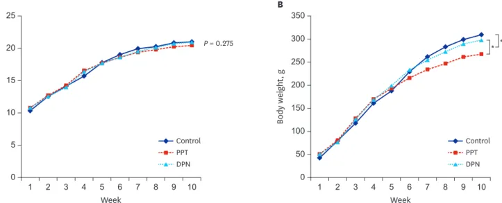
The body lengths of 1-week-old rats in the control, PPT, and DPN groups were 10.33 ± 0.75, 10.79 ± 0.51, and 10.8 ± 0.5 cm (P = 0.275). At 4 weeks of age and before the injections, the body lengths were 15.7 ± 0.68, 16.56 ± 0.53, and 16.39 ± 0.49 cm for each group (P = 0.969);
and at 10 weeks of age and after injections 21.0 ± 0.52, 20.44 ± 0.29, and 20.94 ± 0.4 cm for each group (P = 0.083). There was no significant difference in body length among 3 groups before and after injections (P = not significant).
The body weights of 1-week-old rats in the control, PPT, and DPN groups were 42.52 ± 3.11, 50.85 ± 4.78, and 50.92 ± 4.48 g (P = 0.172). At 4 weeks of age and before injections, the body weights were 161.05 ± 10.71, 169.94 ± 15.3, and 168.54 ± 8.19 g for each group (P = 0.637); and at 10 weeks of age and after injections, 309.46 ± 22.65, 267.49 ± 16.82, and 297.66 ± 16.25 g for each group (P = 0.012). The mean body weight after 5 weeks of injections in the PPT group was significantly lower than that in the other 2 groups (P < 0.05) (Fig. 2).
Bone mineral density
The total body BMD in the control, PPT, and DPN group at 6 weeks of age was 0.101 ± 0.004, 0.108 ± 0.008, and 0.105 ± 0.008 g/cm2 (P = 0.083), and at 10 weeks of age was 0.121 ± 0.006, A
20 25
5
0 1 2 3 5 8 10
15
10
Crown-rump length, cm
Week
4 6
P = 0.275
9 7
Control PPT DPN
Control PPT DPN
B
300 350
100
0 1 2 3 5 8 10
250 200
Body weight, g
Week
4 6
150
50
9 7
* *
Fig. 2. Comparison of auxological changes among 3 groups of rat subjected to treatment with sesame oil, PPT, or DPN. (A) Changes of the crown-rump length in 3 groups. (B) Changes of the body weight in 3 groups.
PPT = propylpyrazole triol, DPN = 2,3-bis (4-hydroxyphenyl) propionitrile.
*P < 0.05.
0.146 ± 0.009, and 0.124 ± 0.009 g/cm2 (P = 0.003). The lumbar vertebral BMD in the control, PPT, and DPN group at 6 weeks of age was 0.093 ± 0.001, 0.099 ± 0.001, and 0.098 ± 0.002 g/cm2 (P = 0.075), and at 10 weeks of age was 0.135 ± 0.006, 0.155 ± 0.009, and 0.138 ± 0.009 g/cm2, respectively (P = 0.038). Therefore, the total body and lumbar vertebral BMD were significantly increased in PPT group than those in the other 2 groups (P < 0.05) after 5 week- injections (Table 1).
Serum hormone levels
There was no significant difference in serum levels of GH and E2 in 6-week-old rats among 3 groups. The serum GH level in 10-week-old rats was 3.36 ± 0.18, 7.29 ± 0.58, and 3.84 ± 0.37 pg/mL in the control, PPT, and DPN groups, respectively. Therefore, the serum GH level in the PPT group was significantly increased compared with those in the other 2 groups (P <
0.05). The serum E2 level in 10-week-old rats was 6.11 ± 0.92, 5.45 ± 1.38, and 5.94 ± 1.23 µg/
mL in the control, PPT, and DPN groups, respectively. There was no significant difference in serum E2 levels among the groups (P = 0.602) (Table 2).
Gh1 expression levels in the pituitary gland
The relative expression level of the Gh1 gene was 0.94 ± 0.14, 1.44 ± 0.66, 1.23 ± 0.2 in the control, PPT, and DPN groups, respectively. The Gh1 expression was significantly increased in the PPT and DPN group (P < 0.05) (Fig. 3).
Quantitative histology
The thicknesses of the proliferative zones were 45.77 ± 1.7, 45.12 ± 2.98, and 41.78 ± 1.2 μm in the control, PPT, and DPN group after injection, respectively, and there was no significant difference among the groups (P = 0.332); those of the hypertrophic zones were 35.48 ± 2.09, 34.4 ± 1.62, and 30.58 ± 1.03 μm, respectively, and there was no significant difference among the groups (P = 0.226). The thicknesses of total epiphyses were 91.25 ± 2.41, 89.51 ± 2.66, and 82.36 ± 2.85 μm, respectively, and there was no significant difference among the groups (P = 0.251) (Fig. 4).
Table 1. The changes of bone mineral density before and after injections in three groups
Age, wk Site, g/cm2 Control PPT DPN P value (one-way ANOVA)
6 Total body 0.101 ± 0.004 0.108 ± 0.008 0.105 ± 0.008 0.083
L-spine 0.093 ± 0.001 0.099 ± 0.001 0.098 ± 0.002 0.075
10 Total body 0.121 ± 0.006 0.146 ± 0.009ab 0.124 ± 0.009 0.003
L-spine 0.135 ± 0.006 0.155 ± 0.009ab 0.138 ± 0.009a 0.038
Data are expressed mean ± standard deviation.
L-spine = lumbar spine, PPT = propylpyrazole triol, DPN = 2,3-bis (4-hydroxyphenyl) propionitrile, ANOVA = analysis of variance.
aP < 0.05 vs. control; bP < 0.05 vs. DPN.
Table 2. The changes of serum hormone levels before and after injections in three groups
Age, wk Hormone Control PPT DPN P value (one-way ANOVA)
6 GH, pg/mL 3.42 ± 0.38 3.99 ± 0.47 3.38 ± 0.63 0.154
Estradiol, µg/mL 6.05 ± 0.98 5.04 ± 1.58 5.65 ± 1.58 0.631
10 GH, pg/mL 3.36 ± 0.18 7.29 ± 0.58ab 3.84 ± 0.37 0.002
Estradiol, µg/mL 6.11 ± 0.92 5.45 ± 1.38 5.94 ± 1.23 0.602
Data are expressed as mean ± standard deviation.
GH = growth hormone, PPT = propylpyrazole triol, DPN = 2,3-bis (4-hydroxyphenyl) propionitrile, ANOVA = analysis of variance.
aP < 0.05 vs. control; bP < 0.05 vs. DPN.
DISCUSSION
Estrogen affects the growth, differentiation, and development of a broad range of target tissues, such as those of the reproductive, skeletal, neuroendocrine, adipogenic, and cardiovascular systems. It is also an important factor in controlling the pubertal growth spurt, growth plate closure, and accretion of BMD of long bones.9-11
Many mechanisms or theories attempting to explain these effects have been suggested. First, estrogen binds to ER in the pituitary somatotrope and activates the GH-IGF-I axis and it is believed to be a major factor for the pubertal growth spurt.12 Second, estrogen is involved in the stimulation of osteoblastogenesis, reduction of mature osteoblast apoptosis, suppression of osteoclastogenesis, and inhibition of osteoclastogenic cytokine production, and these actions are thought to be related with the control of BMD in long bones.13,14 But, the effect of estrogen on the mechanism of growth plate closure is still unclear. There are only a few theories, including apoptosis, autophagy, hypoxia, and transdifferentiation of chondrocytes in the epiphyseal plate that maybe promoted by estrogen.15
P = 0.031 2.0
1.5
1.0
0.5
0
Relative expression
Control PPT DPN
P = 0.045
Fig. 3. Relative expression of Gh1 gene in the control, PPT, and DPN groups of rat after 5 weeks of treatment with sesame oil, PPT, or DPN. The expression was determined using quantitative RT-PCR.
PPT = propylpyrazole triol, DPN = 2,3-bis (4-hydroxyphenyl) propionitrile, RT-PCR = real-time polymerase chain reaction.
60
40
20
0
Thickness, µm
Control PPT DPN
P = 0.332
A 40
30
10 20
0
Thickness, µm
Control PPT DPN
P = 0.226
B 100
40 60 80
20
0
Thickness, µm
Control PPT DPN
P = 0.251
C
Fig. 4. Comparison of the epiphyseal plate thicknesses including proliferative and hypertrophic zones among 3 groups of rat after 5 weeks of treatment with sesame oil, PPT, or DPN. (A) thickness of the proliferative zone (B) thickness of the hypertrophic zone (C) thickness of the total epiphyseal plate.
PPT = propylpyrazole triol, DPN = 2,3-bis (4-hydroxyphenyl) propionitrile.
If we could elucidate the precise process of growth plate closure and delay it without severe side effects, we may increase the final height of children more efficiently.
The most effects of estrogen are mediated by binding to ERs in the classical pathway. ER is a member of the nuclear receptor superfamily and functions as a ligand-inducible transcription factor4. There are two major subtypes, ERα (ESR1) and ERβ (ESR2) that are distributed in various tissues and function in distinct ways in several target tissues. ERα is mainly distributed in the uterus, breast, testis, hypothalamus, liver, heart, and skeletal muscles and ERβ is mainly distributed in the ovary and prostate. Both subtypes are present in bone, epididymis, thymus, adrenal, brain, and other parts of the body.16-18
ER is also present in the epiphyseal plate, and there are 3 distinctive zones according to the distribution of different types of chondrocytes: the resting zone which is composed of stem cells of chondrocytes, the proliferative zone with increasing number of cells, and the hypertrophic zone composed of larger chondrocytes in the growth plate of long bones.19 ERα and ERβ are largely distributed in the resting and proliferative zones, although ERβ is slightly more prominent in the hypertrophic zone, which is involved in the transition of the chondrocyte to osteocyte in the epiphyseal plate.20
Börjesson et al.21 and Chagin et al.22 suggested that low E2 levels increase skeletal growth during the early sexual maturation and the pubertal growth spurt, whereas high E2 levels during late puberty result in growth plate fusion. If a higher E2 serum concentration is needed to activate ERβ than to activate ERα, it can be inferred that the activation of ERβ is essential for the growth plate fusion, and the activation of ERα is more important for the stimulation of the GH-IGF-I axis under low E2 levels. ERβ inhibits bone growth in mouse only when activated through increased estrogen serum levels, and the ERβ activation has the ability to induce growth plate fusion in old female mice. Therefore, we hypothesized that the selective inhibition of the ERβ activation might be a preferred method to delay the growth plate closure with lesser side effect on the pubertal growth spurt or BMD.
To test our hypothesis, we used synthetic ERα or ERβ agonist to stimulate each ER subtype selectively. It has been previously demonstrated that PPT is a potent ERα agonist, with a 400-fold preference for ERα over Erβ.23,24 In contrast, DPN is a selective ERβ agonist with a 70-fold higher affinity to ERβ than to ERα.25,26
In our study, after injections for 5 weeks, there were no significant differences in crown-rump length among 3 groups, but there was a significant decrease in the weight for the PPT group.
This may mean that ERα mediates estrogen's anorexigenic effect or plays a role in suppressing white adipose tissue development in subcutaneous fat, and those effects are consistent with previous studies showing increased adipose tissue in an ERα knockout (KO) female mouse.27,28 We also measured serum GH levels and pituitary Gh1 gene expression in each group. GH secretion was increased in PPT group and Gh1 expression was significantly increased in both PPT and DPN groups, but it was more prominent in the PPT group. This result suggested that the ERα activation is related with the stimulation of GH-IGF-I axis and the pubertal growth spurt, and is in agreement with a previous report by Avtanski et al.29
BMD was increased in PPT group without estrogenic effect after ovariectomy. Therefore, ERα stimulation is believed to be important for bone mineral deposition. This finding is
consistent with the previous studies that the reduced BMD induced by estrogen deficiency by ovariectomy in animal models was recovered by E2 or ERα agonists. Khalid and Krum30 reported that signaling via ERα protects against ovariectomy-induced trabecular bone loss, and ERα activity can be modulated by ERβ in female bones. Lindberg et al.31 found that the ovariectomized wild or double ER knockout mice had the phenotype of increased cortical and trabecular bone dimension after E2 injection and it may mean the bone mineral deposition is mainly ERα mediated. Hertrampf et al.32 reported that the injections of the ERα-specific agonist (16α-LE2) increased BMD and serum bone formation markers but the ERβ-specific agonist (8β-VE2) did not in female rats.
In the studies about the growth and epiphyseal plate fusion of long bones, Chagin et al.33 reported that young adult ERβ−/− mice demonstrated an increased axial- and appendicular- skeletal growth, supporting that ERβ inhibits skeletal growth and has the capacity to mediate growth plate fusion. But, Iravani et al.34 reported that E2- and PPT-treated ovariectomized female mice had shorter tibia and femur bones, and their growth plate and hypertrophic zone height were decreased, which means the ERα is more important for growth plate fusion.
Like these previous reports, some data are conflicting, which could be explained by strain differences or low numbers of animals.
In our study, neither ERα nor ERβ stimulation significantly affected the growth plate thicknesses.
Perhaps, this is because the growth plates do not fuse directly after sexual maturation in rodents or the physiology of longitudinal bone growth is different between humans and rodents.
A few limitations of our study are the low number of individuals in the sample, the failure of getting the 24-hour growth hormone secretion profile and the fact that the physiological mechanism of growth plate senescence may be different between humans and rodents.
In conclusion, we evaluated the growth of the body length and weight, secretion of GH, acquisition of BMD, and fusion of epiphyseal plate after ERα and ERβ stimulation in
ovariectomized female rats. Our study showed that the ERα activation is more important than the ERβ in the pubertal growth spurt with activation of the GH-IGH-I axis. ERα stimulation is also believed to be important for bone mineral deposition and prevention of weight gain.
Therefore, the ERα agonists are thought to be effective for height growth, bone mineral deposition and weight loss.
But, the effects of activation of each ER subtype on bone growth are considered to be complex and mediated by multiple signaling pathways. Therefore, more studies are necessary to elucidate the mechanisms of action of each ER subtype in regulating the pubertal growth spurt and growth plate closure. In addition, in vitro studies on signaling pathways of each ER subtype and in vivo studies in other ERα or ERβ KO animal models are needed.
ACKNOWLEDGMENTS
Authors appreciate that Dr. Sally Radovick gave many supports to KS Shim as a mentor, Prof.
Andrew Wolfe, Sheng Wu, Horacio Novaira, and Dimiter Avtanski helped KS Shim practice animal studies at the Johns Hopkins University Hospital during his visiting scholarship.
We also would like to thank Moon Suk Park for animal care, Min Ja Jung for statistical analysis and Editage (www.editage.co.kr) for English language editing.
REFERENCES
1. Styne DM, Grumbach MM. Chapter 24. Puberty: ontogeny, neuroendocrinology, physiology, and disorders. In: Kronenberg HM, Melmed S, Polonsky KS, Larsen PR, editors. Williams Textbook of Endocrinology. 11th ed. Philadelphia, PA: Saunders Co.; 2008, 969-1166.
2. Krum SA. Direct transcriptional targets of sex steroid hormones in bone. J Cell Biochem 2011;112(2):401-8.
PUBMED | CROSSREF
3. Emons J, Chagin AS, Sävendahl L, Karperien M, Wit JM. Mechanisms of growth plate maturation and epiphyseal fusion. Horm Res Paediatr 2011;75(6):383-91.
PUBMED | CROSSREF
4. Nilsson O, Chrysis D, Pajulo O, Boman A, Holst M, Rubinstein J, et al. Localization of estrogen receptors- alpha and -beta and androgen receptor in the human growth plate at different pubertal stages. J Endocrinol 2003;177(2):319-26.
PUBMED | CROSSREF
5. Stavrou I, Zois C, Chatzikyriakidou A, Georgiou I, Tsatsoulis A. Combined estrogen receptor α and estrogen receptor β genotypes influence the age of menarche. Hum Reprod 2006;21(2):554-7.
PUBMED | CROSSREF
6. van der Eerden BC, Karperien M, Wit JM. Systemic and local regulation of the growth plate. Endocr Rev 2003;24(6):782-801.
PUBMED | CROSSREF
7. Carel JC, Léger J. Clinical practice. Precocious puberty. N Engl J Med 2008;358(22):2366-77.
PUBMED | CROSSREF
8. Image J. Contributors. http://imagej.nih.gov/ij/download.html. Accessed April 15, 2014.
9. Shim KS. The growth and pubertal development in female mice with tissue-specific knock out of estrogen receptor. J Korean Soc Pediatr Endocrinol 2011;16(2):67-72.
CROSSREF
10. MacGillivray MH, Morishima A, Conte F, Grumbach M, Smith EP. Pediatric endocrinology update: an overview. The essential roles of estrogens in pubertal growth, epiphyseal fusion and bone turnover: lessons from mutations in the genes for aromatase and the estrogen receptor. Horm Res 1998;49 Suppl 1:2-8.
PUBMED | CROSSREF
11. Kronenberg HM. Developmental regulation of the growth plate. Nature 2003;423(6937):332-6.
PUBMED | CROSSREF
12. Hiney JK, Ojeda SR, Dees WL. Insulin-like growth factor I: a possible metabolic signal involved in the regulation of female puberty. Neuroendocrinology 1991;54(4):420-3.
PUBMED | CROSSREF
13. Juul A. The effects of oestrogens on linear bone growth. Hum Reprod Update 2001;7(3):303-13.
PUBMED | CROSSREF
14. Ohlsson C, Mohan S, Sjögren K, Tivesten A, Isgaard J, Isaksson O, et al. The role of liver-derived insulin- like growth factor-I. Endocr Rev 2009;30(5):494-535.
PUBMED | CROSSREF
15. Baron J, Klein KO, Yanovski JA, Novosad JA, Bacher JD, Bolander ME, et al. Induction of growth plate cartilage ossification by basic fibroblast growth factor. Endocrinology 1994;135(6):2790-3.
PUBMED | CROSSREF
16. Bord S, Horner A, Beavan S, Compston J. Estrogen receptors α and β are differentially expressed in developing human bone. J Clin Endocrinol Metab 2001;86(5):2309-14.
PUBMED | CROSSREF
17. Ohlsson C, Engdahl C, Börjesson AE, Windahl SH, Studer E, Westberg L, et al. Estrogen receptor-α expression in neuronal cells affects bone mass. Proc Natl Acad Sci U S A 2012;109(3):983-8.
PUBMED | CROSSREF
18. Börjesson AE, Lagerquist MK, Liu C, Shao R, Windahl SH, Karlsson C, et al. The role of estrogen receptor α in growth plate cartilage for longitudinal bone growth. J Bone Miner Res 2010;25(12):2690-700.
PUBMED | CROSSREF
19. Weise M, De-Levi S, Barnes KM, Gafni RI, Abad V, Baron J. Effects of estrogen on growth plate senescence and epiphyseal fusion. Proc Natl Acad Sci U S A 2001;98(12):6871-6.
PUBMED | CROSSREF
20. Zhao C, Dahlman-Wright K, Gustafsson JA. Estrogen receptor β: an overview and update. Nucl Recept Signal 2008;6(1):e003.
PUBMED | CROSSREF
21. Börjesson AE, Lagerquist MK, Windahl SH, Ohlsson C. The role of estrogen receptor α in the regulation of bone and growth plate cartilage. Cell Mol Life Sci 2013;70(21):4023-37.
PUBMED | CROSSREF
22. Chagin AS, Sävendahl L. Oestrogen receptors and linear bone growth. Acta Paediatr 2007;96(9):1275-9.
PUBMED | CROSSREF
23. Stauffer SR, Coletta CJ, Tedesco R, Nishiguchi G, Carlson K, Sun J, et al. Pyrazole ligands: structure- affinity/activity relationships and estrogen receptor-α-selective agonists. J Med Chem 2000;43(26):4934-47.
PUBMED | CROSSREF
24. Kraichely DM, Sun J, Katzenellenbogen JA, Katzenellenbogen BS. Conformational changes and coactivator recruitment by novel ligands for estrogen receptor-α and estrogen receptor-β: correlations with biological character and distinct differences among SRC coactivator family members. Endocrinology 2000;141(10):3534-45.
PUBMED | CROSSREF
25. Frasor J, Barnett DH, Danes JM, Hess R, Parlow AF, Katzenellenbogen BS. Response-specific and ligand dose-dependent modulation of estrogen receptor (ER) α activity by ERβ in the uterus. Endocrinology 2003;144(7):3159-66.
PUBMED | CROSSREF
26. Meyers MJ, Sun J, Carlson KE, Marriner GA, Katzenellenbogen BS, Katzenellenbogen JA. Estrogen receptor-β potency-selective ligands: structure-activity relationship studies of diarylpropionitriles and their acetylene and polar analogues. J Med Chem 2001;44(24):4230-51.
PUBMED | CROSSREF
27. Butler MJ, Hildebrandt RP, Eckel LA. Selective activation of estrogen receptors, ERα and GPER-1, rapidly decreases food intake in female rats. Horm Behav 2018;103:54-61.
PUBMED | CROSSREF
28. Heine PA, Taylor JA, Iwamoto GA, Lubahn DB, Cooke PS. Increased adipose tissue in male and female estrogen receptor-alpha knockout mice. Proc Natl Acad Sci U S A 2000;97(23):12729-34.
PUBMED | CROSSREF
29. Avtanski D, Novaira HJ, Wu S, Romero CJ, Kineman R, Luque RM, et al. Both estrogen receptor α and β stimulate pituitary GH gene expression. Mol Endocrinol 2014;28(1):40-52.
PUBMED | CROSSREF
30. Khalid AB, Krum SA. Estrogen receptors alpha and beta in bone. Bone 2016;87:130-5.
PUBMED | CROSSREF
31. Lindberg MK, Weihua Z, Andersson N, Movérare S, Gao H, Vidal O, et al. Estrogen receptor specificity for the effects of estrogen in ovariectomized mice. J Endocrinol 2002;174(2):167-78.
PUBMED | CROSSREF
32. Hertrampf T, Schleipen B, Velders M, Laudenbach U, Fritzemeier KH, Diel P. Estrogen receptor subtype- specific effects on markers of bone homeostasis. Mol Cell Endocrinol 2008;291(1-2):104-8.
PUBMED | CROSSREF
33. Chagin AS, Lindberg MK, Andersson N, Moverare S, Gustafsson JA, Sävendahl L, et al. Estrogen receptor-β inhibits skeletal growth and has the capacity to mediate growth plate fusion in female mice. J Bone Miner Res 2004;19(1):72-7.
PUBMED | CROSSREF
34. Iravani M, Lagerquist M, Ohlsson C, Sävendahl L. Regulation of bone growth via ligand-specific activation of estrogen receptor alpha. J Endocrinol 2017;232(3):403-10.
PUBMED | CROSSREF