Total Knee Arthroplasty (TKA) is a successful treatment
option that is beneficial to patients with arthritis of the knee joint.1,2) Pain relief, improved knee function, implant longevity, and patient satisfaction are the main goals of TKA.1-4) Compared to the conventional instrumentation technique, computer-navigated TKA has been widely used in the last decade, and reports claim improved accuracy of implant placement and alignment,1-3) leading to improved implant survival5) and reduced failure rates.6) Optimal placement of the implants within 3° of the mechanical axis of the lower limb is important to reduce implant wear and early implant failure.7) Computer-assisted devices have
Comparative Study of Pinless Navigation System versus Conventional Instrumentation in
Total Knee Arthroplasty
Prashant Pawar, DNB, Lokesh Naik, DNB, Dipit Sahu, MS, Vaibhav Bagaria, MS
Department of Orthopaedics, Sir H. N. Reliance Foundation Hospital and Research Centre, Mumbai, India
Background: Optimal placement of the components and achieving a neutral mechanical axis are the main goals of total knee arthroplasty (TKA). Different computerised navigation systems are presently used for these purposes. This aim of this study was to compare the pinless navigation (PNA) TKA performed using iAssist with the conventional instrumented (CIN) TKA in terms of func- tional and radiological outcomes.
Methods: A total of 100 knees operated for TKA by a single surgeon were studied retrospectively for a period of 2 years. Weight- bearing postoperative radiographs of the knees along with scanograms of the lower limbs were used for measurements of compo- nent positioning, mechanical axis alignment, and number of outliers. Oxford knee scoring was used for functional analysis.
Results: No statistically significant difference was seen in the mean mechanical axis alignment (hip-knee-ankle angle), coronal alignment (α and β angles) and sagittal alignment (γ and δ angles) of the femoral and tibial components between the two groups.
Though the percentage of outliers for mechanical axis alignment was lower in the PNA-TKA group than in the CIN-TKA group, the difference was not statistically significant (p = 0.73). The number of outliers for the femoral and tibial component positioning in coronal and sagittal planes was not statistically significantly different between the two groups. No statistically significant differ- ence (p = 0.68) was noted between the two groups with respect to the Oxford Knee Score. The mean surgical time was greater in the PNA-TKA group by 11 minutes, which was statistically significantly longer (p = 0.018). Complications were seen in 6.89% of the cases in the CIN-TKA group, while none in the PNA-TKA group.
Conclusions: The accurate mechanical axis alignment and component positioning can be achieved with the conventional instru- mentation, so the use of PNA system, which adds to the surgical cost, is questionable. Also, equally good short-term functional outcome can be achieved with the conventional instrumentation. The surgeon must be accustomed with the instrumentation of the PNA system, or it adds to the surgical time.
Keywords: Navigation, Alignment, Total knee arthroplasty, Functional outcome, Radiological outcomes
Copyright © 2021 by The Korean Orthopaedic Association
This is an Open Access article distributed under the terms of the Creative Commons Attribution Non-Commercial License (http://creativecommons.org/licenses/by-nc/4.0) which permits unrestricted non-commercial use, distribution, and reproduction in any medium, provided the original work is properly cited.
Clinics in Orthopedic Surgery • pISSN 2005-291X eISSN 2005-4408 Received September 16, 2020; Revised December 6, 2020;
Accepted December 8, 2020
Correspondence to: Vaibhav Bagaria, MS
Department of Orthopaedics, Sir H. N. Reliance Foundation Hospital and Research Centre, Prarthana Samaj, Girgaon, Mumbai 400004, India Tel: +91-22-61305047, Fax: +91-2-223845900
E-mail: [email protected]
been reported to reduce the number of outliers with more than 3° deviation with respect to the neutral mechani- cal axis.8) However, there are also many studies that have failed to show the superiority of computer navigation over the conventional method in terms of alignment and com- ponent positioning.9)
The pinless navigation (PNA) technique for TKA was introduced in the 1990s to increase the accuracy of the cutting jigs, with the aim of improving mechanical alignment, implant survival, and functional outcomes.10) It consists of accelerometers and gyroscopes, which are less bulky and simplify the navigation procedure without the need of inserting tracking pins.11) The current literature comparing the PNA-TKA with conventional instrumented (CIN) TKA is not very extensive. Some series have proved improved lower limb alignment and placement of com- ponents using PNA system.12,13) This aim of this study was to compare PNA-TKA performed using iAssist with CIN- TKA in terms of mechanical axis alignment, component positioning, functional outcomes, surgical time, and com- plications.
METHODS
Patient Selection
A retrospective, observational study was conducted for patients operated between April 23, 2015, and May 17, 2018. The study was approved by the Ethical and Scientific Advisory Committee of Sir H. N. Reliance Foundation Hospital and Research Centre (IRB No. IEC/2017/DNB/
ORTH/01). The waiver of consent was taken from the Ethical Committee to conduct the study. The surgery con- sent was taken from the patient. Patients who underwent primary TKA for osteoarthritis and inflammatory arthritis of the knee were included in the study. Those who had re- vision TKA, required constrained implants, and sustained peri-prosthetic fracture were excluded from the study. The decision for CIN or PNA was according to the choice of the patients, who were informed about both techniques.
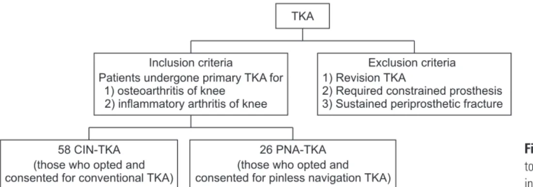
Fifty-eight consecutive consented patients were included in the CIN-TKA group and 26 patients in the PNA-TKA group during the study period (Fig. 1). All the patients were followed for a period of 2 years except for two pa- tients in the CIN-TKA group who died postoperatively on day 2 or 3 months. All the operations were performed by a single surgeon (VB) under spinal anaesthesia using a tour- niquet. A standard medial parapatellar approach for the knee was used in all cases. Arthrotomy was done, which was followed by eversion of the patella and necessary soft- tissue releases to dislocate the knee.
Conventional TKA Technique
CIN-TKA was performed using standard extramedullary jigs for the proximal tibia with the aim of cutting the bone perpendicular to the tibial axis and intramedullary align- ment jigs for the distal femur cut with an aim of achiev- ing 6° of valgus. The anteroposterior (AP) sizing guide was positioned with respect to the Whiteside’s line and an anterior referencing technique was used. The anterior, posterior, anterior chamfer, and posterior chamfer cuts for distal femur were performed with a 4-in-1 cutting guide.
The intercondylar box cut was made depending on the use of posterior cruciate ligament-substituting (PS) implant.
The femoral and tibial trial implants were impacted along with a spacer. Final femoral and tibial components were cemented and denervation of the patella was performed.
PNA-TKA Technique (iAssist)
The iAssist (Zimmer, Warsaw, IN, USA) is an acceler- ometer-based computer-assisted stereotaxic instrument system to assist the surgeon in positioning of orthopaedic implants intraoperatively. The feedback from the ac- celerometer and gyroscopes from the Pods is transmit- ted over a screen via Wi-Fi network. The femur was first prepared in all cases. A 7.9-mm intramedullary spike was impacted with respect to the Whiteside’s line. The femoral reference Pod was then mounted on the spike, and femur registration was done by acquiring 13 stable positions by
Fig. 1. Patient selection flowchart. TKA:
total knee arthroplasty, CIN: conventional instrumented, PNA: pinless navigation.
TKA
Inclusion criteria
Patients undergone primary TKA for 1) osteoarthritis of knee
2) inflammatory arthritis of knee
Exclusion criteria 1) Revision TKA
2) Required constrained prosthesis 3) Sustained periprosthetic fracture
58 CIN-TKA (those who opted and consented for conventional TKA)
26 PNA-TKA (those who opted and consented for pinless navigation TKA)
accelerating and stopping the leg, creating a star-shaped pattern. Audio feedback was generated to confirm the acquisition of each stable position. The femoral resection guide was then attached to the femoral reference Pod and the distal femur cut was adjusted in terms of varus/valgus and flexion/extension using the green and gold screws, respectively; the degree of resection was reflected on the screen (Fig. 2). An appropriate distal femur cut was done with validation of the cut using a validation tool mounted with Pod. The anterior, posterior, anterior, and posterior chamfer cuts were done by conventional methods.
The proximal tibia cuts were performed using an extramedullary guide mounted with Pod. The distal part of the tibial alignment guide was installed over the ankle by firmly gripping the clamps around the malleolus. The proximal spikes were inserted into the tibia, consider-
ing the mechanical axis and rotation of the tibia while continuing to hold the distal clamps firmly around the malleolus. The tibia resection guide was then attached to the proximal part of the extramedullary guide. The tibia registration was done by positioning the leg in abduction, adduction, and neutral position. The proximal tibia cut was adjusted for varus/valgus and flexion/extension with green and gold screws, respectively, on the resection guide, which was reflected on the screen in degrees. An appropri- ate tibia cut was done by validation of the cut as was done for the femur. Then, the proximal tibia was ready to pro- ceed with the next step.
Postoperative Radiological Assessment
As part of the standard institutional protocol, postopera- tive weight-bearing radiographs of the knees were taken in AP and lateral projections along with weight-bearing scanograms of the lower limbs once the patient bore weight comfortably. Component position and lower limb alignment were measured by two independent observers, one was a junior orthopaedic consultant (GS) and the oth- er was an orthopaedic resident (RK). These measurements were repeated after 10 days as described below.
Following measurements were done for coronal alignment. (1) Hip-knee-ankle (HKA) angle on scanogram (Fig. 3): it is measured between the mechanical axis of the femur and the mechanical axis of the tibia.14) It represents the overall alignment of the lower extremity and is usually 180°.2,15) The outliers were recorded as those lying outside
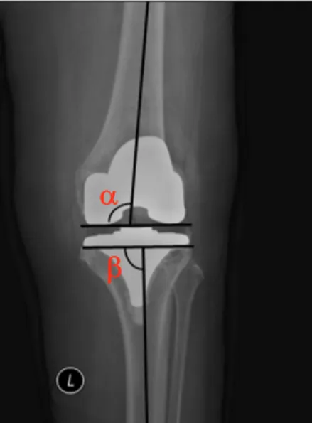
±3 of 180°. (2) α and β angles (femoral and tibial compo- nent coronal alignment, respectively) on an AP radiograph of the knee16) (Fig. 4): the α angle is measured between the Fig. 2. Femoral reference Pod with a cutting jig.
Pod
Cutting jig
Fig. 3. Hip-knee-ankle (HKA) angle measurement.
HKA HKA
Fig. 4. Measurement of α and β angles.
line across the inferior margin of the femoral component and the femoral shaft axis. The femoral component usually is implanted in 5° to 7° of valgus to the anatomical axis of the femur, the amount necessary to re-establish a neutral mechanical axis of the limb.2,17) For this study purpose, the target of the femoral component placement was 6° of valgus and outliers were recorded as those lying outside ± 3 of 6°.
The β angle is measured between the line across the base of the tibial plate and the tibial shaft axis. The tibial com- ponent should be placed in the neutral alignment of 90°.17) The outliers were recorded as those lying outside ± 3 of 90°.
Following measurements were done for sagittal alignment: (1) γ angle (femoral flexion angle) on a lateral radiograph of the knee (Fig. 5): the γ angle is measured between the frontal femoral cortex and the inner frontal
part of the femoral component. The ideal γ angle recom- mended by various studies18,19) varies between 0 and 10°.
The outliers recorded as those lying outside ± 3 of 0°–10°.
(2) σ angle (tibial slope angle) on a lateral radiograph of the knee16,18) (Fig. 5): The σ angle is measured between the line across the base of the tibial plate and the tibial shaft axis. The ideal σ angle is 86°.18) Also, the recommendations vary depending on the implants: PS and cruciate-retaining (CR) types. PS or CR implants (Zimmer-Nexgen or Biom- et-Vanguard) were used in the study. Although calculated for this study, no outlier limit was defined due to the wide recommended range to prevent inaccuracy and confusion.
Functional Assessment
Functional assessment by Oxford Knee Score (OKS) at 2 years was done for all patients (except for the 2 in the CIN- TKA group who died). The data were collected during in person follow-up visit or with video/tele-consultation.
Surgical Time and Complications
Surgical time was measured from the start of incision till closure. Morbidity and mortality along with complica- tions, if any, were noted.
Statistical Analysis
The data of the two groups were compared using the unpaired t-test for continuous variables (age, body mass index, HKA, α , β, γ, σ angles, OKS, and surgical time) and chi-square test for categorical variables (sex). Statistical significance was defined as a p-value of ≤ 0.05. Inter- and intrarater reliability was measured using one way random single-measure intraclass correlation coefficients (ICCs) with associated 95% confidence intervals (CI) to gauge the precisions of the ICCs.
Fig. 5. Measurement of γ and δ angles.
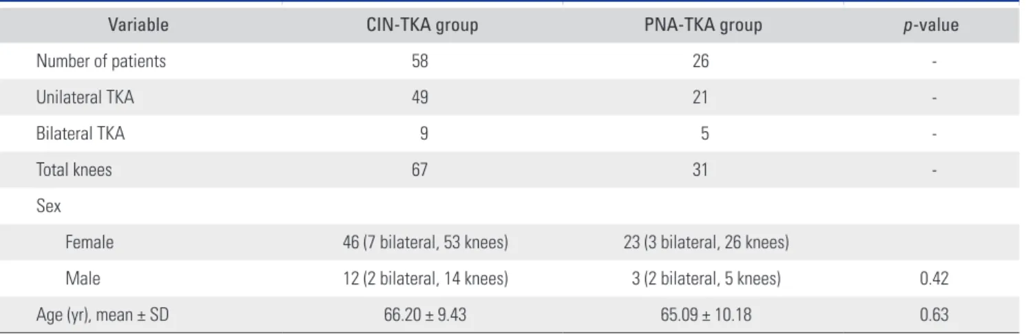
Table 1 . Patient Demographics
Variable CIN-TKA group PNA-TKA group p-value
Number of patients 58 26 -
Unilateral TKA 49 21 -
Bilateral TKA 9 5 -
Total knees 67 31 -
Sex
Female 46 (7 bilateral, 53 knees) 23 (3 bilateral, 26 knees)
Male 12 (2 bilateral, 14 knees) 3 (2 bilateral, 5 knees) 0.42
Age (yr), mean ± SD 66.20 ± 9.43 65.09 ± 10.18 0.63
CIN: conventional instrumented, TKA: total knee arthroplasty, PNA: pinless navigation, SD: standard deviation.
RESULTS
A total of 58 patients were operated with CIN-TKA (9 bilateral cases included) and 26 patients were operated with PNA-TKA (5 bilateral knees included), thus form- ing a 2 to 1 ratio (Table 1). The mean age of the CIN-TKA group was 66.20 ± 9.43 years and that of the PNA-TKA group was 65.09 ± 10.18 years. There was no statistically significant difference between the two groups with respect to age. (Table 1). Radiologically, there was no statistically significant difference in the mean HKA, mean α, β, γ, and σ angles between the two groups. (Table 2). The percentage of outliers for HKA and α, β and, γ angles in the PNA-TKA
group was less than that in the CIN-TKA group, but the difference did not reach statistical significance (Table 3).
The preoperative OKS for the CIN-TKA group and PNA-TKA group improved from 20.67 ± 3.27 and 20.69
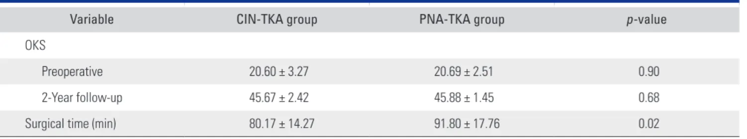
± 2.51, respectively, to 45.67 ± 2.42 and 45.88 ±1.45, re- spectively, at 2 years of follow-up (Table 4). There was no statistically significant difference (p = 0.68) in the mean OKS between the CIN-TKA (45.67 ± 2.42) and PNA- TKA (45.88 ± 1.45) groups at 2 years of follow-up (Table 4). The mean surgical time was shorter in the CIN-TKA group (80.17 ± 14.27) than in the PNA-TKA group (91.80
± 17.76), showing statistically significant difference (p = 0.018) (Table 4).
Complications seen in the CIN-TKA group (6.89%) and PNA-TKA group (0%) were not statistically different (p = 0.17) (Table 5). Regarding the 4 complications in the CIN-TKA group, 1 patient died due to myocardial infarc- tion during the same admission; 1 patient died due to pul- monary embolism 3 months later; 1 patient had anterior femoral cortex perforation while intramedullary drilling; 1 patient had foot drop (ankle dorsiflexion 1/5) on the same day of surgery, which was recovered within 3 months. No complication was noted in the PNA-TKA group. Interob- server correlation was good to excellent for radiological assessment in the CIN-TKA group except for the β angle, which was moderate (0.40). Interobserver correlation was
Table 3. Number of Outliers
Variable CIN-TKA group PNA-TKA group p-value
HKA 12 (17.91) 4 (12.90) 0.73
α 10 (14.92) 3 (9.67) 0.69
β 2 (2.98) 1 (3.22) 0.57
γ 12 (17.91) 5 (16.12) 0.85
Values are presented as number of outliers (%).
CIN: conventional instrumented, TKA: total knee arthroplasty, PNA:
pinless navigation, HKA: hip-knee-ankle.
Table 4. OKS and Surgical Time
Variable CIN-TKA group PNA-TKA group p-value
OKS
Preoperative 20.60 ± 3.27 20.69 ± 2.51 0.90
2-Year follow-up 45.67 ± 2.42 45.88 ± 1.45 0.68
Surgical time (min) 80.17 ± 14.27 91.80 ± 17.76 0.02
Values are presented as mean ± standard deviation.
OKS: Oxford Knee Score, CIN: conventional instrumented, TKA: total knee arthroplasty, PNA: pinless navigation.
Table 2. Radiological Outcome
Variable CIN-TKA group PNA-TKA group p-value HKA (°) 177.63 ± 1.90 177.82 ± 2.17 0.69 α (°) 94.84 ± 1.91 94.95 ± 1.59 0.79 β (°) 90.16 ± 1.17 89.98 ± 1.48 0.55 γ (°) 9.82 ± 2.85 9.87 ± 3.04 0.94 δ (°) 86.98 ± 1.74 86.35 ± 1.57 0.12 Values are presented as mean ± standard deviation.
CIN: conventional instrumented, TKA: total knee arthroplasty, PNA:
pinless navigation, HKA: hip-knee-ankle.
Table 5. Complications
Complication CIN-TKA group PNA-TKA group
Death 2 (myocardial infarction,
1; PE, 1) -
Femoral cortex
perforation while drilling 1 -
Foot drop 1 -
Total, no (%) 4 (6.89) 0
CIN: conventional instrumented, TKA: total knee arthroplasty, PNA:
pinless navigation, PE: pulmonary embolism.
good to excellent for radiological assessment in the PNA- TKA group except for the β angle, which was bad (0.28).
DISCUSSION
Our study found that there was no statistically significant difference in the mean mechanical axis alignment (HKA) and mean coronal (α and β angles) or sagittal (γ and σ angles) component position between the CIN-TKA and PNA-TKA groups. We also found no statistically signifi- cant difference in the number of outliers for HKA and α, β, and γ angles between the CIN-TKA and PNA-TKA groups. Our results are consistent with the study of Chen et al.13) and Maderbacher et al.20) who found no significant difference in the mean mechanical axis alignment (HKA) between conventional and pinless TKAs. In contrast, Liow et al.21) noted significant improvement in the mean mechanical axis alignment with PNA as compared to conventional TKA. In terms of component positioning, our results are in accordance with studies of Liow et al.21) and Keyes et al.,22) who observed no significant difference between the pinless and conventional TKAs. Reducing the number of outliers helps in achieving positive outcomes in maximum patients.23) Some studies have shown a signifi- cant reduction in the number of outliers for mechanical axis alignment of the lower limb and component position- ing with the PNA system as compared to the CIN sys- tem,13,20,21) which is contradictory to our results (Table 3).
The OKS at 2 years was comparable in both the CIN-TKA and PNA-TKA groups with no statistically sig- nificant difference (p = 0.68) in our study. Similar results for OKS have been found in a comparative study con- ducted by Zhu et al.24) Also no significant difference was seen in functional outcome between the conventional and computer navigation groups in a meta-analysis by Zamora et al.25) An increase in surgical time by approximately 11 minutes was noted in the PNA-TKA group as compared to the CIN-TKA group, which was a statistically signifi- cant difference (p = 0.018). However, it did not result in anaesthetic/systemic complications, an increased infection rate, or blood loss. The intraoperative steps such as femur and tibia registration and validation of the femur and tibia cuts add to the surgical time. The steps for registration of femur and tibia need to be done precisely, or they need to be repeated. The surgeon must be well versed with the instrumentation and surgical steps of the PNA system.
The statistically significant increase in surgical time with computer navigation was also noted in other comparative studies by Gothesen et al.2) and Maderbacher et al.20)
The number of complications seen in the CIN-
TKA group was greater than in the PNA-TKA group, but it was not statistically significantly different (p = 0.2). We noted two deaths in the CIN-TKA group due to myocar- dial infraction and pulmonary embolism. Although it was beyond the scope of this study, it has been reported by Kalairajah et al.26) that there are possibly fewer chances of blood loss and systemic embolism with the PNA system as compared to the CIN system, as intramedullary drilling is not required in the PNA system. The steep learning curve and dependence on conventional instrumentation for determining the rotational alignment and implant size of femoral and tibial components are some of the drawbacks of PNA-TKA, which need to be taken into consideration.
Also, as the pods are disposable, each patient operated with PNA-TKA was charged USD 675 extra for the use of PNA- TKA system (p < 0.001), which adds to the financial cost.
To our knowledge, our study was perhaps the first study done in the Indian subcontinent where patients have much varus deformity and also extra-articular deformity of the knee.27,28) Although we did not do a priori sample size calculation, our sample size was comparable to that of previous studies by Maderbacher et al.20) and Liow et al.21) Long-term studies with a large sample size and a multivar- iate analysis would be ideal to determine the true benefit of use of this technology. Short-term follow-up and small sample size are limitations of our study. We aim to further follow up patients for 10 years to assess the functional out- come.
This study demonstrates that the PNA does not result in statistically significant improvement in the (1) mechanical axis alignment of the lower limb (HKA angle);
(2) accuracy of component positioning; or (3) reduction of the number of outliers as compared to the conventional instrumentation. The accurate mechanical axis align- ment and component positioning can be achieved with the conventional instrumentation, so the use of PNA system, which adds to the surgical cost, is questionable.
Also, equally good short-term functional outcome can be achieved with conventional instrumentation. The surgeon must be accustomed with the instrumentation of the PNA system, or it adds to the surgical time.
CONFLICT OF INTEREST
No potential conflict of interest relevant to this article was reported.
ACKNOWLEDGEMENTS
We appreciate contribution of Gaurav Sharma (MS, Junior
Consultant, Sir H. N. Reliance Foundation Hospital) and Rajiv Kulkarni (Diploma Ortho, Orthopedic Resident, Sir H.N. Reliance Foundation Hospital) for this study.
ORCID
Prashant Pawar https://orcid.org/0000-0003-3886-3154 Lokesh Naik https://orcid.org/0000-0001-7693-6559 Dipit Sahu https://orcid.org/0000-0003-1888-4994 Vaibhav Bagaria https://orcid.org/ 0000-0002-3009-3485
REFERENCES
1. Robertsson O, Dunbar M, Pehrsson T, Knutson K, Lidgren L. Patient satisfaction after knee arthroplasty: a report on 27,372 knees operated on between 1981 and 1995 in Swe- den. Acta Orthop Scand. 2000;71(3):262-7.
2. Gothesen O, Espehaug B, Havelin LI, et al. Functional outcome and alignment in computer-assisted and conven- tionally operated total knee replacements: a multicentre parallel-group randomised controlled trial. Bone Joint J.
2014;96(5):609-18.
3. Huang TW, Peng KT, Huang KC, Lee MS, Hsu RW. Differ- ences in component and limb alignment between computer- assisted and conventional surgery total knee arthroplasty.
Knee Surg Sports Traumatol Arthrosc. 2014;22(12):2954-61.
4. Insall JN, Binazzi R, Soudry M, Mestriner LA. Total knee arthroplasty. Clin Orthop Relat Res. 1985;(192):13-22.
5. Delp SL, Stulberg SD, Davies B, Picard F, Leitner F. Com- puter assisted knee replacement. Clin Orthop Relat Res.
1998;(354):49-56.
6. Stulberg SD, Loan P, Sarin V. Computer-assisted navigation in total knee replacement: results of an initial experience in thirty-five patients. J Bone Joint Surg Am. 2002;84(Suppl 2):S90-8.
7. Mainard D, Guillemin F, Cuny C, Mejat-Adler E, Galois L, Delagoutte J. Quality of life assessment one year after total hip or knee arthroplasty. Rev Chir Orthop Reparatrice Ap- par Mot. 2000;86(5):464-73.
8. Abdel MP, Oussedik S, Parratte S, Lustig S, Haddad FS. Cor- onal alignment in total knee replacement: historical review, contemporary analysis, and future direction. Bone Joint J.
2014;96(7):857-62.
9. Kim YH, Kim JS, Choi Y, Kwon OR. Computer-assisted sur- gical navigation does not improve the alignment and orien- tation of the components in total knee arthroplasty. J Bone Joint Surg Am. 2009;91(1):14-9.
10. Schlatterer B, Linares JM, Cazal J, Merloz P, Plaweski S;
Computer Assisted Orthopedic Surgery - France (CAOS - France). Posterior tibial slope accuracy with patient-specific cutting guides during total knee arthroplasty: a preliminary study of 50 cases. Orthop Traumatol Surg Res. 2015;101(6
Suppl):S233-40.
11. Mannan A, Smith TO, Sagar C, London NJ, Molitor PJ. No demonstrable benefit for coronal alignment outcomes in PSI knee arthroplasty: a systematic review and meta-analysis.
Orthop Traumatol Surg Res. 2015;101(4):461-8.
12. Chen JY, Chin PL, Tay DK, Chia SL, Lo NN, Yeo SJ. Func- tional outcome and quality of life after patient-specific instrumentation in total knee arthroplasty. J Arthroplasty.
2015;30(10):1724-8.
13. Chen JY, Chin PL, Tay DK, Chia SL, Lo NN, Yeo SJ. Less outliers in pinless navigation compared with conventional surgery in total knee arthroplasty. Knee Surg Sports Trau- matol Arthrosc. 2014;22(8):1827-32.
14. Moreland JR, Bassett LW, Hanker GJ. Radiographic analysis of the axial alignment of the lower extremity. J Bone Joint Surg Am. 1987;69(5):745-9.
15. Chin PL, Yang KY, Yeo SJ, Lo NN. Randomized control trial comparing radiographic total knee arthroplasty implant placement using computer navigation versus conventional technique. J Arthroplasty. 2005;20(5):618-26.
16. Petersen TL, Engh GA. Radiographic assessment of knee alignment after total knee arthroplasty. J Arthroplasty.
1988;3(1):67-72.
17. Gromov K, Korchi M, Thomsen MG, Husted H, Tro- elsen A. What is the optimal alignment of the tibial and femoral components in knee arthroplasty? Acta Orthop.
2014;85(5):480-7.
18. Dyrhovden GS, Gothesen O, Lygre SH, et al. Is the use of computer navigation in total knee arthroplasty improving implant positioning and function? A comparative study of 198 knees operated at a Norwegian district hospital. BMC Musculoskelet Disord. 2013;14:321.
19. Kim YH, Park JW, Kim JS, Park SD. The relationship be- tween the survival of total knee arthroplasty and postopera- tive coronal, sagittal and rotational alignment of knee pros- thesis. Int Orthop. 2014;38(2):379-85.
20. Maderbacher G, Schaumburger J, Keshmiri A, et al. Pinless navigation in total knee arthroplasty: navigation reduced by the maximum? Int Orthop. 2015;39(3):455-60.
21. Liow MH, Goh GS, Pang HN, Tay DK, Lo NN, Yeo SJ.
Computer-assisted stereotaxic navigation improves the ac- curacy of mechanical alignment and component position- ing in total knee arthroplasty. Arch Orthop Trauma Surg.
2016;136(8):1173-80.
22. Keyes BJ, Markel DC, Meneghini RM. Evaluation of limb alignment, component positioning, and function in primary total knee arthroplasty using a pinless navigation tech- nique compared with conventional methods. J Knee Surg.
2013;26(2):127-32.
23. Longstaff LM, Sloan K, Stamp N, Scaddan M, Beaver R.
Good alignment after total knee arthroplasty leads to faster rehabilitation and better function. J Arthroplasty.
2009;24(4):570-8.
24. Zhu M, Chen JY, Chong HC, et al. No difference in func- tional outcomes after total knee arthroplasty with or with- out pinless navigation. J Knee Surg. 2018;31(7):649-53.
25. Zamora LA, Humphreys KJ, Watt AM, Forel D, Cameron AL. Systematic review of computer-navigated total knee ar- throplasty. ANZ J Surg. 2013;83(1-2):22-30.
26. Kalairajah Y, Cossey AJ, Verrall GM, Ludbrook G, Spriggins AJ. Are systemic emboli reduced in computer-assisted knee surgery? A prospective, randomised, clinical trial. J Bone Joint Surg Br. 2006;88(2):198-202.
27. Joshi R, Ganguli N, Carvalho C, de Leon F, Pope J. Varus and valgus deformities in knee osteoarthritis among dif- ferent ethnic groups (Indian, Portuguese and Canadians) within an urban Canadian rheumatology practice. Indian J Rheumatol. 2010;5(4):180-4.
28. Saibaba B, Dhillon MS, Chouhan DK, Kanojia RK, Prakash M, Bachhal V. significant incidence of extra-articular tibia vara affects radiological outcome of total knee arthroplasty.
Knee Surg Relat Res. 2015;27(3):173-80.