www.ophthalmology.org 1139 대한안과학회지 2010년 제 51 권 제 8 호 J Korean Ophthalmol Soc 2010;51(8):1139-1141 pISSN: 0378-6471 eISSN: 2092-9374 DOI : 10.3341/jkos.2010.51.8.1139
= 증례보고 =
속눈썹에 의해 발생한 눈물샘 결석
권용대⋅임지원
한림대학교 의과대학 춘천성심병원 안과학교실
목적: 속눈썹에 의해 유발된 눈물샘 결석의 증례를 보고하고자 한다.
증례요약: 31세 여자 환자가 우안 상이측 결막 부위에 6개월 이상 지속되는 이물감과 결막 충혈, 통증을 주소로 내원하여 시행한 이학 적 검사상, 외안각 부위에 종괴가 관찰되었다. 절제 생검에서 속눈썹을 싸고 있는 눈물샘 결석이 발견되었으며, 절제 후 1년간의 경과 관찰 기간 중 합병증이나 재발이 없었다.
결론: 드물지만 속눈썹이 눈물샘 결석의 생성에 한 요인이 될 수 있다.
<대한안과학회지 2010;51(8):1139-1141>
■ 접 수 일: 2010년 4월 8일 ■ 심사통과일: 2010년 6월 29일
■ 책 임 저 자: 임 지 원
강원도 춘천시 교동 153 한림대학교 춘천성심병원 안과
Tel: 033-240-5176, Fax: 033-255-5210 E-mail: jiwoneye@hallym.or.kr
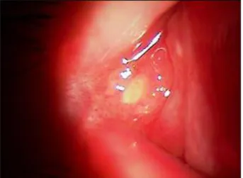
Figure 1. Non-tender, yellowish mass in the lateral canthal area of the right eye.
코눈물관을 비롯한 눈물 배출 기관에서 발생한 결석은 이전에 많은 보고가 있었으나, 눈물샘에 발생한 결석은 매 우 드문 것으로 알려져 있다.1-4눈물샘에 발생한 결석은 증 상이 없을 수도 있으나, 대부분의 경우 통증, 결막 충혈, 눈 물흘림, 광시증, 가려움증 등을 유발하며, 심한 경우에는 안 구돌출도 일으킬 수 있다.1-5
결석의 발생 원인은 대부분 명확한 기전이 밝혀져 있지 않지만, 만성 염증, 눈물 내의 높은 칼슘이나 인의 농도, 세 균 감염 등이 원인으로 의심되고 있으며, 드물게 속눈썹에 의해 눈물샘 결석이 유발되었다는 국외 몇몇 보고가 있
다.1,4,6저자들은 속눈썹에 의해 발생한 눈물샘 결석을 경험
하여 이를 보고하고자 한다.
증례보고
31세 여자 환자가 6개월간 우안 이물감과 상이측 결막 의 지속적인 충혈과 통증을 주소로 만성 결막염 진단하에 타 병원에서 국소 점안 항생제와 점안 스테로이드를 하루 4회 점안하는 치료를 받았으나 호전되지 않아 본원으로 전 원 의뢰되었다. 이학적 검사상 우안 외안각 근처 상안검 부 위에 작은 종괴가 관찰되었다(Fig. 1). 추가로 시행한 안와 전산화단층촬영상 눈물샘 주위에 다른 안와 내 구조물을 침범하지 않는 경계가 지어진 종괴가 보여, 에피네프린을
첨가한 2% 리도카인을 종괴 주위에 주사한 후, 절제를 시 행하여 명확하게 경계가 지어져 있고 가운데 속눈썹이 있 는 노란색의 딱딱한 종괴를 적출하였다(Fig. 2). 해부병리 조직검사상 중심부에 속눈썹을 포함하고 있는 크기 6×6×3 mm의 무정형의 여포성 결석으로 진단되었고, 만성 염증을 나타내는 림프구가 관찰되었다(Fig. 3). 세균 도말 염색과 배양검사에서 특이소견은 발견되지 않았다. 종괴 제거 후 환자 증상은 소실 되었으며, 1년의 경과관찰 기간 중 재발 이나 합병증은 보이지 않았다.
고 찰
눈물 배출 기관에 발생하는 결석은 흔하지만, 눈물샘에 만 발생하는 결석은 매우 드물다.7 눈물샘 결석은 다른 눈
www.ophthalmology.org 1140
- 대한안과학회지 2010년 제 51 권 제 8 호 -
Figure 2. Macroscopic view of an eyelash (arrow) removed from the center of the mass and the stone fragments excised from the lateral canthal area.
A B
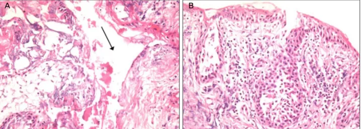
Figure 3. Histopathologic examination of a mass (H&E stain, ×200). (A) Eosinophilic amorphous and granular material with a area of cystic wall (removed stone)(arrow). (B) Non-specific chronic inflammatory cells infiltrated. There is no ductule or glands.
물 배출 기관에 발생하는 결석과 달리 남자에서 호발하지 만 본 증례는 여자에서 발생하였다.1,8 눈물샘 결석의 병인 에 관해서는 명확한 가설은 없지만, Duke-Elder5는 눈물샘 상피세포의 잔존물이 유발한다고 하였고, Baker and Bartley4은 눈물층의 과도한 알부민이 원인이라 하였으며, 세균의 증식이 원인으로 보고된 바도 있다.6 또한 눈썹 성 분에 의해 발생한 눈물 배출 기관 결석도 보고되었음을 생 각할 때 눈물샘 결석은 먼저 눈물샘 소관이 여러 원인에 의 해서 막힘이 선행되고, 단백질을 비롯한 부유물이 축적된 후 이들 성분이 핵으로 작용하여 결석을 형성하고 마지막 으로 이들 결석 주위에 세균의 증식이 일어나는 것으로 생 각된다.9
본 증례에서는 절제 생검 시 결석의 중앙에 눈썹이 묻혀 있는 형태로 있었고, 해부병리조직검사에서 결석을 싸고 있 는 막에 상피세포가 보였으며, 세균 도말 염색에서 세균이 나 진균에 의한 소견은 관찰되지 않은 것으로 보아 눈썹에 의해 눈물샘 소관의 정체가 일어난 후 눈썹을 핵으로 한 결 석 형성이 일어났을 것으로 보인다. 본 증례에서는 결석의 성분 분석은 시행하지 않았으며, 생검 시 온전한 형태의 결 석으로 얻지 못하고 부스러진 형태로 제거된 점이 병인의 분석에 제한점이다.
눈물샘 결석이 흔한 질환은 아니지만, 만성적으로 가쪽 결막에 충혈과 이물감이 지속될 때 감별 질환으로 고려되 어야 하며, 결석의 제거로 쉽게 치료가 가능하다. 평상시 외 래 검사에서 흔히 발견되는 눈물층이나, 결막낭 내 탈락된 속눈썹은 눈물샘 결석의 한 원인이 될 수 있다.
참고문헌
1) Zafar A, Jordan DR, Brownstein S, Faraji H. Asymptomatic lac- rimal ductule dacryolithiasis with embedded cilia. Ophthal Plast Reconstr Surg 2004;20:83-5.
2) Baratz KH, Bartley GB, Campbell RJ, Garrity JA. An eyelash ni- dus for dacryoliths of the lacrimal excretory and secretory systems.
Am J Ophthalmol 1991;111:624-7.
3) Jay JL, Lee WR. Dacryolith formation around an eyelash retained in lacrimal sac. Br J Ophthalmol 1976;60:722-5.
4) Baker RH, Bartley GB. Lacrimal gland ductule stones.
Ophthalmolgy 1990;97:531-4.
5) Duke-Elder S, ed. System of ophthalmology. Vol. XIII The ocular adenexa part 2: lacrimal, orbital and para-orbital diseases. St.
Louis: CV Mosby Co., 1974:672.
6) Mawn LA, Sanon A, Conlon MR, Nerad JA. Pseudomonas da- cryoadenitis secondary to a lacrimal gland ductule stone. Ophthal Plast Reconstr Surg 1997;13:135-8
7) Halborg J, prause JU, Toft PB, et al. Stones in the lacrimal gland: a
www.ophthalmology.org 1141
=ABSTRACT=
An Eyelash-induced Lacrimal Gland Stone
Yong Dae Kwon, MD, Ji Won Lim, MD
Department of Ophthalmology, Chuncheon Sacred Heart Hospital, Hallym University College of Medicine, Chuncheon, Korea
Purpose: To report a case of lacrimal gland stone initiated by an eyelash.
Case summary: A 31-year-old woman presented with foreign body sensation, pain, and conjunctival injection in the lateral palpebral conjunctiva of her right eye over 6 months in duration. The physical examination revealed a small, firm nodule at the lateral canthal area. The excisional biopsy was performed, and the mass was a concretion that contained an eyelash in the center. During one-year follow-up, the patient showed no signs of recurrence or complication after excision.
Conclusions: Although rare in occurence, eyelashes may be the initial nidus for lacrimal gland stone formation.
J Korean Ophthalmol Soc 2010;51(8):1139-1141 Key Words: Eyelash, Lacrimal gland stone
Address reprint requests to Ji Won Lim, MD
Department of Ophthalmology, Hallym University Sacred Heart Hospital
#153 Gyo-dong, Chuncheon 200-060, Korea
Tel: 82-33-240-5176, Fax: 82-33-255-5210, E-mail: jiwoneye@hallym.or.kr
- 권용대⋅임지원 : 속눈썹에 의해 발생한 눈물샘 결석 -
rare condition. Acta Ophthalmol 2009;87:672-6.
8) Marthin JK, Lindegaard J, Prause JU, Heegaard S. Lesions of the lacrimal drainage system: a clinicopathological study of 643 biopsy specimens of the lacrimal drainage system in Denmark
1910-1999. Acta Ophthalmol Scand 2005;83:94-9.
9) Garfin SW. Etiology of dacryocystitis and epiphora. Arch oph- thalmol 1942;27:167-88.