INTRODUCTION
Caffeine is one of the most widely consumed neuroactive drugs, coming mostly from everyday beverages such as cof- fee and tea; it exhibits a variety of central effects including stimulation of locomotor behavior (1). Caffeine exerts multi- ple effects at the cellular level on the central nervous system (CNS); for instance, antagonizes adenosine receptor (2), inhi- bits GABA receptor-mediated effect (3), and inhibits phos- phodiesterase (4). In electrophysiological studies, caffeine at high concentrations triggered Ca2+release via ryanodine re- ceptors (5, 6), and it is known as a classic pharmacological agonist for activating Ca2+-induced Ca2+release. It was also reported that high-concentration caffeine administration has some deleterious effects, induces various morphological ano- malies on cultured rat embryonic cells, and modifies the rate of neural cell proliferation in certain regions of the brain (7, 8).
Apoptosis, also known as programmed cell death, is a form of cell death that occurs in several pathological situations in multicellular organisms and constitutes part of a common me- chanism of cell replacement, tissue remodeling, and removal of damaged cells (9). Apoptosis is a complex process charac- terized by cell shrinkage, chromatin condensation, internu- cleosomal DNA fragmentation, and formation of “apoptotic bodies” (10). The caspase family of aspartate-specific cysteine proteases is emerging as the central executioner of apoptosis.
Of particular interest is caspase-3, which is activated in a vari- ety of cell types during apoptosis (11).
In the present study, to investigate whether caffeine induces apoptosis in the CNS, 3-(4,5-dimethylthiazol-2-yl)-2,5-di- phenyltetrazolium bromide (MTT) assay, 4,6-diamidino-2- phenylindole (DAPI) staining, terminal deoxynucleotidyl transferase (TdT)-mediated dUTP nick end labeling (TUN EL) assay, flow cytometric analysis, DNA fragmentation assay, and caspase-3 enzyme assay were performed on SK-N-MC human neuroblastoma cells.
MATERIALS AND METHODS Drugs and reagents
Caffeine and DAPI were obtained from Sigma Chemical Co.
(St. Louis, MO, U.S.A.). The MTT assay kit and the TUNEL assay kit were purchased from Boehringer Mannheim GmbH (Mannheim, Germany). The DNA fragmentation assay kit was obtained from TaKaRa (Shiga, Japan) and the caspase-3 assay kit was from CLONTECH (Palo Alto, CA, U.S.A.).
Cell culture
SK-N-MC human neuroblastoma cells were purchased from Mi-Hyeon Jang, Min-Chul Shin,
In-Sug Kang*, Hyung-Hwan Baik*, Yong-Ho Cho*, Jong-Phill Chu�, Ee-Hwa Kim�, Chang-Ju Kim
Departments of Physiology, Biochemistry*, and Parasitology�, College of Medicine, Kyung Hee University, Seoul; Department of Meridianology�, College of Oriental Medicine, Semyung University, Jechon, Korea
Address for correspondence Chang-Ju Kim, M.D.
Department of Physiology, College of Medicine, Kyung Hee University, 1 Hoigi-dong, Dongdaemoon-gu, Seoul 130-701, Korea Tel : +82.2-961-0282, Fax : +82.2-964-2195 E-mail : changju@khu.ac.kr
674
Caffeine Induces Apoptosis in Human Neuroblastoma Cell Line SK-N-MC
Caffeine is one of the most widely consumed neuroactive drugs, coming mostly from everyday beverages such as coffee and tea. To investigate whether caffeine induces apoptosis in the central nervous system, 3-(4,5-dimethylthiazol-2-yl)-2,5- diphenyltetrazolium bromide (MTT) assay, 4,6-diamidino-2-phenylindole (DAPI) staining, terminal deoxynucleotidyl transferase (TdT)-mediated dUTP nick end labeling (TUNEL) assay, flow cytometric analysis, DNA fragmentation assay, and caspase-3 enzyme assay were performed on SK-N-MC human neuroblastoma cells. Cells treated with caffeine at concentrations as high as 10 mM exhibited several characteristics of apoptosis. In addition, caffeine was shown to increase the caspase-3 activity. These results suggest that high-dose of caffeine induces apoptosis in human neuroblastoma cells, probably by increasing the caspase-3 enzyme activity.
Key Words : Caffeine; Apoptosis; Neuroblastomas; Caspases
Received : 3 March 2002 Accepted : 10 June 2002
the Korean Cell Line Bank (KCLB, Seoul, Korea) and cul- tured according to the previously reported method (12). Cells were cultured in Dulbecco’s Modified Eagle Medium (DMEM) (Gibco BRL, Grand Island, NY, U.S.A.) supplemented with 10% heat-inactivated FBS (Gibco BRL, Grand Island, NY, U.S.A.) at 37℃in 5% CO2and 95% O2in a humidified cell incubator, and the medium was changed every 2 days.
MTT cytotoxicity assay
Cell viability was determined using the MTT assay kit as per the manufacturer’s protocol. In order to determine the cytotoxicity of caffeine, cells were treated with caffeine at con- centrations of 0.1 mM, 1 mM, and 10 mM for durations of 12 hr, 24 hr, and 36 hr. Cultures of the control group were left untreated. Ten microliters of the MTT labeling reagent was added to each well, and the plates were incubated for 4 hr. One hundred microliters of the solubilization solution was then added to each well, and the cells were incubated for another 12 hr. The absorbance was then measured with a microtiter plate reader (Bio-Tek, Winooski, VT, U.S.A.) at a test wavelength of 595 nm and a reference wavelength of 690 nm. The optical density (O.D.) was calculated as the dif- ference between the absorbance at the reference wavelength and that at the test wavelength. Percent viability was calcu- lated as “(O.D. of drug-treated sample/control O.D.)×100”. DAPI staining
DAPI staining was performed according to the previously described protocol (13). Cells were first cultured on 4-cham- ber slides (Nalge Nunc International, Naperville, IL, U.S.A.).
After treatment with caffeine, cells were collected and fixed by incubation in 4% PFA for 30 min. Following washing in PBS, the cells were incubated in 1 g/mL DAPI solution for 30 min in the dark. The cells were then observed with a fluorescence microscope (Zeiss, Oberkochen, Germany).
TUNEL staining
For in situ detection of apoptotic cells, TUNEL assay was performed using ApoTag�peroxidase in situ apoptosis detec- tion kit. SK-N-MC cells were cultured on 4-chamber slides at a density of 2×10⁴cells/chamber. After treatment with caffeine, the cells were washed with PBS and fixed by incubat- ing in 4% paraformaldehyde (PFA) for 10 min at 4℃. The fixed cells were then incubated with digoxigenin-conjugat- ed dUTP in a terminal deoxynucleotidyl transferase (TdT)- catalyzed reaction for 60 min at 37℃in a humidified atmo- sphere and were then immersed in stop/wash buffer for 10 min at room temperature. The cells were then incubated with anti- digoxigenin antibody conjugated with peroxidase for 30 min.
DNA fragments were stained using 3,3′-diaminobenzidine (Sigma Chemical Co.) as the substrate for the peroxidase.
Flow cytometric analysis
For flow cytometric analysis, after treatment with caffeine, cells were collected and fixed by incubation with 75% ethanol in PBS at -20℃for 1 hr. Afterwards, the cells were incubat- ed with 100 g/mL RNase and 20 g/mL propidium iodide (Sigma Chemical CO.) in PBS for 30 min at 37℃and were analyzed using FACScan (Becton Dickinson, San Jose, CA, U.S.A.).
DNA fragmentation
For detection of apoptotic DNA cleavage, DNA fragmen- tation assay was performed using ApopLadder EXTMDNA fragmentation assay kit (TaKaRa, Shiga, Japan). Cells were first treated with caffeine as mentioned earlier and were col- lected in Eppendorf tubes. The cells were then centrifuged and were lysed with 100 L of lysis buffer per tube. The lysate was incubated with 10 L of 10% SDS solution and 10 L of Enzyme A at 56℃for 1 hr and then at 37℃for 1 hr fol- lowing the addition of 10 L of Enzyme B. Afterwards, 70 L of precipitant was added, and the resultant pellet was re- suspended in TE buffer. DNA fragmentation was visualized by electrophoresis on a 2% agarose gel containing ethidium bromide.
Caspase-3 enzyme activity assay
Caspase enzyme activity was measured using ApoAlert� caspase-3 assay kit according to the manufacturer’s protocol.
First, after treatment with caffeine, cells were lysed in 50 L of chilled Cell Lysis Buffer. Fifty microliters of 2×reaction buffer (containing DTT) and 5 L of the appropriate conju- gated substrate at a concentration of 1 mM were added to each lysate. The mixture was incubated in a water bath at 37
℃for 1 hr, and the absorbance was measured with a micro- titer plate reader at a test wavelength of 405 nm.
Statistical analyses
Results are expressed as mean±standard error mean (SEM).
The data were analyzed by one-way ANOVA followed by Scheffe’s post-hoc test using SPSS. Differences were consid- ered statistically significant at p<0.05.
RESULTS Effect of caffeine on cell viability
As shown in Fig. 1, the viabilities of cells incubated with caffeine at a concentration of 0.1 mM for durations of 12 hr, 24 hr, and 36 hr were 96.06±1.85%, 101.56±2.93%, and 96.35±1.02% of the control value, respectively. The viabili-
. .
ties of cells incubated with caffeine at a concentration of 1 mM for durations of 12 hr, 24 hr, and 36 hr were 89.70±2.26%, 88.16±2.46%, and 81.14±1.76% of the control value, re- spectively. The viabilities of cells incubated with caffeine at a concentration of 10 mM for durations of 12 hr, 24 hr, and 36 hr were 63.01±0.10%, 50.11±0.80%, and 26.19± 0.30% of the control value, respectively. These results show that caffeine at concentrations as high as 10 mM has a cyto- toxic effect on SK-N-MC cells and this cytotoxic effect was increased in a time-dependent manner.
Morphological changes induced by caffeine
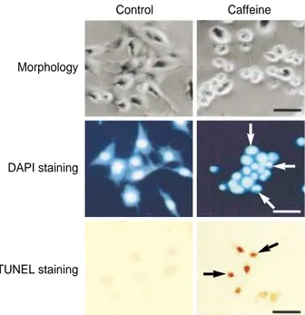
The morphological changes induced by caffeine were ex- amined by phase-contrast microscopy. Cells treated with caf- feine at a concentration of 10 mM for 24 hr were seen to have detached from the dish, and cell rounding, cytoplasmic bleb- bing, and irregularities in shape were observed. In the DAPI assay, cells were observed via fluorescence microscopy follow- ing treatment with DAPI, which specifically stains the nuclei.
The assay revealed the presence of nuclear condensation, DNA fragmentation, and perinuclear apoptotic bodies upon caf- feine treatment at a concentration of 10 mM for 24 hr. To further confirm the induction of apoptosis by caffeine in SK- N-MC cells, 10 mM caffeine-treated cells were analyzed via TUNEL assay. As shown in Fig. 2, TUNEL-positive cells were shown to be stained dark brown under the light micro- scope, and nuclear condensation was observed.
Cell cycle distribution change
Flow cytometric analysis revealed an increase in the sub- G1 phase fraction, from 38.26% to 42.07% and 52.83% and a decrease in the G1 phase fraction, from 43.39% to 38.26%
and 32.10%, in cells treated with caffeine at concentrations of 1 mM and 10 mM for 24 hr, respectively (Fig. 3).
Characterization of apoptosis via examination of DNA fragmentation
In order to ascertain the induction of apoptosis by caffeine, DNA fragmentation, reflecting the endonuclease activity characteristic of apoptosis, was assessed. Caffeine treatment resulted in the formation of definite fragments, which could be seen via electrophoresis as a characteristic ladder pattern (Fig. 4).
Caspase-3 enzyme activity analysis
Caspase-3 enzyme activity was measured using DEVD pep- tide-nitroanilide (pNA). After 24 hr of exposure to caffeine at concentrations of 1 mM, and 10 mM, the concentration of DEVD-pNA cleavage product at the end of an 18-hr reac- tion was increased from 11.46±0.54 pM to 15.34±0.48
Fig. 2.Characterization of caffeine-induced cell death in SK-N-MC cells. Cells were cultured without caffeine (control) or with 10 mM caffeine for 24 hr in each case. Top: photomicrograph of phase- contrast microscope. Cell shrinkage, irregularity in cellular shape, and cellular detachment are seen in the nicotine-treated cultures.
Middle: SK-N-MC cells stained with DAPI. White arrows indicate condensed nuclei. Bottom: SK-N-MC cells stained via TUNEL assay. Black arrows indicate where condensed and marginated chromatin have been labeled. All scale bars represent 100 m.
Morphology
DAPI staining
TUNEL staining
Control Caffeine
Relative viability (% control)
120
100
80
60
40
20
0
Control 0.1 1 10
Fig. 1. Cytotoxic effects of caffeine. Human neuroblastoma cell line SK-N-MC cells were incubated with caffeine at various con- centrations and duration prior to the determination of cellular via- bility through MTT assay. Results are represented as mean± standard error for two independent experiments, each with a minimum of three cultures. *represents p<0.05 compared to the control.
Concentration of caffeine (mM) 12 hr incubation
24 hr incubation 36 hr incubation
* *
** *
*
*
Fig. 4.Electrophoretic examination of the genomic DNA of SK-N- MC cells. Genomic DNA was extracted and analyzed via elec- trophoresis on 2% agarose gels containing ethidium bromide. A, control group; B, 1 mM caffeine-treated group; C, 10 mM caffeine- treated group.
pM and to 18.56±0.66 pM, respectively. After 36 hr of ex- posure to caffeine at concentrations of 1 mM, and 10 mM, the concentration of DEVD-pNA cleavage product at the end of an 18-hr reaction was increased from 15.41±0.34 pM to 18.45±0.52 pM and to 20.14±0.26 pM, respectively (Fig. 5).
DISCUSSION
At the molecular level, caffeine interferes with cell division (14) and induces disturbances in early neurogenesis in mouse
embryo cultures (8). In electrophysiological studies, caffeine at high concentrations (e.g. 1-10 mM) was shown to be nec- essary in order to initiate calcium release from intracellular stores via a caffeine-sensitive ryanodine receptor (5, 6), and increased intracellular calcium is known to induce cell death in the CNS (15). In addition, caffeine has been shown to atten- uate G2 delay produced by DNA-damaging agents and to augment the cytotoxicity of these agents in a number of cell lines in vitro (16). In embryonic cell culture system, caffeine
1 mM caffeine
M A B C
10 mM caffeine Control
Fig. 3.Flow cytometric analyses. An increase in the sub-G1 phase fraction and a decrease in the G1 phase fraction were observed following caffeine treatment.
pmol pNA/min/mg of protein
30
20
10
0
A B C D E
24 hr incubation 36 hr incubation
Fig. 5. Result of caspase-3 enzyme assay. Caffeine increases the caspase-3 enzyme activity. Results are presented as mean± standard error mean. *represents p<0.05 compared to the 24 hr incubated control group. **represents p<0.05 compared to the 36 hr incubated control group. #represents p<0.05 compared to the 24 hr incubated 10 mM caffeine-treated group. ##represents p<0.05 compared to the 36 hr incubated 10 mM caffeine-treat- ed group. As the positive control, 1 M stausporine was treated.
DEVD-fmk is a caspase inhibitor. The rate of DEVD-pNA cleavage was measured at 405 nm. A, Control; B, 1 M stausporine-treated group; C, 1 mM caffeine-treated group; D, 10 mM caffeine-treated group; E, 10 mM caffeine- and DEVD-fmk-treated group.
** ** **
*
*
#
##
Counts
DNA content
Counts
DNA content
Counts 350280210140700
0 200 400 600 800 1000
DNA content
350280210140700
0 200 400 600 800 1000
350280210140700
0 200 400 600 800 1000
induced various morphological anomalies (7). However, it has not yet been reported whether caffeine actually induces apoptotic cell death in the CNS. In the present study, the effect of caffeine on cells of the neuroblastoma cell line SK- N-MC was investigated.
Assessment of cell viability in the present study via MTT assay confirmed that caffeine exerts cytotoxic effects on SK- N-MC cells at high concentrations. In addition, caffeine at concentration as high as 10 mM was shown to cause charac- teristic changes in the morphology of SK-N-MC cells. Apop- totic bodies, the presence of which is a stringent morphologi- cal criterion for apoptosis, were seen in cultures treated with caffeine at high concentration upon DAPI staining. It has been reported that cells undergoing apoptosis exhibit cyto- plasmic blebbing, nuclear shrinkage, chromatin condensa- tion, irregularity in shape, and retraction of processes (11).
In addition, DNA strand breaks are known to occur during the process of apoptosis, and such breaks in the DNA mole- cules can be detected via TUNEL assay (17). In the present study, TUNEL-positive cells, indicative of the occurrence of apoptosis, were observed among caffeine-treated cells. It is also known that apoptosis involves the activation of endonu- cleases and that this activation results in the cleavage of ge- nomic DNA into well-defined fragments which appear as a characteristic ladder pattern upon agarose gel electrophore- sis (18). To provide further evidence supporting the involve- ment of apoptosis in caffeine-induced cytotoxicity, DNA frag- mentation assay was performed. Caffeine-treated cells present- ed with the distinctive ladder pattern characteristic of apop- tosis. Furthermore from flow cytometric analysis, increased apoptosis and decreased DNA synthesis were observed. In ad- dition, caspases, a family of cysteine proteases, are known to form integral parts of the apoptotic pathway; in particular, caspase-3, when activated, has many cellular targets that, when severed and/or activated, produces the morphologic features of apoptosis (11). In the present study, an increase in the caspase-3 enzyme activity was observed in cells exposed to caffeine.
Based on these results, it is possible that caffeine at high concentrations induces apoptotic death in neuroblastoma cells by increasing the caspase-3 enzyme activity.
REFERENCES
1. Dassesse D, Ledent C, Parmentier M, Schiffmann SN. Acute and chronic caffeine administration differentially alters striatal gene ex- pression in wild-type and adenosine A2Areceptor-deficient mice.
Synapse 2001; 42: 63-76.
2. Daly JW, Hide I, Muller CE, Shamim M. Caffeine analogs: struc-
ture-activity relationships at adenosine receptors. Pharmacology 1991; 42: 309-21.
3. Lopez F, Miller LG, Greenblatt DJ, Kaplan GB, Shader RI. Interac- tion of caffeine with the GABAAreceptor complex: alterations in receptor function but not ligand binding. Eur J Pharm 1989; 172:
453-9.
4. van Staveren WCG, Markerink-van Ittersum M, Steinbusch HWM, de Vente J. The effects of phosphodiesterase inhibition on cyclic GMP and cyclic AMP accumulation in the hippocampus of the rat. Brain Res 2001; 888: 275-86.
5. Tsai TD, Barish ME. Imaging of caffeine-inducible release of intra- cellular calcium in cultured embryonic mouse telencephalic neurons.
J Neurobiol 1995; 27: 252-65.
6. Hoesch RE, Weinreich D, Kao JPY. A novel Ca2+influx pathway in mammalian primary sensory neurons is activated by caffeine. J Neu- rophysiol 2001; 86: 190-6.
7. Iwase T, Arishima K, Ohyama N, Inazawa K, Iwase Y, Ikeda Y, Shirai M, Yamamoto M, Somiya, H, Eguchi Y. In vitro study of ter- atogenic effects of caffeine on cultured rat embryos and embryonic cells. J Vet Med Sci 1994; 56: 619-21.
8. Marret S, Gressens P, Van-Maele-Fabry G, Picard J, Evrard P. Caf- feine-induced disturbances of early neurogenesis in whole mouse embryo cultures. Brain Res 1997; 773: 213-6.
9. DeLong MJ. Apoptosis: a modulator of cellular homeostasis and disease states. Ann N Y Acad Sci 1998; 842: 82-90.
10. Kaufmann SH, Hengartner MO. Programmed cell death: alive and well in the new millennium. Trends Cell Biol 2001; 11: 526-34.
11. Cohen GM. The executioners of apoptosis. Biochem J 1997; 326:
1-16.
12. Jang MH, Shin MC, Kim YJ, Chung JH, Yim SV, Kim EH, Kim Y, Kim CJ. Protective effects of Pueariae flos against ethanol-induced apoptosis on human neuroblastoma cell line SK-N-MC. Jpn J Pharm 2001; 87: 338-42.
13. Yim SV, Kim KW, Kim CJ, Chung JH. Serotonin induces apopto- sis in PGT- pineal gland tumor cells. Jpn J Pharm 2000; 84: 71-4.
14. Schlegel R, Pardee AB. Caffeine-induced uncoupling of mitosis from the completion of DNA replication in mammalian cells. Science 1986;
232: 1264-6.
15. Orrenius S, Nicotera P. The calcium ion and cell death. J Neural Trans Suppl 1994; 43: 1-11.
16. Janss AJ, Levow C, Bernhard EJ, Muschel RJ, McKenna WG, Sut- ton L, Phillips PC. Caffeine and staurosporine enhance the cytotox- icity of cisplatin and camptothecin in human brain tumor cell lines.
Exp Cell Res 1998; 243: 29-38.
17. Qiao L, Hanif R, Sphicas E, Shiff SJ, Rigas B. Effect of aspirin on induction of apoptosis on HT-29 human colon adenocarcinoma cells.
Biochem Pharm 1998; 55: 53-64.
18. Eastman A, Barry MA. The origins of DNA breaks: a consequence of DNA damage, DNA repair or apoptosis? Cancer Invest 1992; 10:
229-40.