INTRODUCTION
Melasma is a relatively common acquired symmetric hyper- melanosis characterized by irregular light- to gray-brown ma- cules and patches on sun-exposed areas (1). The most common sites of involvement are the cheeks, forehead, upper lip, nose, chin, and occasionally the forearms. Women are predominant- ly affected, although it occasionally occurs in men. Melasma occurs more commonly in Asian females, who account for one- third of all the women in the world, than in Caucasians (2).
Melasma is often distributed in one of three clinical pat- terns, i.e., centrofacial, malar, and mandibular (3). Melasma can also be divided into three types based on Wood's light examination of the skin (1). The epidermal type has increased melanin predominantly in the basal and suprabasal epider- mis with accentuation by Wood's lamp. The dermal type has melanin-laden macrophages in a perivascular distribution in superficial and deep dermis, with no Wood's lamp accentu- ation. The mixed type has both elements and appears as a deep brown color, with Wood's lamp accentuation of only the epidermal component. Various factors and causes are re-
sponsible for the pathogenesis of melasma, but a genetic pre- disposition and solar radiation (UV radiation and visible light) may be the two most important factors (4).
Many therapeutic agents are available but are often unsat- isfactory. At present hydroquinone is the most frequently used for treatment and has been used alone and in combination with corticosteroids and retinoic acid. Although the thera- peutic effect of hydroquinone and retinoic acid in white and black populations is well known, the effect among Asian pop- ulations remains controversial (5-7). Higher concentrations of hydroquinone increase not only the efficacy but also the risk of untoward effects such as local irritation, contact der- matitis, leukoderma, and occasionally ochronosis (7, 8).
Thus, it is necessary to develop a more effective and less irritating alternative treatment for better management of melasma. Recently, it has been demonstrated that lincomycin (LM) and linoleic acid (LA) can inhibit melanogenesis in in vitro tests (9, 10). The purpose of this study was to investi- gate the clinical efficacy of a new type of depigmenting agent, topical application of LM and LA in combination with beta- methasone valerate (BV), in Korean patients with melasma.
Mu-Hyoung Lee, Hyun-Jin Kim, Dong-Ju Ha, Jong-Hyun Paik, Hong-Yong Kim*
Department of Dermatology, College of Medicine, Kyunghee University, Seoul; Chonbuk National University*, Chonju, Korea
Address for correspondence Mu-Hyoung Lee, M.D.
Department of Dermatology, Kyunghee University, 1 Hoeki-dong, Dongdaemun-gu, Seoul 130-702, Korea
Tel : +82.2-958-8512, Fax : +82.2-969-6538 E-mail : mhlee@khmc.or.kr
*This work was supported by grant from the Ministry of Health and Welfare, Korea.
518
Therapeutic Effect of Topical Application of Linoleic Acid and Lincomycin in Combination with Betamethasone Valerate in Melasma Patients
Melasma is an acquired symmetric hypermelanosis characterized by irregular light- to gray-brown macules and patches on sun-exposed areas. Many therapeutic agents are available but are unsatisfactory. Recently, it has been demonstrated that lincomycin (LM) and linoleic acid (LA) can inhibit melanogenesis in vitro. Our purpose was to investigate the clinical efficacy of topical application of LM and LA in combination with betamethasone valerate (BV) in melasma patients. Forty- seven Korean female adults with clinically diagnosed melasma were enrolled in a 6-week, double-blind, randomized clinical trial. Patients were treated with one application of the vehicle (group A), 2% LM mixed with 0.05% BV (group B), or 2% LM mixed with 0.05% BV and 2% LA (group C) on the face every night. Deter- mination of efficacy was based on the Melasma Area and Severity Index (MASI) score and objective assessment (no effect, mild, moderate, or excellent) at inter- vals of 2 weeks until the end of the study at 6 weeks. After 6 weeks, in comparison with the pre-treatment MASI score, the average MASI score of group C decreased to 68.9%, compared with 98% in group A (p<0.05) and 85.4% in group B. There was no statistically significant difference between group A and group B. Seven patients (43.7%) in group C revealed more than moderate improvement in objec- tive assessment, compared with none in group A and two patients (12.5%) in group B. There were no significant side effects. Topical application of linoleic acid is considered to be effective in the treatment of melasma patients.
Key Words : Lincomycin; Linoleic Acid; Melanosis; Melasma
Received : 5 February 2002 Accepted : 17 April 2002
MATERIALS AND METHODS Patients
The study included forty-seven Korean female adults (mean age, 39.7 yr; range, 28-54) with a clinical diagnosis of melas- ma. The subjects were randomized according to the order they were enrolled into the study. Fifteen women (mean age, 39.7 yr; range, 31-52) were treated with the vehicle (group A), sixteen (mean age, 39.8 yr; range, 28-47) with 2% LM mixed with 0.05% BV (group B), and sixteen (mean age, 39.7 yr; range, 30-54) with 2% LM mixed with 0.05% BV and 2% LA (group C). Prior to enrollment into the study, none of the patients had used systemic or topical agents for at least 1 month or 2 weeks, respectively. Patients with a histo- ry of systemic illness and nursing or pregnant women were excluded. All patients signed informed consent forms, ap- proved by the institutional review board of Kyunghee Uni- versity Hospital.
Treatment
Patients were instructed to apply the ointments to the pig- mented areas of melasma lesions once daily at night. Patients were advised to avoid sun-exposure and to use topical sun- screen with sun protection factor of at least 15 during the entire period of the treatment. This study started after the summer months in all patients to avoid seasonal effect.
Clinical evaluation
At baseline, patients were questioned as to the age of onset and family history of melasma. A clinical pattern of melasma was assigned to each patient as centrofacial, malar, or mandi- bular. Wood's light was used to determine the melasma type as epidermal, dermal, or mixed. Determination of treatment efficacy was based on the Melasma Area and Severity Index (MASI) scores and objective assessment before treatment and at intervals of 2 weeks until the end of 6 weeks' treatment.
The MASI system was developed by Kimbrough-Green et al.
(11) and calculated by the following equation.
MASI=0.3 (DF+HF) AF+0.3 (DMR+HMR) AMR+0.3 (DML+HML) AML+0.1 (DC+HC) AC
where D is darkness, H is homogenesity, A is area, F is fore- head, MR is right malar, ML is left malar, C is chin, and the values 0.3, 0.1=respective percentages of the total facial area.
Objective assessment was grouped into four categories: (1) no effect (no visible changes of pigmentation); (2) mild (de- crease of visible pigmentation, but there is still some visible border); (3) moderate (marked decrease of visible pigmenta- tion, but there is still some visible border); and (4) excellent (a complete loss of visible abnormal pigmentation), which was a modification of the objective assessment developed by Jimbow (12). Two dermatologists evaluated the MASI score
and objective assessment for each photograph independently.
A mean of the MASI scores by each investigator was calcu- lated.
Photography
Color photographs of patients were taken at baseline and after 2 and 6 weeks of treatment. The en face view and the left and right oblique profiles of each patient were taken using a reproduction ratio of 1:4.
A Nikon N70 camera with AF micro 105 mm lens, SB 23 Nikon flash with Canfield twin flash clinical system, and two 5×2 ft umbrellas at 45-degree angle were used against a green unit background. The photograph was taken in the exactly same location each time, in a room without natural light and lit only by fluorescent light on the ceiling.
Statistical analysis
The non-parametric Wilcoxon rank sum test was used for comparing clinical changes before and after 6 weeks of treat- ment in MASI scores.
RESULTS
Forty-six patients completed the 6 weeks' treatment. One patient in group C dropped out at 4 weeks because of non- compliance. For patient demographics, see Table 1.
Clinical outcomes
After 6 weeks, in comparison with pre-treatment MASI score, the average MASI score of group C decreased to 68.9%,
Sex (M/F) 0/15 0/16 0/16
Age (yr) 39.7 (31-52) 39.8 (28-47) 39.7 (30-54) Duration (yr) 8.5 (1-23) 6.8 (1-20) 10.2 (1-30)
<1 1 3 0
1-5 5 4 5
>5 9 9 11
Pattern (No.)
Malar 11 12 13
Centrofacial 4 4 3
Mandibular 0 0 0
Type (No.)
Epidermal 12 13 15
Mixed 3 3 1
Dermal 0 0 0
Family history (No.) 8 7 11
C (n=16) B (n=16)
A (n=15) Variable
Table 1.Baseline characteristics of the melasma patients
A: Vehicle (Hydrophilic ointment); B: 2% Lincomycin+0.05% Betametha- sone valerate; C: 2% Lincomycin+0.05% Betamethasone valerate+2%
Linoleic acid.
compared with 98% in the vehicle-treated group A (p<0.05) and 85.4% in group B. There was no statistically significant difference between group A and group B (Fig. 1). As early as 4 weeks after treatment in group C, four (25%) out of 16 patients showed an improvement of more than 50% of the MASI score, none between 30 and 50%, 6 patients (37.5%) between 10 and 29%, and 5 patients (31.3%) less than 10%
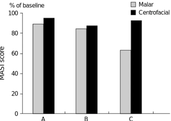
(data not shown). Patients with negative family history of melasma showed a greater decrease of the MASI score com- pared to those with positive family history in group C after 6 weeks of treatment (p<0.05) (Fig. 2). Patients with malar pattern rather than centrofacial pattern revealed a greater de- crease of the MASI score (Fig. 3). Patients with a shorter dura- tion of melasma showed a greater decrease of the MASI score (Fig. 4). Patients with the epidermal type revealed a greater decrease of the MASI score (Fig. 5). However, the differences
shown in Fig. 3, 4, and 5 had no statistical significance. Results of the objective assessment showed that seven patients (43.7
%) revealed more than moderate improvement in group C, compared with two patients (12.5%) in group B and none in group A (Fig. 6). Visible changes in the pigmentation of melasma lesions could be seen as early as 2 weeks after topi- cal application in group C. These patients in group C showed an almost complete loss of melasma lesions after 6 weeks of treatment (Fig. 7).
Side effects
In all three groups, there were no significant side effects during the entire period of treatment.
MASI score
100
80
60
40
20
0
A B C
% of baseline * p <0.05
Fig. 1. Changes of MASI score after 6 weeks of treatment.
A: Vehicle, B: 2% Lincomycin+0.05% Betamethasone valerate, C: 2% Lincomycin+0.05% Betamethasone valerate+2% Linoleic acid.
MASI score
100
80
60
40
20
0 A B C
Positive Negative
*p <0.05
*
% of baseline
Fig. 2. Changes of MASI score according to family history after 6 weeks of treatment.
A: Vehicle, B: 2% Lincomycin+0.05% Betamethasone valerate, C: 2% Lincomycin+0.05% Betamethasone valerate+2% Linoleic acid.
MASI score
100
80
60
40
20
0 A B C
Malar Centrofacial
% of baseline
Fig. 3. Changes of MASI score according to the pattern of melas- ma after 6 weeks of treatment.
A: Vehicle, B: 2% Lincomycin+0.05% Betamethasone valerate, C: 2% Lincomycin+0.05% Betamethasone valerate+2% Linoleic acid.
MASI score
100
80
60
40
20
0
A B C
<1 yr 1-5 yr
% of baseline
Fig. 4. Changes of MASI score according to the duration of melas- ma after 6 weeks of treatment.
A: Vehicle, B: 2% Lincomycin+0.05% Betamethasone valerate, C: 2% Lincomycin+0.05% Betamethasone valerate+2% Linoleic acid.
>5 yr
DISCUSSION
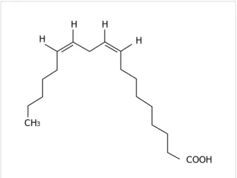
Lincomycin is elaborated by an actinomycete, Streptomyces lincolensis; it was the first lincosamide antibiotic to be used clinically. Lincomycin inhibits melanogenesis post-transcrip- tionally and abrogates glucocorticoid-induced melanogenesis on the transcriptional level in B16 melanoma cells (9). Fatty acids have been shown to have remarkable regulatory effects on melanogenesis in cultured B16F10 murine melanoma cells. Unsaturated fatty acids, such as oleic acid, linoleic acid, or -linolenic acid, decrease melanin synthesis and tyrosinase activity, while saturated fatty acids, such as palmitic acid or stearic acid, increase them (10, 13). Linoleic acid (Fig. 8) is a new bleaching agent and inhibits melanogenesis by accel- eration of proteolytic degradation of tyrosinase in B16 murine melanoma cells (14). It also accelerates the turn over of the
MASI score
100
80
60
40
20
0
A B C
Epidermal Mixed
% of baseline
Fig. 5. Changes of MASI score according to the type of melas- ma after 6 weeks of treatment.
A: Vehicle, B: 2% Lincomycin+0.05% Betamethasone valerate, C: 2% Lincomycin+0.05% Betamethasone valerate+2% Linoleic acid.
Number of person
16 14 12 10 8 6 4 2 0
A B C
Mild
Fig. 6. Objective assessment of melasma patients after 6 weeks of treatment.
A: Vehicle, B: 2% Lincomycin+0.05% Betamethasone valerate, C: 2% Lincomycin+0.05% Betamethasone valerate+2% Linoleic acid.
No Moderate Excellent
Fig. 8.Chemical structure of linoleic acid.
COOH H
H H H
CH3
Fig. 7.Clinical photographs of responses of melasma to 2% lincomycin mixed with 0.05% betamethasone valerate and 2% linoleic acid.
A 35-yr-old woman with melasma (A) before treatment and (B) after 6 weeks of treatment. A 40-yr-old woman with melasma (C) before treatment and (D) after 6 weeks of treatment.
A B C D
stratum corneum, which results in the faster desquamation of melanin pigment from the epidermis. Topical application of linoleic acid to UV-stimulated hyperpigmented dorsal skin of brownish guinea pigs resulted in a pigment-lightening effect (15).
There are still many arguments about the application of topical steroids for the treatment of melasma. Kligman &
Willis (16) failed to find any beneficial effects from applying topical corticosteroid alone. On the contrary, topical steroids such as betamethasone, dexamethasone, clobetasol propionate, and hydrocortisone can be effectively used for depigmentation in combination with other depigmenting agents or alone (17- 19). Corticosteroids have been shown to exert their antimeta- bolic effects by decreasing epidermal turnover (20). This in turn may also affect the melanocyte by decreasing its secre- tory function. Fluorinated steroids are generally more potent than non-fluorinated steroids, but the risk of adverse effects such as acne, itching, atrophy, and telangiectasias increases with the use of potent steroids. In cases where steroids are used in addition to peeling or depigmenting agents, less po- tent steroids should be used to minimize the side effects. Our patients did not show any steroid-induced and treatment- induced complications during the study period.
In this study, the 2% LM mixed with 0.05% BV and 2%
LA yielded a better outcome. It is postulated that linoleic acid should cause the pigmentary lightening effect in melasma patients, because 2% LM mixed with 0.05% BV and 2% LA was clearly superior to the 2% LM mixed with 0.05% BV.
Contrary to in vitro test results, topical lincomycin did not have a lightening effect in melasma patients. We could not also exclude the permissive role of these components. Patients without a family history of melasma showed a statistically greater improvement in the group C, but the duration, pat- tern, and type were not significant factors. The effect of ther- apy was characterized primarily by a progressive lightening of the site treated, followed by a reduction of the size of the lesion, i.e., some parts of the lesion resolved completely, while other parts had a still discernible outline. The response rate of more than moderate improvement (43.7%) after 6 weeks treatment of 2% LM mixed with 0.05% BV and 2% LA was somewhat lower than the improvement rate ranging from 65% to 73% in other reports using topical tretinoin and/or hydroquinone (11, 21). While other reports evaluated the efficacy during a 10-week period, this study employed a 6- week study period. This may explain the lower response rate in this study. The formula containing tretinoin and/or hydro- quinone induces irritation dermatitis in many cases (22), which hampers the use of this formula. Other investigators have reported increased pigmentaion in Asian patients on daily tretinoin and hydrocortisone; the increased pigmenta- tion was presumably caused by retinoid dermatitis, with resultant postinflammatory hyperpigmentation (17). The formula used in this study did not show any side effects including irritation.
In conclusion, the effect of treatment with 2% LM mixed with 0.05% BV and 2% LA was superior to that with 2%
LM mixed with 0.05% BV in melasma patients. Topical appli- cation of the formula containing linoleic acid in this study is considered to be effective in the treatment of melasma patients without apparent side effects.
REFERENCES
1. Grimes PE. Melasma. Etiologic and therapeutic considerations. Arch Dermatol 1995; 131: 1453-7.
2. Sivayathorn A. Melasma in orientals. Clin Drug Invest 1995; 10 (suppl 2): 34-63.
3. Sanchez NP, Pathak MA, Sato S, Fitzpatrick TB, Sanchez JL, Mihm MC Jr. Melasma: a clinical, light microscopic, ultrastruc- tural, and immunofluorescence study. J Am Acad Dermatol 1981;
4: 698-710.
4. Pathak MA. Clinical and therapeutic aspects of melasma: an over- view. In: Fitzpatrick TB, Wick MM, Toda K, eds. Brown Melanoder- ma. Tokyo, Japan: University of Tokyo Press; 1986: 161-72.
5. Garcia A, Fulton JE Jr. The combination of glycolic acid and hydro- quinone or kojic acid for the treatment of melasma and related con- ditions. Dermatol Surg 1996; 22: 443-7.
6. Balina LM, Graupe K. The treatment of melasma. 20% azelaic acid versus 4% hydroquinone cream. Int J Dermatol 1991; 30: 893-5.
7. Pathak MA, Fitzpatrick TB, Kraus EW. Usefulness of retinoic acid in the treatment of melasma. J Am Acad Dermatol 1986;15: 894-9.
8. Findlay GH, Morrison JG, Simson IW. Exogenous ochronosis and pigmented colloid milium from hydroquinone bleaching creams. Br J Dermatol 1975; 93: 613-22.
9. Kim DG, Kim HY, Kim MY, Lee MY, You KR. Lincomycin abro- gates dexamethasone-enhanced melanogenesis in B16 melanoma cells.
Pigment Cell Res 1998; 11: 143-50.
10. Ando H, Itoh A, Mishima Y, Ichihashi M. Correlation between the number of melanosomes, tyrosinase mRNA levels, and tyrosinase activity in cultured murine melanoma cells in response to various melanogenesis regulatory agents. J Cell Physiol 1995; 163: 608-14.
11. Kimbrough-Green CK, Griffiths CE, Finkel LJ, Hamilton TA, Bulen- go-Ransby SM, Ellis CN, Voorhees JJ. Topical retinoic acid (treti- noin) for melasma in black patients. A vehicle-controlled clinical trial.
Arch Dermatol 1994; 130: 727-33.
12. Jimbow K. N-Acetyl-4-S-Cysteaminylphenol as a new type of depig- menting agent for the melanoderma of patients with melasma. Arch Dermatol 1991; 127: 1528-34.
13. Shono S, Toda K. Phenotype Expression on Pigment cells. Tokyo, Japan: University of Tokyo Press: 1981: 263-8.
14. Ando H, Funasaka Y, Oka M, Ohashi A, Furumura M, Matsunaga J, Matsunaga N, Hearing VJ, Ichihashi M. Possible involvement of pro- teolytic degradation of tyrosinase in the regulatory effect of fatty acids on melanogenesis. J Lipid Res 1999; 40: 1312-6.
15. Ando H, Ryu A, Hashimoto A, Oka M, Ichihashi M. Linoleic acid and alpha-linolenic acid lightens ultraviolet-induced hyperpigmen- tation of the skin. Arch Dermatol Res 1998; 290: 375-81.
16. Kligman AM, Willis I. A new formula for depigmenting human skin.
Arch Dermatol 1975; 111: 40-8.
17. Tadaki T, Watanabe M, Kumasaka K, Tanita Y, Kato T, Tagami H, Horii I, Yokoi T, Nakayama Y, Kligman AM. The effect of topical tretinoin on the photodamaged skin of the Japanese. Tohoku J Exp Med 1993; 169: 131-9.
18. Neering H. Treatment of melasma (chloasma) by local application of a steroid cream. Dermatologica 1975; 151: 349-53.
19. Kanwar AJ, Dhar S, Kaur S. Treatment of melasma with potent top- ical corticosteroids. Dermatology 1994; 188: 170.
20. Hennings H, Elgio K. Hydrocortisone: inhibition of DNA synthesis and mitotic rate after local application to mouse epidermis. Virchows Arch B Cell Pathol 1971; 8: 42-9.
21. Gano SE, Garcia RL. Topical tretinoin, hydroquinone, and betametha- sone valerate in the therapy of melasma.Cutis 1979; 23: 239-41.
22. Kang WH, Chun SC, Lee S. Intermittent therapy for melasma in Asian patients with combined topical agents (retinoic acid, hydroquinone and hydrocortisone): clinical and histological studies. J Dermatol 1998; 25: 587-96.