pISSN 2288-9272 eISSN 2383-8493 J Oral Med Pain 2014;39(4):119-126 http://dx.doi.org/10.14476/jomp.2014.39.4.119
Dental Age Estimation in Adults:
A Review of the Commonly Used Radiological Methods
Hye-Mi Jeon 1 , Seok-Min Jang 1 , Kyung-Hee Kim 2 , Jun-Young Heo 1 , Soo-Min Ok 1 , Sung-Hee Jeong 1 , Yong-Woo Ahn 1
1 Department of Oral Medicine, School of Dentistry, Pusan National University, Yangsan, Korea
2 Department of Oral Medicine, Busan Paik Hospital, Inje University College of Medicine, Busan, Korea
Received September 24, 2014 Revised October 7, 2014 Accepted October 20, 2014
This review provides an overview of the most commonly used dental age estimation techniques which focus on radiological methods in Korean adults. The literature from 1995 through July 31, 2014, was searched, using PubMed, for publications in English language. In PubMed, the keywords ‘tooth’ OR ‘dental’ AND ‘pulp’ AND ‘age estimation’ were searched. Inclusion criteria was comprised of the following: the subjects were living adults and dental radiography (ex- cluded computed tomography [CT] and cone-beam CT) was used to measure the pulpal size.
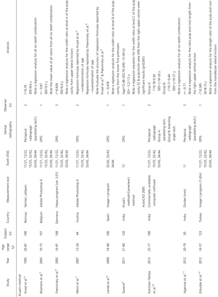
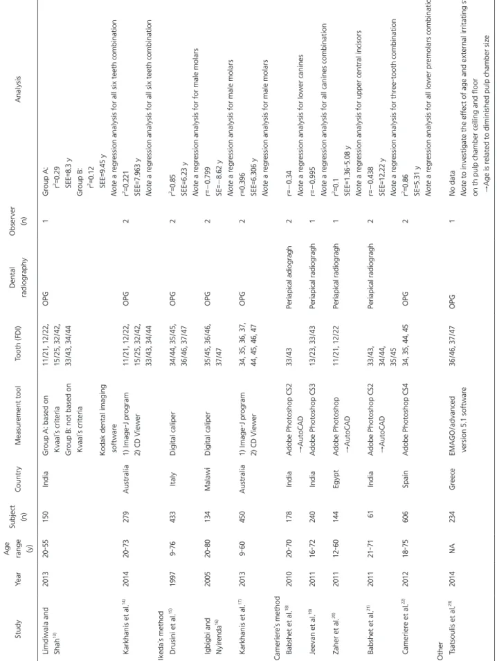
Twenty articles that met the criteria were selected. The method of age estimation using dental radiographs for measuring pulp and tooth size was represented in all studies. The methods were assorted into three categories generally; Kvaal’s, Ikeda’s and Cameriere’s methods. Those meth- ods had certain limitations such as large error range and low correlation coefficient depending on populations, type of employed teeth and particular method. Various techniques and many studies have been published for age estimation from human teeth using dental radiographs, but those techniques showed various predictability and reliability. Therefore, future studies on larger samples with well-distributed age group using not only existing techniques but new techniques are necessary for deriving convincing results.
Key Words: Adult; Age determination by teeth; Radiography; Secondary dentin
Correspondence to:
Yong-Woo Ahn
Department of Oral Medicine, School of Dentistry, Pusan National University, 20, Geumo-ro, Mulgeum- eup, Yangsan 626-787, Korea Tel: +82-55-360-5233 Fax: +82-55-360-5234 E-mail: ahnyongw@pusan.ac.kr
pulp area gradually becomes smaller because of continu- ous secondary dentin deposition, so the measurement of this reduction can be used as an indicator of dental age es- timation. Secondary dentin has been studied using various methods for age estimation and one method used was by X-rays. 2) Dental radiographs have been used relatively re- cently for age estimation methods in living persons due to convenient, simple, and can be examined directly in living subjects, without tooth extraction and destruction. 3) During the last decade, numbers of articles were published exam- ining the various methods applied and comparing the pre- cision and reproducibility of each one of them. The objec- tive of the present study was to systematically review the
INTRODUCTION
The forensic literature has provided several methods for estimating age in adults, both dead and living. Several methods of age estimation are based on the study of teeth, because teeth have the benefit to be preserved long after other tissue, even bone, and dental maturity has low vari- ability. 1) Some dental age estimation methods apply various forms of tooth modification including attrition, root dentin transparency, tooth cementum annulation, racemization of aspartic acid, and apposition of secondary dentin. Among these, analysis of the apposition of secondary dentin is the currently available nondestructive method. With aging the
JOMP Journal of Oral Medicine and Pain
Copyright Ⓒ 2014 Korean Academy of Orofacial Pain and Oral Medicine. All rights reserved.
CC