마황 추출물의 나노 입자화를 통한 항암 활성 증진
정향숙*·김승섭*·오성호*·정명훈*·최운용*·서용창*·나천수***·곽형근****·이현용*,**†
*강원대학교 생물소재공학과, **강원대학교 생명공학연구소, ***생명의나무, ****스카이007
Enhancement of Anticancer Activities of Ephedra sinica Stapf Extracts by Nano-encapsulation
Hyang Suk Jeong
*, Seoung Seop Kim
*, Sung Ho Oh
*, Myoung Hoon Jeong
*, Woon Yong Choi
*, Yong Chang Seo
*, Chun Soo Na
***, Hyeong Geun Kwak
****and Hyeon Yong Lee
*,**†*
Dpartment of Biomaterials Engineering, Kangwon National University, Chuncheon 200-701, Korea.
**
Research Institute of Bioscience and Biotechnology, Kangwon National University, Chuncheon 200-701, Korea.
***
Tree of Life, Seodun-dong, Kwonsun-gu, Suwon 441-853, Korea.
****
SKY007, Daejeon-dong, Kangneung 210-340, Korea.
ABSTRACT : This study showed the increase of antitumor activities of water soluble E. sinica extract by nano-encapsula- tion process with lecithin. Five groups of lecithin only group (LO), lecithin nano-encapsulated E. sinica group (LE), E. sinica only group (EO), one negative control group (NCO) and positive control group (PCO) were set for several anticancer exper- iment and fed into Sarcoma-180 injected mice. The cytotoxicity of LE on the human normal kidney cell (HEK293) showed 14.8% lower than 19.2% of EO and 18.4% of LO. Growth of human liver carcinoma cell and human stomach carcinoma cell as representative of digestive system in vitro was inhibited up to about 85.1% and 87.3%, in adding 1.0 ㎎ / ㎖ of LE, which values 15% higher than that from conventional EO. The survival rates of each mice group were 40%, 63%, 48%, 33% and 100%, respectively after 40 days of injecting Sarcoma-180. The increment of their body weights of the extract feed- ing groups was suppressed down to 10~15%, compared to the negative control. The nano-particles also reduced the hyper- trophy of the internal organs such as spleen and liver down to 15~20%, compared to those as the other groups. Among them, LE effectively reduced the size of tumor form to 20%. From these results, in vitro and in vivo antitumor activities of E. sinica could be enhanced by using nano-encapsulation process with lecithin because of better permeation into the cancer cells by confocal observations.
Key Words : Nano-encapsulation, Ephedra sinica Stapf, Antitumor Activities, Lecithin
서 언
마황은 중국 북부 , 몽골 등지에 주로 분포하는 길이가 30~70 ㎝ 인 한약재로서 ephedrine, pseudoephedrine, ephed- roxane, norephedrine 등 체액성 면역과 관련된 물질에 의해
교감신경계 자극에 영향을 주는 것으로 알려져 있다 . 주로 천 식치료 및 열병을 다스리는데 쓰이고 있으며 (Lee et al ., 2005) 발한 , 해열 , 항염증에 효과가 있다고 보고되었다 (Song and Lee, 2006). 이외에도 최근에는 마황의 미백활성과 관련된 연구 (Kim, 2008) 및 알러지성 피부염 억제 효과에 대해 보 고되어진 바 있다 (Shin and Kim, 2005).
한편 , 한약재들은 알려진 뛰어난 효능에 비해 체내에서 그
역할을 다하지 못하는 것이 사실이다 . 최근 한약재의 유용성 분을 체내 특정 부분에 표적화 하여 응용하고자 하는 시도
(Li et al ., 2009) 와 함께 나노기술을 이용한 약물전달시스템
(DDS : Drug Delivery System) 이 세포치료분야에서 혁신적인
변화를 가져올 것으로 전망됨에 따라 (Youn et al ., 2008), 전
달체에 대표적인 항종양제인 독소루비신을 봉입하여 효능 및 효과를 최적화하는 연구 (Lee and Yoo, 2008) 및 복분자와 같은 천연물을 고분자로 포집한 후 나노 입자화하여 면역 활 성을 평가하는 연구 (Han et al ., 2009) 등이 활발하게 진행 되고 있다 .
나노입자 (10
−9m) 는 인체에 쉽게 침투할 수 있기 때문에 순
환을 통하여 각기 다른 신체 부위로 전달되며 , 특히 100 ㎚
†
Corresponding author: (Phone) +82-33-250-6455 (E-mail) [email protected]
Received 2010 April 3 / 1st Revised 2010 May 7 / 2nd Revised 2010 May 20 / Accepted 2010 May 26
이하의 소수성 표면을 갖는 리포솜은 분자성 약물의 운반을
용이하게 하여 세포에 대한 친화성이 증가하게 된다 (Mertins
et al. , 2005). 하지만 인체의 내피세포는 인지질로 구성되어
있어 수용성 성분이 침투하기 어려운 구조로 되어 있다 . 따라 서 수용성 물질의 세포내 침투 효율 향상을 위해서 활성물질 을 레시틴의 유상이 포집한 리포솜 형태로 제조하고 이를 생 체에 적용시켜 높은 침투력 및 생체활용성을 나타내고자 하는 연구가 많이 진행되고 있다 (Kim and Kwak, 2004).
본 연구에서는 식품과 천연물 중 암의 발생을 억제 또는 지연하거나 항암보조기능 성분들이 다수 포함되어 있다는 것 을 밝혀낸 다양한 연구 결과 (Jin et al., 2008; Ha et al.,
2009) 를 근거로 보통 열수추출 방법을 통해 얻은 마황 추출
물을 생체적합성 고분자 소재인 레시틴으로 나노 입자화하여
in vitro 및 in vivo 활성을 비교함으로써 항암효과를 알아보 았다 . 최근 마황의 각성 작용으로 인해 마황이 뛰어난 항암
활성 등의 유용한 기능을 가짐에도 불구하고 그 이용이 제한 적이다 (Marchei et al., 2006; Deventer et al., 2007). 이 에 본 연구에서는 적은 양의 추출물로도 세포 수준에서 최적 의 효과를 낼 수 있는 나노의 가치를 재평가하고 , 더 나아가 이러한 항암 효과를 극대화시킬 수 있는 기능성 소재와 관련 된 분야에 활용하기 위한 기초 자료로 이용하고자 마황 나노 포집물의 다양한 분석 연구 및 세포 투과 관찰 실험을 수행 하였다 .
재료 및 방법
1. 실험재료 및 추출방법
본 실험에 사용된 마황은 경동시장에서 2009 년 국내산을 건 시료로 구입하여 음지의 상온에서 보관하면서 사용하였다 . 마
황 50 g 을 수직 환류 냉각기에 부착된 추출 flask 에 시료 중
량에 대하여 10 배의 증류수를 추출용매로 사용하여 60 ℃에서
24 시간 추출하였다 . 얻어진 추출물을 감압여과장치 (Rotary Vacuum Evaporator N-N series, Eyela, Germany) 로 여과하 여 농축을 하였고 , 동결건조를 한 후에 파우더를 얻어 실험에 사용하였다 (Jeong et al ., 2009).
2. 나노입자 제조 및 확인
나노 입자화를 통한 항암활성 증진효과의 탐색을 위해 천연 항암 효과가 기대되는 마황 추출물 (Park et al., 2004) 을 이 용해 nanoparticle 을 제조하였다 . 우선 마황 60 ℃ 물 추출 시 료 50 ㎎을 1 ㎎ / ㎖의 농도로 증류수에 녹였다 . 그리고 레시 틴 (L- α -phosphatidylcholine, Sigma, USA) 을 둥근바닥플라스 크에 넣어 chloroform (Sigma, USA) 으로 녹였고 , 이후 감압
회전상태에서 유기용매를 모두 날려 multilayer 를 형성시켰다 .
완전히 건조되어 다층의 layer 가 형성된 둥근바닥플라스크에
액상의 마황 추출시료를 넣고 , 초음파 분산기 VCX500 (Sonics & Materials Inc., USA) 를 이용하여 상온에서 2 시간
동안 균질화시켜 수용성 나노입자를 제조하였다 (Fallouh et al , 1986). 본 실험에서 레시틴만 나노 입자화 한 것은 LO 로 ,
마황 나노 추출물 레시틴 나노 입자는 LE 로 , 마황 추출물은
EO 로 , 음성대조군은 증류수로써 NCO 로 , 양성대조군은 대표 적인 항암제인 Taxol (Sigma, USA) 로써 PCO 로 각각 표기하 였다 (Kim and Lee, 2001; Sandoval et al, 2002).
나노 입자의 분포 및 확인을 위해 사용된 장치는 TEM (Transmission Electron Microscopy) 이고 , 나노 입자의 크기를 알아보기 위해 사용된 장치는 DLS (Dynamic Light Scattering, Benthos, USA) 이다 . TEM 은 나노입자 한 개를 크게 확대하여 관찰하는 것도 가능하기 때문에 입자의 모양 , 입자의 크기 등 을 결정하는데 효과적으로 사용될 수 있다 . TEM 사진을 찍기 위해서 phosphotungstic acid solution 을 이용하여 negative
staining 을 하였다 . 나노 입자가 충분히 염색이 된 후 ,
formvar/carbon 으로 코팅된 grid 에 얇게 펴서 말린 뒤 EF- TEM (LEO 912AB OMEGA, Carl Zeiss, Germany), 120 kV 에서 관찰 하였다 (Saxena et al ., 2005).
3. 시약
세포배양에 필요한 배지로 RPMI 1640 을 사용하였고 , 그 밖
에 배양에 필요한 시약으로 hepes buffer (Sigma, USA) 와
fetal bovine serum (Gibco, USA), gentamycin sulfate (Sigma, USA), trypsin-EDTA (Sigma, USA) 를 사용하였다 . 4. 세포주 및 실험동물
실험에 이용된 세포주로는 추출물 및 나노입자의 정상 세포
에 대한 독성을 알아보기 위해 인간 신장 세포인 HEK293
(Kidney normal, Human, ATCC, USA) 를 이용하였고 , 세포
수준의 항암 활성을 평가하기 위해 인간 유래 간암 세포인
Hep3B (Hepatoma adenocarcinoma, Human, ATCC, USA),
인간 유래 위암 세포인 AGS (Stomach adenocarcinoma, Human, KCLB, Korea) 를 이용하였다 . 모든 세포는 RPMI 1640 배지에서 10% heating-inactivated FBS (fetal bovine serum) 으로 적응시켜 배양하여 실험에 이용하였다 .
In vivo 실험에 사용된 실험동물은 오리엔트바이오
(KOREA) 로부터 4 주령의 ICR (Female, 22~24 g) 마우스를 구입하여 온도 20~25 ℃ , 습도 55 ± 10% 가 유지 가능한 사육 실에서 각 군별로 8 마리씩 일주일간 안정기를 취한 후 사용하 였다 . 12 시간 간격으로 점등하였으며 , 멸균된 고형 사료와 식 수를 자유 급식하였다 . 마우스에 접종된 세포주는 Sarcoma- 180 (ATCC, USA) 로 복강 내에서 6~7 일 간격으로 계대 배양
하여 사용하였다 .
5. 나노입자의 정상세포독성 및 항암 활성 측정
마황 나노입자의 정상세포독성 및 항암 활성 측정을 위해 사용된 SRB (Sulforhodamine B) assay 는 세포 단백질을 염 색하여 세포의 증식이나 독성을 측정하는 방법이다 . 실험에 사 용된 세포주는 인간 신장 유래 정상세포인 HEK293 과 소화계
장기 유래의 대표적인 암인 간암세포 (Hep3B), 위암세포
(AGS) 였다 . 세포의 농도가 4~5×10
4cells/ ㎖ 가 되도록
NucleoCounter
TM(Cell counting device, New Brunswick, USA) 를 이용해 세포수를 세었고 적정 농도로 배지희석을 한
후 , 96well plate 의 각 well 에 100 ㎕ 씩 분주하였다 . 24 시간 동안 배양 (37 ℃ , 5% CO
2incubator) 한 후 , 각각의 시료를 최종농도 0.2, 0.4, 0.6, 0.8, 1.0 ㎎ / ㎖가 되게 100 ㎕ 씩 세 포가 배양된 배지에 첨가 분주하여 48 시간 동안 다시 배양하 였다 . 배양이 완료된 후에 상층액을 aspirator 를 이용해 제거하 였고 , 차가운 10% (w/v) TCA (trichloroacetic acid) 100 ㎕
를 가하여 4 ℃에서 1 시간 동안 방치하여 세포가 더 이상 자라
지 못하게 하였다 . 증류수로 5 회 반복 세척하여 TCA 를 제거 하고 실온에서 plate 를 건조한 뒤 각 well 에 1% (v/v) acetic acid 에 녹인 0.4% (w/v) SRB 용액을 100 ㎕ 씩 첨가하고 상 온에서 30 분 동안 염색시켰다 . 결합되지 않은 SRB 염색액은
1% acetic acid 로 5 회 정도 세척 , 건조시킨 후에 10 mM Tris buffer 100 ㎕를 첨가하여 염색액을 녹여냈다 . Micro reader (Molecular Devices, Thermo max, USA) 를 이용하여 540
㎚에서 흡광도를 측정하였다 . Selectivity 측정은 SRB assay 를 이용하여 정상세포 (HEK293) 에 대한 각 sample 농도에서 세
포독성을 측정하고 , SRB assay 를 이용하여 각 암세포주의 생
육억제활성을 측정한 후 각 농도에서의 세포 독성에 대한 암 세포 생육 억제 활성의 비로 selectivity 를 계산하였다 (Dol and Peto, 1981).
6. Sarcoma-180 복수암 및 고형암에 대한 in vivo 항암
효과 측정
Sarcoma-180 세포를 10% heating inactivated FBS 를 함유
한 RPMI 1640 의 기본 배지에서 배양하였다 . Cell scraper (Biocompare, UK) 로 배양 flask 로부터 cell 을 분리한 후
2×10
6cells/ ㎖의 농도로 조절하여 200 ㎕를 마우스 복강에
1 ㎖ syringe 를 이용하여 피하이식 하였다 . 상기의 방법으로 마 우스 복강 내에서 6~7 일 간격으로 계대배양을 통해 보존된
sarcoma-180 세포를 취하여 복수암 실험군 마우스의 복강과
고형암 실험군 마우스의 왼쪽 대퇴부 근육에 200 ㎕ 씩 피하
이식 하였다 . 종양세포 이식 후 21 일 동안 시료의 농도를 100
㎎ / ㎖로 조절하여 매일 동일한 시간에 0.5 ㎖ 씩 경구 투여
하였다 . 양성대조군인 Taxol 은 이미 효능이 알려진 약물이므
로 추출물의 농도 대비 20 배 희석된 5 ㎎ / ㎏의 농도로 제조
하여 마황 시료와 동일한 조건에서 매일 1 회분씩 나누어 투여 하였다 (Bárdos et al., 2003). 40 일간 3 일 간격으로 복수암 유발 마우스의 체중을 측정하였고 , 생존율 (survival rate) 을 측 정하였으며 , 고형암 유발 마우스에서 종양 세포 이식 40 일 후 희생시켜 고형암과 비장 및 간의 무게를 측정하여 체중에 대 한 장기의 중량을 측정하였다 (Jung et al., 2002; Kim et al., 2007).
S : Mean survival days of treated mouse C : Mean survival days of control mouse
C : The average tumor weight of control / The average body weight of control
S : The average tumor weight of treated group / The average body weight of treated group
7. Confocal 현미경으로 세포 투과 관찰
나노시료 첨가에 의한 면역세포 B cell (Raji) 의 분포양상을
공초점 레이저 주사 현미경 LSM510 META NLO (Carl
Zeiss Jena GmbH, Germany) 으로 관찰하였다 . 나노입자 제조 를 위해 초음파 분산을 시작할 때 , dye 로 FITC (fluorescein isothiocyanate) 를 넣어줌으로써 fluorescence tagging 하였다 . 1 ㎖의 면역 B 세포 2 × 10
8cells/ ㎖을 CO
2농도 5%, 30 ℃의
incubator 에서 배양한 후 , FITC 로 염색된 나노시료 (1 ㎎ / ㎖ ) 100 ㎕와 1 시간 동안 반응시켰다 (Lee et al., 2007).
8. 통계처리
SPSS program (ver. 12.0, SPSS Inc., Chicago, IL, USA)
의 T-test 로 검정하였으며 모든 data 는 평균 ± 표준오차 (Mean ± standard error) 로 나타내었다 .
결과 및 고찰
1. 나노입자의 확인
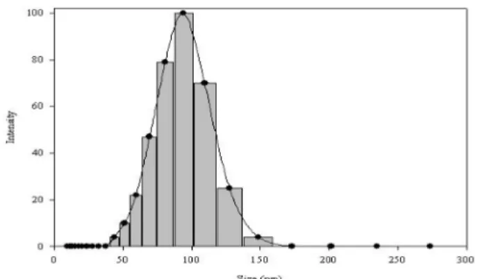
Fig. 1 과 같이 레시틴으로 포집한 마황 추출물 나노입자 LE
는 50~200 ㎚ 의 범위로 형성된 것을 알 수 있다 . 평균
200 ㎚ 인 세포 간극의 크기를 고려한다면 제조된 나노입자는 암세포에 효과적으로 적용 가능한 제형이라고 생각된다 . 이는
Selectivity = 암세포 생육 억제 활성
정상 세포의 세포 독성
Survival rate (%) = S − C C × 100
Tumor growth inhibition ratio (%) = C C − S × 100
기존 대장암 세포의 나노입자 투과를 물리학적 접근을 통해 밝혀낸 연구 결과와 비교해 의미가 있다 (Lochner et al., 2003). 또한 나노입자가 모두 깨지지 않고 70% 이상 그대로 피크를 이루며 detection 된 결과를 바탕으로 , 이는 기존의 복 분자 추출물을 나노 입자화 하여 항산화 효과 및 미백 활성을 측정했던 결과와 비교해 (Jeong et al., 2009) 천연물이 나노 입자화 공정을 통해 적은 양으로도 높은 생리 활성 증진 효과 를 낼 수 있음을 알 수 있다 . Fig. 2 는 TEM 으로 촬영한 마 황 레시틴 나노입자의 모습이다 . 구형의 나노 입자들은
negative staining 에 의해 하얗게 보인다 . 염색에 사용한
phosphotungstic acid 는 수용성 부분을 염색시키므로 지질이 포함되어 있는 나노 리포솜 바깥부분은 염색이 되지 않아 하 얗게 보인다 . 제조된 나노 입자는 수용성 추출물을 유상이 포
집한 W/O 형태의 리포솜으로 구성된다 . 2. 나노입자의 정상세포독성
실험에 사용된 마황 나노 시료의 농도는 0.2, 0.4, 0.6, 0.8
그리고 1.0 ㎎ / ㎖로 조절하여 정상세포에 대한 세포독성과 각 암세포에 대한 성장 효과를 검토하였다 . Fig. 3 은 HEK293 정 상 세포에 대한 독성을 나타낸 것으로 최고 농도인 1.0 ㎎ / ㎖ 에서 LE 가 14.8% 로 EO 의 19.2%, LO 의 18.4% 와 비교했을
때 가장 낮은 세포 독성을 보였다 . 레시틴으로 나노 입자화한
경우 , 마황 추출물군의 세포독성보다 4% 낮은 세포 독성을 나 타냈다 . 전반적으로 20% 이하로 나타난 마황 시료의 세포 독
성 값은 HEK293 세포를 이용하여 천연알칼로이드의 세포 독
성을 연구한 결과인 40% 와 비교했을 때 (Ji et al ., 2008) 세 포 수준에서 큰 영향을 주지 않으며 , 경미한 독성을 보이는 것으로 사료된다 .
3. 나노입자의 in vitro 항암 활성
Fig. 4 의 (A) 와 (B) 는 인간 간암 세포 유래 Hep3B 와 인간 위암 세포 유래 AGS 에 대한 항암 활성을 나타낸 것이다 . 1.0
㎎ / ㎖의 농도에서 간암세포와 위암세포 모두에서 나노 입자 화한 LE 가 각각 85.1%, 87.3% 로 가장 높은 항암 활성을 나 타냈다 . 마황 열수 추출물 EO 가 간암세포에서 67.2%, 위암세
포에서 70.6% 의 저해활성을 나타낸 것과 비교했을 때 , 레시틴
으로 나노 입자화한 군에서 16~17% 감소된 암세포 생육을
나타낸 것은 나노 입자화를 통해 시료의 세포 투과가 용이해 졌고 (Parag et al. , 2009), 마황 고유의 유용성분의 방출이 효과적으로 제어되었기 때문인 것으로 사료된다 (Antina et al. , 2005). 약 60% 의 암세포 생육 저해 효과를 보이는 기존 의 다른 한약재 추출물 가시오갈피 , 복분자 등과 비교했을 때 (Park et al. , 2004) 역시 나노 입자화를 통해 항암 활성이
20% 이상 향상된 것을 확인할 수 있다 . 한편 , 양성대조군으로 사용한 taxol 의 경우 30 µ M 를 첨가하였을 때 , 간암에서는
Fig. 1.Size distribution of nanoparticles with lecithin using
dynamic light scattering (DLS).
Fig. 2.
TEM micrograph of nanoparticles from the
E. sinicaextract by lecithin-encapsulation. Scale of bar is 50~
200
㎚.
Fig. 3.
Cytotoxicity of
E. sinicaextracts by nano-encapsulation on normal cell line, HEK293. Results are expressed as mean
±S.D. of data obtained from three independent experiments. Each values were compared with control at
p
< 0.05.
†
LO : lecithin only nanoparticles, EO :
E. sinicaonly extracts,
LE : lecithin
E. sinicaextracts by nano-encapsulation
98.5% 의 항암 효과를 위암에서는 96.2% 의 억제 효과를 나타 내는 것으로 보고되었다 (Sandoval et al, 2002).
4. S-180 복수암 및 고형암에 대한 in vivo 항암 효과
복수암 유발에 따른 복수의 형성을 확인하기 위한 지표로
Table 1 에서와 같이 체중 측정 결과를 이용하였다 . EO 및
LE 실험군이 S-180 복수암 주사 후 최장 40 일까지 생존하였 기 때문에 전체 마우스 군에 시료를 매일 경구투여하며 3 일 간격으로 40 일까지 마우스의 체중과 수명을 측정하였다 . 증류
수를 경구 투여한 마우스 군의 최종 체중이 48.8 ± 0.5 g 으로
나타난 것과 대조적으로 LO 군에서는 44.0 ± 0.4 g, EO 군에서 는 42.6 ± 0.2 g, LE 군에서는 39.1 ± 1.3 g 로 나타나 전반적으로 체중 감소를 보였다 . 특히 , LE 를 경구 투여 하였을 때 음성대
Fig. 4.
Inhibition ratio of growth of (A) Hep3B (H, hepatoma adenocarcinoma) and (B) AGS (A, stomach adenocarcinoma) in adding the nanoparticles from
E. sinicaextracts (bar chart, %). The line chart means selectivities of (A) Hep3B and (B) AGS against normal cell line, HEK293. Results are expressed as mean
±S.D. of data obtained from three independent experiments. Each values were compared with control at
p< 0.05.
†
LO : lecithin only nanoparticles, EO :
E. sinicaonly extracts, LE : lecithin
E. sinicaextracts by nano-encapsulation
Table 1.
The effect of treatment with 100
㎎/
㎖of samples on body weight (g) and survival rate (%) in female ICR mice.
Group** Inintial (1day) Final (40 day) Body weight (g) Survival rate (%)
LO 28.4 ± 0.5 44.0 ± 0.4* 40
EO 27.2 ± 0.6 42.6 ± 0.2 48
LE 29.0 ± 1.2 39.1 ± 1.3 63
NCO 28.6 ± 0.4 48.8 ± 0.5 33
PCO 29.0 ± 1.1 35.6 ± 0.1 100
*Mean ± S.D. of data obtained from three independent experiments.
**Each values were compared with control at
p< 0.05.
**LO : lecithin only nanoparticles, EO :
E. sinicaonly extracts, LE : lecithin
E. sinicaextracts by nano-encapsulation,
NCO : negative control by D.W,
PCO : positive control by taxol (5
㎎/
㎖)
조군과 비교해서 15% 이상 복수 형성이 지연된 것으로 미루
어 보아 , 마황 추출물의 나노 입자가 마우스의 복강에 효과적
으로 침투해 종양의 형성을 더디게 진행시킨 것으로 사료된다
(Twan, 2010).
Fig. 5 는 S-180 을 마우스 복강에 투여한 후 40 일까지 마우 스의 생존율을 측정한 결과이다 . 본 실험 기간인 40 일 동안의 마우스의 생존 상황을 뚜렷하게 가시화 위해 그래프로 표현하 였다 (Ha et al., 2009). 음성대조군에서 16 일째부터 생존율의
감소가 일어나 33% 의 생존율을 나타낸 것과 대조적으로 , 마
황을 나노 입자화한 LE 군에서는 63% 로 생존율의 감소가
30% 정도 더디게 진행되었다 . Taxol 을 투여한 양성대조군의
경우 실험이 종료되는 40 일 째까지 100% 의 생존율을 나타냈
다 (Jagetia and Nayak, 1996). 본 실험 결과를 통해 복수암 에 효과적으로 작용하는 마황 레시틴 나노입자의 기능을 확인 하였고 , 이번 연구 결과를 기반으로 나노입자의 항암 기작에
대한 더욱 심도있는 연구가 진행될 필요가 있을 것으로 사료
된다 .
나노입자를 경구 투여한 LE 군에서의 고형암 성장 억제 효
과 및 면역 장기에 미치는 영향을 알아보고자 종양세포 이식
40 일 후 고형암 및 장기의 무게를 측정한 결과를 Table 2 에 나타냈다 . 나노입자를 경구 투여한 LE 에서 마우스 간의 무게 는 1.92 ± 0.14 g 으로 , 비장의 무게는 0.51 ± 0.15 g 으로 양성대 조군과 비교했을 때 가장 적은 차이를 보였다 . 경구 독성이 있는 추출물을 섭취하였을 때 간 , 비장과 같은 면역 장기의
비대화가 일어나기 때문에 (Ma et al., 2010) 면역 기능이 약
한 사람들에게는 더욱 치명적이다 . 따라서 나노입자의 섭취 시 장기의 비대화가 일어나지 않은 것으로 보아 , 향후 나노 단위 로 포집한 천연물이 항암소재로서 높은 부가가치를 가질 수 있을 것으로 사료된다 .
5. Confocal 현미경으로 세포 투과 관찰
Fig. 6 은 Confocal 현미경을 이용해 마황 레시틴 나노입자의
모습을 실시간 10 분 간격으로 찍은 사진으로서 , 나타난 면역
Fig. 6.
Confocal microscope photographs of cancer cell line (Hep3B) containing nanoparticles of
E. sinicaevery 10 min. (A) 0 min, (B) 10 min and (C) 20 min.
Fig. 5.
The survival ratio (%) treated with different samples on the growth of Sarcoma-180 inoculated intraperitoneally.
†
LO : lecithin only nanoparticles, EO : E. sinica only extracts, LE : lecithin E. sinica extracts by nano-encapsulation, NCO : negative control by D.W,
PCO : positive control by taxol (5
㎎/
㎖)
Table 2.