말채나무의 항염증 효과
이상현․윤광로․이 은․차윤엽1*
상지대학교 보건과학대학 제약공학과, 1 : 한의과대학 한방재활의학과
Anti-Inflammatory Effect of Cornus Walteri
Sang Hyun Lee, Kwang Ro Yoon, Eun Lee, Yun Yeop Cha1*
Department of Pharmaceutical Engineering, College of Health Sciences,
1 : Department of Oriental Rehabilitation Medicine, College of Oriental Medicine, Sangji University
This research seeks a basis for developing new anti-inflammatory medicine by investigating
Cornus Walteriextract for its anti-infla mmatory effects. After the injection of LPS in to rats with
Cornus Walteriextract, its anti-inflammatory effects were compared among the treatment groups. The plasma concentration of IL-1β, IL-6 and TNF- α peaked at 5h after LPS injection, and the values of the
Cornus Walteriextract groups were lower than those of the control group. In the increment of concentration of these cytokines at 2h and 5h after LPS injection, the
Cornus Walterigroups were lower than that of control group. The plasma concentration of IL-10 peaked at 5h after LPS injection, and the values of the
Cornus Walteriextract groups were higher than those of the control group. In the increment of cytokines concentration at 2h and 5h after LPS injection, the
Cornus Walterigroups were higher than that of control group. Liver cytokines measurement was done at 5h after LPS injection. The concentration of liver IL-1β and IL-6 in the
Cornus Walterigroups was lower than that of the control group. The concentrations of liver TNF-α, and IL-10 showed no significant differences among all the treatment groups. In the studies of lipopolysaccharide-exposed Raw 264.7 cells, the concentration of IL-1β, IL-6 and TNF-α in the lipopolysac charide-exposed cells groups was higher than that of control group (normal group). However, in lipopolysaccharide-exposed cells groups, they showed lower values than those of control group and these values showed a tendency to decrease in the
Cornus Walterigroups. The concentration of IL-10 in the lipopolysaccharide-exposed cells groups was higher than that of control group (normal group), and among the lipopolysaccharide-exposed cells groups, all
Cornus Walteriextract groups showed higher values than single lipopolysaccharide-exposed cells groups. This studies have shown that
in vitroand
in vivo Cornus Walteri extractsare significantly more sensitive to inflammatory cytokines and LPS induced lethality. We conclude that the
Cornus Walteriextracts have an functional material for inflammatory activities.
Key words : inflammatory effect,
Cornus Walteri, LPS shock, cytokines, Raw 264.7 cells
* 교신저자 : 차윤엽, 강원도 원주시 상지대길 80, 상지대학교 부속 한방병원
․E-mail : omdcha@sangji.ac.kr, ․Tel : 033-741-9260
․접수 : 2011/08/31 ․수정 : 2011/09/20 ․채택 : 2011/11/01
서 론
염증제어는 질병 치료에서 가장 기본적으로 수행되는 임상 적 대응 중의 하나이며, 염증반응의 정도에 따라 염증성 질환자 및 수술환자들의 사망률은 크게 달라질 수 있다 1) .
염증반응(inflammation)은 병원균들의 감염, 화학적 자극 및 상처 등과 같은 외부자극에 대한 생체조직의 방어기작으로 각종 병원균들과 생체이물질들에 의해 활성화된 면역세포 혹은 염증
매개인자(pro-inflammatory mediators)들의 발현에서부터 시작 된다 2) .
그러나 생체 내에서 염증반응의 경과시간 및 반응의 정도가 과도할 경우에는 염증매개물질들은 발적, 발열, 종창, 동통 및 기 능장애 등의 증상을 일으키며 3) , 질병의 상태를 악화시키거나 새 로운 질병을 유도하기도 한다 4) . 따라서 염증반응의 적절한 제어 기술은 임상현장에서 대단히 중요하다 1) .
현재 임상현장에서 염증반응의 제어를 위해 다양한 약물들
이 개발되어 사용되고 있다. 그러나 대부분의 항염증제 들은 장
기복용 할 경우 출혈성 소화관궤양, 신장 기능저하 및 혈압상승
등의 부작용과 함께 심근경색 및 혈전형성 등의 순환기계 질환
도 유발할 수 있다는 것이 보고되었으며 5) , 이와 같은 부작용으로 인해 수술환자 및 염증성 질환자들에 대해 제한적으로 사용되고 있다. 따라서 효능이 우수하고 부작용이 없는 새로운 항염증제의 개발이 절실히 필요하다.
최근 들어 다양한 분야에서 새로운 항염증제 개발을 위한 많은 연구들이 수행되었다 6-8) . 특히 마황 9) , 감국 10,11) 및 하고초 12) 등을 비롯한 천연물들을 이용한 연구결과들은 새로운 항염증제 의 개발가능성을 보여주었으며, 보다 더 광범위하게 한약재 및 천연물들을 대상으로 하여 체계적인 기초연구가 필요함을 인식 시켜 주었다.
말채나무( Cornus Walteri Wanger)는 층층나무과에 속하는 낙엽교목으로 한방에서는 잎을 모엽이라 하여 과실과 함께 칠창
13) 및 강래지장약 14) 으로 사용하기도 한다. 민간에서는 잎을 지사 제로 사용하였으며 15) , 함유성분으로는 gallic acid 와 isoquercitrin을 비롯하여 미네랄 성분이 다량 함유되어 있어 이 뇨작용에 효과가 있고, 비만 치료에 탁월한 효과가 있음이 보고
되었다 13,15) . 또한 수피에서 8종의 페놀성 화합물이 내재하고 있
음이 밝혀졌으며 16) , 말채나무 추출물이 α-amylase활성을 저해하 여 식후 혈당 상승을 억제함이 보고되었다 17) . 이와 갈은 연구결 과들을 참고해 보면 말채나무에는 생체기능의 여러 분야에 영향 을 줄 수 있는 많은 생리활성물질들이 내재하고 있을 가능성을 시사해준다.
Lipopolysaccharide(LPS)는 병원균의 내독소로서 그람음성 세균의 막 구조물로 다당류, 인지질 및 소량의 단백질로 구성되 어 있으며, 여러 종류의 염증세포 및 조직구성 세포들이 생산하 는 cytokine들의 생산을 촉진시켜 18) , 염증반응을 연구하는 실험 모델로 많이 응용된다 19) .
따라서 본 연구는 항염증효과가 우수하고, 부작용이 없는 새 로운 항염증제를 개발하기위한 기초연구의 일환으로, 말채나무 추출물을 급여한 흰쥐에게 LPS에 의한 급성기 염증반응을 유발 시킨 후, 혈액 및 간장의 전염증성 cytokines들의 생산량과 혈액 내 생물학적 수치들을 조사하였으며, 한편으로는 말채나무 추출 물을 처리한 Raw 264.7 cell에 LPS를 처리한 후, 처리군 별 전염 증성 cytokines들의 생산량을 조사하여, in vivo 및 in vitro 에서 말채나무 추출물의 항염증효과를 검토하였다.
재료 및 방법
1. 시험동물 및 시험군
평균체중이 185.39±8.21 g의 Sprague-Dawley계 수컷 32두 를 1주일간 시험식이에 적응시킨 후, 평균체중이 유사하게 대조 군[생리식염수 100 mg/kg, body weight(BW)], 처리 1군(말채나 무 추출액 100 mg/kg, BW), 처리 2군(말채 추출액 200 mg/kg, BW) 및 처리 3군(말채나무 추출액 300 mg/kg, BW)으로 나누 어, 각 처리군당 8두 씩 임의 배치했다.
2. 식이 및 물
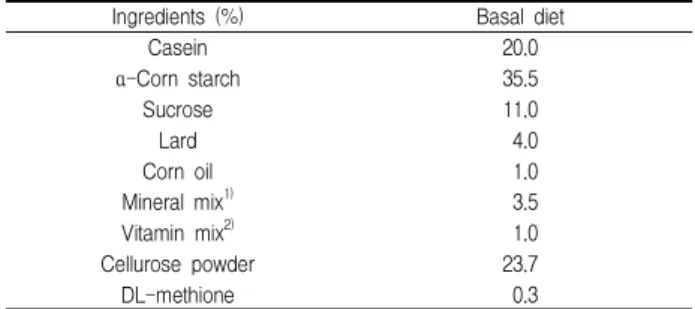
식이(Table 1) 및 물은 시험기간 6주 동안 자유 급여하였다.
Table 1. Composition of experimental diet
Ingredients (%) Basal diet
Casein 20.0
α -Corn starch 35.5
Sucrose 11.0
Lard 4.0
Corn oil 1.0
Mineral mix 1) 3.5
Vitamin mix 2) 1.0
Cellurose powder 23.7
DL-methione 0.3
1)Mineral mix. (g/kg diet) : CaCO3, 29.29; CaHPO4․2H2O, 0.43; KH2PO4, 34.30; NaCl, 25.06; MgSO4․7H2O, 9.98; Feric citrate hexahydrate, 0.623; CuSO4․5H2O, 0.516; MnSO4․ H2O, 0.121; ZnCl2, 0.02; KI, 0.005; (NH4)6MO7O24․4H2O, 0.0025.2)Vitamin mix (mg/kg diet) : Thiamine-HCl, 12; Riboflavin, 40; Pyrodoxin-HCl, 8; Vitamin-B12, 0.005; Ascorbic acid, 300; D-biotin, 0.2; Menadione, 52; Folic acid, 2; D-calcium pantothenate, 50;
P-aminobenzoic acid, 50; Nicotinic acid, 60; Cholin choloride, 2000 (IU/kg diet); Rethinyl acetae, 5000 (IU/kg diet); Cholecalciferol, 250 (IU/kg diet).
3. 말채나무 추출물 및 급여
시중에서 구입한 양질의 말채나무(한국 강원도, 2010년산) 수피 1.2 kg (건조중량)을 수조에서 냉각수 환류하에서 EtOH로 5시간씩 3회 추출하고, 여과, 감압 농축하여 EtOH 추출물 148 g 을 얻었다. 말채나무 추출물의 투여는 매일 오후 5시경에 죤대를 이용하여 경구 투여하였으며, 대조군은 동일한 방법으로 생리식 염수를 투여했다.
4. LPS 처리
LPS 처리는 6주간의 사양기간이 종료된 후, 5 mg/kg의 수 준으로 각 처리군 모두 동일하게 복강 주사하였다.
5. 혈액 및 간장 채취
혈액 채취는 시험 최종일에 LPS 처리 직전(0h), LPS 처리 후 2시간(2h) 및 5시간 (5h)에 각 처리군 별로 심장 천자법에 의해 채혈했다. 간장 채취는 LPS 처리 후 5시간에 혈액 채취가 끝난 후 적출하였다.
6. Raw 264.7 세포배양과 cytokines 정량용 시료 채취
마우스 대식세포인 Raw 264.7 cells은 한국세포주은행(서울) 에서 구입하였으며, Dulbecco's modified Eagle's medium(DMEM)에 10% fetal bovine serum(FBS), penicillin(100 U/ml) 및 streptomycin(100 μg/ml)이 첨가된 배지 를 사용하여 37℃, 5% CO 2 incubator에서 배양하였다. 시험과정 의 모든 cells는 80-90%의 confluency에서 실험하였고, 20 passages를 넘기지 않은 cell만 사용하였다. 세포배양은 4 well dish에 1x10 6 /ml의 cells을 분주하고(1x10 6 /ml), 말채나무 추출물 을 각 처리 별 농도 (0 μg/ml, 10 μg/ml, 30 μg/ml, 100 μg/ml)로 처치한 다음, 1시간 후에 각각 LPS 1μg/ml를 처치하였으며, LPS 처치 후 6시간 후에 시료를 채취하여 Cytokines을 측정하였다.
7. Cytokines 정량
혈장 cytokine 정량용 시료는 채혈 직후, 혈장을 분리하여
-80℃에 냉동 보관하였다. 간장 cytokine 정량용 시료는 1 g의
간장을 채취하여 5 ml의 cold phosphate buffered saline (PBS,
pH 7.4, containing a protease inhibitors cocktail)과 함께 혼합하 여 얼음위에서 분쇄(homogenized)하였다. 분쇄혼합물을 4℃, 15,000 rpm, 15분간 원심분리한 후, 상층부를 0.45 ㎛ 필터로 여 과하고, 다시 원심분리해서 상층부를 -80℃에 냉동 보관하였다.
Raw 264.7 cells들이 생산한 cytokines 정량을 위해 배양액 을 4℃, 15,000 rpm, 15분간 원심분리한 후, 상층부를 0.45 ㎛ 필 터로 여과하고, 다시 원심분리해서 상층부를 -80℃에 냉동 보관 하였다.
Cytokine(IL-1β, TNF-α, IL-6 및 IL-10)정량은 시판 Kit(Biosource International, USA)를 이용했다. TNF-α의 최저 측정농도는 0.7 pg/ml이며, 다른 cytokine들은 3-8 pg/ml이다.
간장 cytokines 정량은 5 ml의 PBS에 생 간장 1 g를 혼합한 조 정액으로 측정하였으며, pg/mg 단위로 나타내었다.
8. 통계처리
실험결과는 SPSS package를 이용하여 one-way ANOVA 검정을 수행하였으며, 각 처리군간의 유의성 검정은 Duncan's multiple range test 에 의해 P<0.05 수준에서 실시했다.
결과 및 고찰
본 연구에서 급성기 염증반응을 유발시키기 위하여 흰쥐에 게 LPS를 처리했다. LPS는 병원균의 내독소로서 처리량과 처리 후 시간의 경과에 따라 생체 내의 염증반응 상태는 상당히 달라 진다. 따라서 본 연구에서는 여러 연구자들의 실험결과를 참고하 여 rat의 내독소 쇼크를 검토하기 위한 시료채취 시간으로 2h 및 5h이 적당하다고 생각되어 결정하였다 19,20) . 또한 LPS 처리농도 는 5 mg/kg으로 하였는데 이 수준은 단시간에 rat와 마우스에 내독소의 쇼크를 주어 간장과 혈액내의 cytokine농도를 높인다 는 다른 연구자의 실험결과를 참고했다 6,21,22) .
1. Plasma cytokines 1). Plasma IL-1β
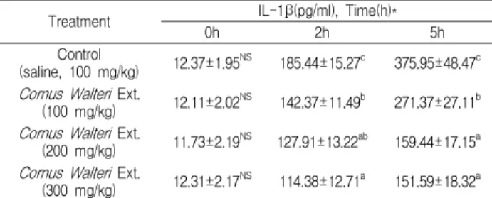
말채나무 추출물이 LPS를 투여한 흰쥐의 Plasma IL-1β 농 도에 미치는 영향을 Table 2 에 나타내었다.
Plasma IL-1β의 농도는 LPS 처리 후 2시간부터 증가하여, 5시간째에도 계속적으로 증가하는 경향을 나타내었다. 이러한 결과는 LPS 처리 후 4-6시간에 혈장 IL-1β농도의 피크가 나타 났다는 다른 연구자의 결과 19) 와 유사하였고, LPS 처리 후 2시간 째 및 5시간째 모두에서 말채나무 추출물의 투여량이 증가함에 따라 Plasma IL-1β 농도는 하락하는 경향을 나타내었다.
IL-1β는 monocyte, macrophage. β-cell, dendritic cell, endothelial cell, neutrophil 및 hepatocyte에서 분비되며, TNF-α, IL-2, IL-6와 함께 pro-inflammatory cytokine으로서 여러 면역 학적 작용들과 연관되어 있다. 특히 IL-1β는 T-cell의 activity를 활성화한다 23) . 또한 급성기 염증반응에서 급격히 증가한다 19) 고 알려져 있다.
이러한 점을 고려해 보면 본 실험의 결과에서 말채나무 추
출물의 투여량이 증가함에 따라 Plasma IL-1β 농도가 하락하는 경향을 나타내어 말채나무 추출물이 염증반응 완화에 긍정적으 로 작용하고 있음을 시사해준다.
Table 2. Effect of Cornus Walteri Ext. on plasma IL-1β concentration in lipopolysaccharide-exposed rats.
Treatment IL-1β(pg/ml), Time(h)*
0h 2h 5h
Control
(saline, 100 mg/kg) 12.37±1.95 NS 185.44±15.27 c 375.95±48.47 c Cornus Walteri Ext.
(100 mg/kg) 12.11±2.02 NS 142.37±11.49 b 271.37±27.11 b Cornus Walteri Ext.
(200 mg/kg) 11.73±2.19 NS 127.91±13.22 ab 159.44±17.15 a Cornus Walteri Ext.
(300 mg/kg) 12.31±2.17 NS 114.38±12.71 a 151.59±18.32 a
*: 0h, 2h and 5h after LPS injection. a,b,c: Values are expressed as compared with vehicle.*a-c with different letters are significantly different at p<0.05 by Duncan's multiple range test.NS: Not significantly different (P>0.05).
Table 3. Effect of Cornus Walteri Ext. on plasma IL-6 concentration in lipopolysaccharide-exposed rats.
Treatment IL-6(pg/ml), Time(h)*
0h 2h 5h
Control
(saline,100 mg/kg) 21.09±3.72 NS 295.72±37.15 c 631.53±51.88 c Cornus Walteri Ext.
(100 mg/kg) 20.19±3.21 NS 212.83±31.11 b 527.11±42.75 b Cornus Walteri Ext.
(200 mg/kg) 22.47±3.75 NS 185.39±28.61 ab 495.97±35.94 b Cornus Walteri Ext.
(300 mg/kg) 21.89±4.02 NS 171.29±23.58 a 421.33±35.58 a
*: 0h, 2h and 5h after LPS injection. a,b,c: Values are expressed as compared with vehicle.*a-c with different letters are significantly different at p<0.05 by Duncan's multiple range test.NS: Not significantly different (P>0.05).
2) Plasma IL-6
말채나무 추출물이 LPS를 투여한 흰쥐의 Plasma IL-6 농도 에 미치는 영향을 Table 3에 나타내었다.
IL-6는 단핵구를 포함한 여러 종류의 세포에서 분비되며, 다
양한 기능을 가지고 있는 cytokine이다. 숙주의 방어, 면역반응,
신경세포의 기능, hema topoiesis등에서 중요한 기능을 하는 물
질이다. IL-6의 생산은 mitogenic 또는 antigenic 자극, LPS, 칼
슘, cytokines, 바이러스 등에 의해 상승된다. 이러한 IL-6가 생
체내에서 과잉 생산되면 수종의 악성종양, 자가면역질환, 그 외
의 감염성 질환 등 여러 가지 질환을 유발한다 24) . 본 연구에서
Plasma IL-6 (Table 3)의 농도가 LPS 처리 후 2시간째 및 5시간
째 모두에서 높은 수치로 증가하는 경향을 보여, LPS에 의한 염
증반응의 정도가 시간이 경과함에 따라 증가하였음을 시사했으
며, 이와 같은 양상은 Mathiak 등 19) 이 LPS로 염증반응을 유발시
켰을 때 혈장 IL-6의 농도는 LPS처리 후 4-6시간에서 최고치를
나타내었다고 보고한 결과와 유사했다. 또한 각 처리군 별
Plasma IL-6의 농도는 LPS 처리 후 2시간째 및 5시간째 모두에
서 대조군보다 말채나무 추출물 처리군 들이 낮은 값을 나타내
었으며, 말채나무 추출물의 투여량이 증가함에 따라 하락하는 경
향을 보였다. 이러한 결과는 말채나무 추출물에 내재하는 기능성
물질이 생체 내의 전염증성 cytokine 들의 생산체계에 어떤 영향
을 주고 있음을 시사해 주며, 이러한 결과가 염증매개물질의 생
Table 6. Effects of Cornus Walteri Ext. on liver cytokines concentration in lipopolysaccharide-exposed rats.
Treatment IL-1β(pg/mg) IL-6(pg/mg) TNF-α(pg/mg) IL-10(pg/mg)
Control(saline, 100 mg/kg) 24.93±2,59 b 16.33±2.38 c 1.95±0.73 NS 1.39±0.99 a
Cornus Walteri Ext. (100 mg/kg) 22.82±3.65 ab 10.97±2.35 b 1.61±0.85 NS 1.91±0.78 ab
Cornus Walteri Ext. (200 mg/kg) 17.91±2.62 a 8.31±2.09 a 1.64±0.77 NS 2.17±0.73 ab
Cornus Walteri Ext. (300 mg/kg) 18.55±2.17 a 7.19±2.26 a 1.59±0.82 NS 2.54±0.81 b
a,b: Values are expressed as compared with vehicle.*a-b with different letters are significantly different at p<0.05 by Duncan's multiple range test.NS: Not significantly different (P>0.05).
산에 영향을 주어 염증반응을 완화하는 데에 긍정적으로 작용할 것으로 생각된다.
3) Plasma TNF-α
말채나무 추출물이 LPS를 투여한 흰쥐의 Plasma TNF-α 농 도에 미치는 영향을 Table 4에 나타내었다. LPS 처리 후 2시간째 에 모든 처리 군에서 Plasma TNF-α농도가 급격하게 증가하였고, 5시간까지 높은 수치를 유지했다. TNF-α는 면역반응과 염증반응 을 유도하는 염증유도매개 사이토카인으로 단핵세포와 대식세포 에서 생산된다 25) . 세포의 성장과 분화, apoptosis 및, necrosis 등 에 관여하며 혈관 투과성을 증가 시키고, 미생물 감염 시에 생산 량이 중가하고, 식세포의 사이토카인 분비를 유도하여 미생물에 대한 숙주세포의 항상성을 유지하는 중요한 방어기전을 담당한 다 26) . 종양발생시 apoptosis를 유도하여 종양 발생의 감시 기전으 로 이용되기도 하고, 호중구 조절, MHC 발현의 조절, β세포 증 식, angiogenesis 유도 및 대식세포 활성 기능을 가지고 있다 27) . 그러나 다량의 TNF-α 발현은 심근 수축력 감소, 혈압강하, 대사 과정의 손상을 유발하기도 한다 28) . 또한 T cell의 수를 감소시켜 방어체계를 무너뜨리며 2) , 광범위의 pathogenic 상태를 일으키고 간장 내에서 간세포의 사멸을 일으키는 것으로 알려져 있다 29) .
본 실험의 결과에서는 Plasma TNF-α농도가 2시간째에 급격 하게 증가하였으나, 말채나무 추출물 처리군 들은 모두가 대조군 보다 증가량이 적었다. 또한 말채나무 추출물 투여량이 증가함에 따라 2시간째 및 5시간째 모두에서 그 증가량이 감소하는 경향 을 나타내었다.
이와 같은 결과는 말채나무 추출물에 내재하는 기능성 물질 들이 염증반응 과정에서 주변조직에 나타날 수 있는 부정적 환 경을 개선하는데 기여할 수 있을 것으로 생각된다.
Table 4. Effect of Cornus Walteri Ext. on plasma TNF-α concentration in lipopolysaccharide-exposed rats.
Treatment TNF-α(pg/ml), Time(h)*
0h 2h 5h
Control
(saline, 100 mg/kg) 17.45±3.27 NS 821.41±44.75 c 1097.11±63.15 b Cornus Walteri Ext.
(100 mg/kg) 18.04±4.13 NS 739.28±31.55 b 873.47±50.69 a Cornus Walteri Ext.
(200 mg/kg) 16.55±3.98 NS 673.74±31.68 ab 808.92±48.51 a Cornus Walteri Ext.
(300 mg/kg) 16.37±3.09 NS 635.45±35.29 a 827.11±39.58 a
*: 0h, 2h and 5h after LPS injection. a,b,c: Values are expressed as compared with vehicle.*a-c with different letters are significantly different at p<0.05 by Duncan's multiple range test.NS: Not significantly different (P>0.05).
4) Plasma IL-10
말채나무 추출물이 LPS를 투여한 흰쥐의 Plasma IL-10 농도 에 미치는 영향을 Table 5에 나타내었다. Plasma IL-10 (Table 5)
농도는 LPS 처리 후, 2시간째 및 5시간째 모두에서 증가하는 경 향을 보였다. 그러나 각 처리군 별 증가량은 대조군에 비교하여 말채나무 처리군 들의 증가량이 높았으며, 말채나무 추출물의 투 여량이 증가함에 따라 Plasma IL-10의 농도도 증가했다. IL-10 은 IL-1β 나 IL-6 및 TNF-α 의 분비를 조절하여 면역 조절작용에 관 여하는 것으로 알려져 있으며 30) , pro-inflammatory cytokine들 이 과량 분비되어 IL-10 과 균형을 이루지 못할 경우, 숙주의 생 존력에 크게 영향을 미치므로 31) 염증 반응에서의 IL-10의 분비는 매우 중요하다.
본 실험의 결과에서 LPS처리 후 2시간째 및 5시간째 모두에 서 Plasma IL-10 농도가 대조군보다 말채나무 추출물 처리군 들 이 높았다. 이와 같은 결과는 증가된 IL-10이 IL-1β, IL-6 및 TNF-α의 분비를 조절하여 염증반응 완화에 영향을 주었을 것 으로 생각된다.
Table 5. Effect of Cornus Walteri Ext. on plasma IL-10 concentration in lipopolysaccharide-exposed rats.
Treatment IL-10(pg/ml), Time(h)*
0h 2h 5h
Control
(saline, 100 mg/kg) 18.53±3.71 NS 31.95±4.73 a 77.35± 9.43 a Cornus Walteri Ext.
(100 mg/kg) 21.39±3.88 NS 40.61±4.38 ab 87.43±11.71 a Cornus Walteri Ext.
(200 mg/kg) 20.61±3.52 NS 47.59±4.11 b 115.38± 9.77 b Cornus Walteri Ext.
(300 mg/kg) 19.54±3.41 NS 59.27±4.85 c 126.49±10.58 b
*: 0h, 2h and 5h after LPS injection. a,b,c: Values are expressed as compared with vehicle.*a-c with different letters are significantly different at p<0.05 by Duncan's multiple range test.NS: Not significantly different (P>0.05).
2. Liver cytokines 1) IL-1β 및 IL-6의 농도
LPS 처리 후 5시간째의 간장 IL-1β 및 IL-6 (Table 6)의 농도 는 말채나무 추출물 처리군 들이 대조군보다 낮은 경향을 나타 내었으며, 말채나무 추출물 투여량이 증가함에 따라 감소하는 경 향을 나타내었다. LPS shock시에 간장의 Kuffer cell에서 방출되 는 IL-1β의 생물학적인 특성은 IL-6 와 비슷하다. 이들 두 가지 cytokine은 서로 협조 하여 염증 및 면역반응을 일으키는 것으로 알려져 있다 32) . IL-6는 IL-1β, TNF-α 및 바이러스 등의 자극으로 kupper cell로부터 합성된다 33,34) . 또한 정상세포는 IL-6 및 IL-1β 를 합성하지 않지만, 바이러스 감염 시에 이들의 합성이 쉽게 유
도된다 35,36) . 본 연구에서 liver IL-1β 및 IL-6 농도가 말채나무 추
출물 처리군 들에서 하락하는 경향을 나타내었다. 이러한 결과는
말채나무 추출물이 LPS shock를 받은 간장세포의 IL-1β의 생산
에 일차적으로 영향을 주었고, 그 후, IL-6의 생산에도 영향을 주
었을 것으로 생각된다. 따라서 추후, 말채나무의 염증반응의 작
용기작에 대한 연구가 세포실험에서 보다 더 구체적으로 검토되 어야 할 것으로 생각된다.
2) TNF-α 농도
간장 TNF-α는 LPS shock에 의해 간장 Kuffer cell로부터 방출되어 간장 세포의 사멸을 일으키고 29) , LPS 처리 후 2시간째 에 급격하게 증가하는 것으로 보고되었다 37) . 본 실험에서 LPS 처 리 후 5시간째의 간장 TNF-α 농도 (Table 6)는 대조군과 말채나 무 추출물 처리 군들 간에 유의한 차이를 나타내지 않았다. 이와 같은 결과는 LPS 처리 후 시간의 경과에 따라 간장에서 생산된 TNF-α가 혈액으로 유입되어 소량으로 잔류 한 것에 기인한 것으 로 생각된다.
3) IL-10의 농도
간장이 주요 공급원인 항염증 사이토카인 IL-10은 macrophages, Kupffer cells, T와 B lymphocytes 및 hepatocytes 등에서 생성된다 38) . 점막의 T-cell 활성화와, metalloproteinase, extracellurlar matrix의 결손을 하향 조절하여 점막 손상을 억제하고, 활성화된 대식세포나 단핵구의 TNF-α, IL-1β, IL-6 및, IL-8의 합성을 억제한다 30,39) . 본 실험에서 IL-10의 농도는 말채나무 추출물 300 mg/kg 처리군을 제외한 나머지 처 리군에서 대조군과 유의한 차이를 나타내지 않았다. 그러나 말채 나무 추출물 처리군에서 대체로 증가하는 양상을 나타내었다. 이 러한 결과는 간장에서 생산된 IL-10이 혈류로 유출된 결과로 생 각된다.
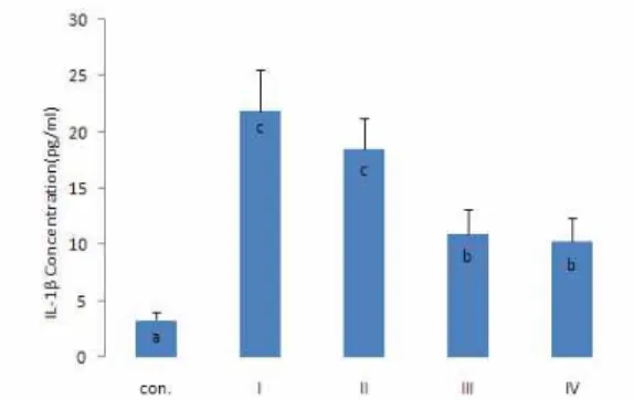
Fig. 1. Effect of Cornus Walteri Ext. on IL-1β concentration in lipopolysaccharide induced Raw 264.7 macrophages.
a,b,c: Values are expressed as compared with vehicle.*a-cBars with different letters are significantly different at p<0.05 by Duncan's multiple range test. Con.:Control. Ⅰ: LPS (1 μ g/ml) Ⅱ: LPS (1 μg/ml)+10 μg/ml,Cornus Walteri Ext. Ⅲ: LPS (1 μg/ml)+30 μg/ml, Cornus Walteri Ext. Ⅳ: LPS (1 μg/ml)+100 μg/ml, Cornus Walteri Ext.3. Raw 264.7 macrophages cytokines
Raw 264.7 macrophages를 이용한 in vitro실험에서 말채나 무 추출물이 LPS shock시의 전염증성 cytokines의 생산에 미치 는 영향을 Fig. 1, 2, 3 및 4에 나타내었다.
IL-1β, IL-6 및 TNF-α의 농도 (Fig. 1-3)는 LPS처리군 모두가 LPS를 처리하지 않은 대조군보다 높은 수치를 나타내었다. LPS 처리 군 간에서는 말채나무 추출물 처리군이 LPS 단일처리군보 다 낮은 값을 나타내었으며, 말채나무 추출물 첨가량이 증가함에 따라 cytokine의 농도가 감소하는 경향을 보였다.
IL-10의 농도는 LPS 처리군 모두가 LPS를 처리하지 않은 대
조군보다 높은 경향을 보였으나, LPS 단일처리군과 10 μg/ml 말 채나무 추출물 처리군은 대조군과 유의한 차이를 나타내지는 않 았다. 그러나 30 μg/ml 및 100 μg/ml 말채나무 추출물처리군 들 은 대조군 및 LPS 단일처리군보다 유의하게 높은 수치를 나타내 었다. 이와 같은 결과는 in vivo 실험의 결과들과 잘 부합되었다.
Fig. 2. Effect of Cornus Walteri Ext. on IL-6 concentration in lipopolysaccharide induced Raw 264.7 macrophages.
a,b,c,d: Values are expressed as compared with vehicle.*a-dBars with different letters are significantly different at p<0.05 by Duncan's multiple range test. Con.:Control. Ⅰ: LPS (1 μ g/ml) Ⅱ: LPS (1 μg/ml)+10 μg/ml,Cornus Walteri Ext.Ⅲ: LPS (1 μg/ml)+30 μg/ml, Cornus Walteri Ext. Ⅳ: LPS (1 μg/ml)+100 μg/ml, Cornus Walteri Ext.Fig. 3. Effect of Cornus Walteri Ext. on TNF-α concentration in lipopolysaccharide induced Raw 264.7 macrophages.
a,b: Values are expressed as compared with vehicle.*a-bBars with different letters are significantly different at p<0.05 by Duncan's multiple range test. Con.:Control. Ⅰ: LPS (1 μ g/ml) Ⅱ: LPS (1 μg/ml)+10 μg/ml,Cornus Walteri Ext. Ⅲ: LPS (1 μg/ml)+30 μg/ml, Cornus Walteri Ext. Ⅳ: LPS (1 μg/ml)+100 μg/ml, Cornus Walteri Ext.Fig. 4. Effect of Cornus Walteri Ext. on IL-10 concentration in
lipopolysaccharide induced Raw 264.7 macrophages.
a,b: Values are expressed as compared with vehicle.*a-bBars with different letters are significantly different at p<0.05 by Duncan's multiple range test. Con.:Control. Ⅰ: LPS (1 μ g/ml) Ⅱ: LPS (1 μg/ml)+10 μg/ml,Cornus Walteri Ext. Ⅲ: LPS (1 μg/ml)+30 μg/ml, Cornus Walteri Ext. Ⅳ: LPS (1 μg/ml)+100 μg/ml, Cornus Walteri Ext.결 론
말채나무의 항염증효과를 알아보기 위하여 말채나무 추출물 을 급여한 흰쥐에게 LPS 처리에 의해 급성기 염증반응을 유발시 킨 후, 혈액 및 간장의 전염증성 cytokines 농도를 조사하였다.
또한 말채나무 추출물을 처리한 Raw 264.7 세포에 LPS shock를 가한 후 말채나무 추출물이 각종 전염증성 cytokines 생산량에 미치는 영향을 처리군 간에 비교 검토하였다.
그 결과, 말채나무 추출물을 급여하고, LPS 처리에 의해 급 성기 염증반응을 유발시킨 rat를 이용한 in vivo 실험과 in vitro 실험에서 말채나무추출물은 전염증성 cytokines들로 분류되는 IL-1β, IL-6 및 TNF-α의 생산을 하락시키는 경향을 보였으며, 반 면에 IL-10의 생산을 촉진하는 경향을 보였다.
이상의 결과들을 종합해 보면 말채나무에는 항염증 효과에 긍정적으로 관여하는 기능성물질이 내재하고 있음을 시사해 준다.
참고문헌