Ⅰ. INTRODUCTION
Tooth avulsion constitutes 0.5-16% of all traumatic injuries in permanent anterior teeth1). Successful re-
plantation of an avulsed tooth may depend on the presence of viable cells in periodontal ligament, which can proliferate on denuded root surface2,3). Therefore, in case of the periodontal injury by tooth avulsion, maintenance of viable periodontal ligament fibroblasts (PDLFs) would be one of the most impor- tant factors for the good prognosis.
During extra alveolar period, storage medium could be used not only to preserve the PDLFs, but also to
The Effect of EGF, T3 and HB-EGF on Human Periodontal Fibroblasts
Eun-Kyoung Hong�, Jeong-Heon Cha�, Yun Tae Kim*, Byung-Jai Choi�, Seong-Oh Kim�
Department of Pediatric Dentistry�, Department of Oral Biology�,
College of Dentistry and Oral Science Research Center, Yonsei University, Daewoong Life Science Institute*
Viable cells of periodontal ligament would be an important factor for the successful replantation of an avulsed tooth. Therefore, it is critical to choose the storage medium for the preservation of traumatically avulsed teeth. Growth factors and hormones could be considered for the therapeutic application of the main- tenance of viable periodontal ligament fibroblasts (PDLFs). Epidermal growth factor (EGF) has been sug- gested as an important player for the regeneration and wound healing process on other tissues. Therefore, EGF was evaluated for the therapeutic application on avulsed teeth. In addition, the synergic effect of EGF with tri-iodothyronine (T3) and heparin-binding epidermal growth factor-like growth factor (HB-EGF).
The cell proliferation of PDLFs was determined by MTT assay and increased dose-dependently up to 10 ng/ml in the presence of EGF. Maximum cellular growth was shown at the concentration of 10 ng/ml EGF. Also, EGF promoted the wound healing of PDLFs examined by in vitro wound healing assay.
Combined effects of EGF with T3 or HB-EGF on the proliferation of PDLFs were also studied.
Interestingly, EGF showed the synergic effect on the proliferation of PDLFs with T3 and HB-EGF. To find out the mechanism of the synergic effect of EGF and T3, the effect of T3 on the expression of en- dogenous EGF receptor was determined by RT-PCR. The result was that T3 enhanced the expression of EGF receptor in PDLFs. It suggested that EGF might be a good choice for a therapeutic application, which can be used as combination with T3 and HB-EGF.
Key words: Epidermal growth factor, Tri-iodothyronine, Heparin-binding epidermal growth factor-like growth factor, Periodontal ligament fibroblasts, Tooth avulsion
Abstract
교신저자 : 김 성 오 서울시 서대문구 신촌동 134 연세대학교 치과대학병원 716호
Tel: 02-2228-3171 Fax: 02-392-7420 E-mail: ksodds@yuhs.ac
stimulate their growth. Polypeptide growth factors are a class of natural biological mediators that regu- late the proliferation, differentiation, migration, and matrix synthesis of cells4). The growth factors studied for proliferation and wound healing nowadays are epidermal growth factor (EGF), platelet-derived growth factor (PDGF), transforming growth factor (TGF) and insulin-like growth factors (IGF I & II).
Recently, it has been reported that polypeptide growth factors enhanced the formation of bone, dentin, and collagen fibers in periodontal wounds5). Lynch et al. demonstrated that short-term applica- tion of a combination of PDGF-B and IGF could en- hance the formation of periodontal attachment appa- ratus by 5-10 folds during the early phase of wound healing after surgery6). Therefore, growth factors could be used as a locally acting therapeutic agent to preserve and regenerate PDLFs after avulsive dental injury.
EGF is the first tissue-derived peptide enhancing proliferation and differentiation of cells. EGF was ex- tracted from submaxillary glands, and induced preco- cious tooth eruption and premature eyelid opening when injected into newborn animals7). In human, most of circulating EGF is associated with blood platelets, synthesized by megakaryocytes, and re- leased in the process of blood coagulation. Locally EGF is also secreted from parotid and submandibular glands, and released directly into saliva8).
Hormones systemically regulate growth and metab- olism of body whereas growth factors locally take ef- fect on tissue. In the developing animal and human, thyroid hormones play a critical role in development and differentiation of tissues and organs9,10). Also, ex- ogenous tri-iodothyronine (T3) injection raised the turnover rate of cells in the periodontal ligament is well known that T3 interact with other hormones such as growth hormones and steroid hormones11).
Heparin-binding epidermal growth factor-like growth factor (HB-EGF) was first identified as a 20- 22 KD glycoprotein in conditioned medium of macrophage-like cells12). Similar to other EGF-family growth factors, HB-EGF binds to the epidermal growth factor receptor (EGFR), thereby inducing its phosphorylation and stimulates DNA synthesis in target cells13). HB-EGF plays an important role in myogenesis, mucosal repair of stomach, protecting
the small bowl from ischemic injury, pancreatic de- velopment, vascular remodeling, renal cell repair and proliferation, liver regeneration and wound healing14). The purpose of this study was to evaluate the ef- fect of EGF alone and combination with T3 or HB- EGF on the proliferation of PDLFs and the effect of EGF on the wound healing and therefore to evaluate EGF for a candidate of therapeutical application of for the preservation and regeneration of PDLFs in a traumatically avulsed tooth.
Ⅱ. MATERIALS AND METHODS 1. Experimental materials
1) EGF
Easyf�(Daewoong Pharm Co. Ltd. Korea) was dis- solved with 10% phosphate buffered saline (PBS) to a concentration of 10-6g/ml for the stock solution. It was then diluted in PBS to several appropriate con- centrations just before the experiment.
2) T3
T3 (Sigma-Aldrich Co) was dissolved in 1 N NaOH to a concentration of 1 mg/ml. It was diluted with 10% phosphate buffered saline (PBS) makin g 10-5M stock solution and stored at -20℃. It was further di- luted to several appropriate concentrations just be- fore experiment.
3) HB-EGF
HB-EGF(R & D Co.) was dissolved with 10% PBS to a concentration of 5 ㎍/ml for the stock solution and stored at -20℃. It was then diluted to several appropriate concentrations for the experiment.
4) Periodontal ligament fibroblasts (PDLFs)
Human PDLFs were obtained and cultured from the explant tissue of human healthy periodontal liga- ment taken from several first premolars that were extracted for orthodontic reasons.
2. Experimental methods
1) Cell culture
After removing calculus and plaque in mouth, the premolar was extracted and rinsed 3 times with
Hanks’balanced salt solution (HBSS) to remove the blood clot of root surface. The periodontal ligament tissues from the middle third of the roots were minced, put in culture dishes and incubated in α- MEM with 10% fetal bovine serum (FBS) and an- tibiotics of 100 mg/ml streptomycin, 0.5 mg/ml am- photericin-B, and 100 unit/ml penicillin at 37 ℃ in a humidified atmosphere of 5% CO2-95% air. The me- dia was changed every 3 days until dense single lay- er was gained. After cells reached confluence, they were trypsinized with 0.25% trypsin-EDTA in PBS for subculture. All the experiments were done within 5-10 cellular passages.
2) Bioassay of EGF
3-(4,5-dimethyl-thiazole-2-yl)-2,5-diphenyl tetra- zolium bromide (MTT) assay, was used to evaluate the viable cell proliferation. MTT assay is based on the principle of tetrazolium salt being reduced by mi- tochondrial reducing enzyme (succinate dehydroge- nase) so that the toxicity of viable cells and cellular differentiations could be evaluated. The reduced tetrazolium salt is converted into colored water-in- soluble formazan salt, and it can be evaluated spec- trophotometrically once the MTT-formazan is dis- solved in an organic solvent. About 2000 PDLFs were seeded and grown on the 96 well plate in 200 ㎕ α-MEM/2% FBS for 1day. EGF was diluted to sever- al appropriate concentrations just before experiment.
Then the culture media was replaced with 200 ㎕ α- MEM containing EGF at 0.1, 0.5, 1, 10 and 100 ng/ml. Cell proliferation was measured by MTT as- say after 3 days and 5 days of culture period. 50 ㎕ of MTT solution (5mg/ml) was added per well then incubated at 37 ℃ in incubator for 4 hrs. The purple formazan product was dissolved in 150 ㎕ of di- methylsulfoxide (DMSO) and incubated for 30 min- utes at 37 ℃ in CO2 incubator. Optical density was
measured on an ELISA reader at 570 nm.
3) In Vitro Wound Healing Assay
Human PDL cells were grown to confluence on 12- well tissue culture plates. The cells were then starved in serum-free α-MEM overnight. The mono- layer was scratch-wounded with yellow tip. After be- ing washed once with PBS, the cells were refeded with serum-free α-MEM in the presence of EGF.
Wound healing was photographed at 24 hr after the wounding scratch using Olympus CKX41(Olympus, Japan) inverted microscope system.
4) Bioassay of mixture (EGF, T3 and HB-EGF) EGF, T3 and HB-EGF was diluted to several ap- propriate concentrations just before experiment. The MTT assay was proceeded as described above 2).
Combination of factors with several concentrations is listed in Table 1.
5) RT-PCR
About 6×10⁵ PDLFs were plated on 100×15mm (100π) dishes in α-MEM/2% FBS and were incubated overnight to allow attachment prior to the addition of T3. After 24hrs of incubation period, α-MEM solu- tions containing (a) 0.1 nM T3, (b) 10 nM T3 were replaced in experimental group and were replaced in control group with same media without T3. After 1 day of incubation, cells of 2 groups were harvested by Trizol. And then DNA was removed with chloro- form and salted out with isopropanol and 70%
ethanol. After centrifuging, mRNA pellet was dried then dissolved in diethyl pyrocarbonate (DEPC) wa- ter. They were stored at -70℃. After mRNA was quantified by spectrophotometry, cDNA was synthe- sized by reverse trascriptase with oligo dT primer.
cDNA was used as a template for PCR to amplify the specific product. The sequences of each set of primers
Table 1.The groups of combination of EGF with T3 or HB-EGF
Group Combinations
Group 1 0.01, 0.1 and 1 ng/ml EGF + 0.1 nM T3 Group 2 0.01, 0.1 and 1 ng/ml EGF + 10 nM T3 Group 3 0.01, 0.1 and 1 ng/ml EGF + 0.1 nM HB-EGF Group 4 0.01, 0.1 and 1 ng/ml EGF + 0.01 nM HB-EGF
are listed below:
EGFR P1, 5’-ATGCGACCCTCCGGGACGGCCG-3’
EGFR P2, 5’-CCTTCAGTCCGGTTTTATTTGC-3’
PCR was proceeded with AccuPower� RT PreMIX tube. RT-PCR products were separated by 1%
agarose gel electrophoresis in 0.5% tris-acetate (TAE) buffer and stained with ethidium bromide. All data were quantitated relative to β-actin genes. Data analysis was performed with Spot Denso Analysis of Frog 2000 software (Alpha Innotech, San Leandro, CA, USA) .
6) Statistical analysis
Statistical analysis was done with statistical soft- ware (SAS version 8.1). Absorbance of each concen- tration was tested by the analysis of Tukey’s multi- ple comparison tests at 95% confidence level.
Ⅲ. RESULT
1. Effect of EGF on PDLFs proliferation
The purpose of this study was to evaluate the ef- fect of EGF on the proliferation of PDLFs. In the treatment of EGF, there was different cellular prolif- eration between control and 10 ng/ml ; control and 100 ng/ml on the 3rd day with statistical significance (p<0.05). In addition, the proliferation of PDLFs in 10 ng/ml EGF on the 3rd day was increased up to 32.3% when compared with control, and the prolifer- ation of PDLFs in 10 ng/ml EGF on the 5th day was increased up to 41.3% when compared with control
(Fig. 1). The MTT assay suggested that maximum cellular proliferation was reached at the concentra- tions of 10 ng/ml EGF on the 3rdand the 5thday.
2. Effect of EGF on PDLFs wound healing
The wound healing effect of EGF was determined byin vitro wound healing assay to measure the abili- ty of PDLFs migration to close the wounded gap. A scratch wound in PDLFs was more closed in the presence of 10 ng/ml EGF, suggesting that EGF pro- motes the wound healing of PDLFs (Fig. 2).
3. Effects of combination of EGF with T3 or HB- EGF on PDLFs proliferation
In this study, the effects of combination of EGF with T3 or HB-EGF on PDLFs proliferation were
Fig. 2. Healing of scratch wound in cultured PDL cells. PDL cells seeded on 12-well plates. After reaching confluence, the cells were starved with serum free α-MEM overnight and injured with yellow tip. Cells were incubated in serum free α-MEM for 24h, in serum free α-MEM containing EGF. A; no treatment (control cells), B; 10 ng/ml EGF.
Fig. 1. The proliferation of PDLFs with several EGF concentrations.
A B
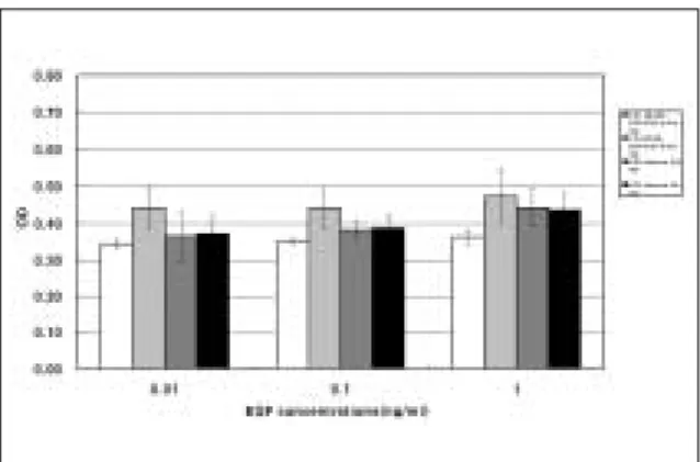
evaluated by MTT assay. The combination of EGF at three different concentrations (0.01, 0.1 and 1 ng/ml) with 0.1 nM T3 on the 5th day, showed higher value of MTT assay than that treated with EGF alone (Fig. 3). However, there was no statistical sig- nificance. The proliferation of PDLFs was increased more in the group of combination compared with the single treatment.
The combination of 10 nM T3, and EGF at every concentration on the 3rd and the 5th day, showed higher value of MTT assay than that treated with EGF alone (Fig. 4). The proliferation of PDLFs was increased dose-dependently in the group of combined treatment, except for the 1 ng/ml EGF on the 5th day.
The combination of 0.01 nM HB-EGF and EGF at every concentration on the 5th day, showed higher
value of MTT assay than that treated with EGF alone (Fig. 5). The proliferation of PDLFs was in- creased dose-dependently in the combined treatment, except for 0.1 nM HB-EGF on the 5thday.
The proliferation of PDLFs was increased in the group of combination compared with the single treat- ment. The combination of 0.1 nM HB-EGF and EGF at every concentration on the 5th day, showed higher value of MTT assay than that treated with EGF alone (Fig. 6). The proliferation of PDLFs was in- creased dose-dependently in the combined treatment on the 5thday.
3. RT-PCR
Using RT-PCR, it was tested if the addition of T3 had an effect on mRNA expression of endogenous
Fig. 3. The proliferation of PDLFs with the combination of 0.1 nM T3 and several different concentrations of EGF.
Fig. 4. The proliferation of PDLFs with the combination of 10 nM T3 and several different concentrations of EGF.
Fig. 5. The proliferation of PDLFs with the combina- tion of 0.01 nM HB-EGF and several different concentrations of EGF.
Fig. 6.The proliferation of PDLFs with the combination of 0.1 nM HB-EGF and several different concentrations of EGF.
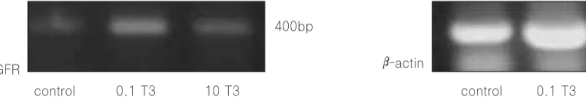
EGFR in PDLFs (Fig. 7).
All results were analyzed as relative units and standardized to the amount of β-actin mRNA. In the treatment of 0.1 nM T3, EGFR expression was in- creased up to 5.6 folds and in the case of 10 nM T3, EGFR expression was increased up to 2.5 folds when compared with control (Table 2). Therefore, T3 en- hanced the expression amount of EGFR.
Ⅳ. DISCUSSION
Periodontal ligament is a dense connective tissue between root cementum and alveolar bone that an- chors the tooth and maintains the structural integri- ty of mineralized tissues15). Fibroblasts in the peri- odontal ligament have multipotential heterogenous ability that can differentiate into either cemento- blasts or osteoblasts16). An avulsion may cause the damage of attachment apparatus in periodontal liga- ment. Thus it is important to maintain the viable cell of periodontal ligament attached to the avulsed tooth for the successful replantation. Ideally, the tooth should be taken good care immediately after the injury in an effort to preserve the viability of PDLFs, and thereby to optimize healing process and minimize root resorption. Therefore, the storage con- ditions should be designed to maximize preservation of PDLFs during the transportation to dental office when immediate replantation of an avulsed tooth is impossible.
The optimal storage medium should be able to pre-
serve the viability, mitogenicity and clogenic capacity of injured PDLFs and their progenitors. Numerous studies have shown that storage conditions and types of storage media affect the viability of PDLFs.
Recently, three media have been introduced to pre- serve avulsed teeth : Hanks’Balanced Salt Solution (HBSS), Viaspan, and fibroblast culture media (α- MEM)17). Because of the limitation of available con- ventional storage media to obtain successful regener- ation of injured PDL tissues, the application of growth factors have been attempted in clinical use to accelerate the proliferation of regeneration of PDLFs, which may locally help tissue repair.
Thyroid hormone modulates the transduction of signals from several cytokines and growth factors in human cell lines. Also, thyroid hormone enhances the effects of EGF on mitogen activated protein ki- nase (MAPK) activation18,19). And the effect of EGF on immediate-early gene expression was enhanced by thyroid hormone. Physiologic levels of thyroid hor- mone may enhance the autocrine/paracrine effects of EGF on cells19).
In this study, the effects of EGF alone and the synergic effect of EGF with T3 or HB-EGF on the proliferation of PDLFs and the effect of EGF on the wound healing were examined to evaluate the poten- tial application of EGF as a storage media or a pre- replantation conditioner of avulsed tooth. Cell prolif- eration of PDLFs was increased dose-dependently in the presence of EGF up to a concentration of 10 ng/ml. Therefore, the optimal concentration of EGF Fig. 7. The expression amount of EGFR using RT-PCR.
control 0.1 T3 10 T3 control 0.1 T3 10 T3 EGFR
400bp
β-actin
Table 2.The ratio of EGFR between control and experimental group
EGFR / b-actin (ratio of area) Experimental group / control group
Control 0.157 1
0.1 nM T3 0.877 5.6
10 nM T3 0.394 2.5
in the cellular growth was reached at 10 ng/ml. It suggested that EGF could be a Good candidate for a therapeutic application.
In the group 1 and 2, the combination of EGF with T 3 or HB-EGF on the 5th day, showed increased val- ue of MTT assay compared with the group of EGF alone in spite of the low concentration (Fig. 3-6).
The positive synergic effect of EGF with T3 or HB- EGF may make EGF better candidate for the thera- peutic application. Since EGF and HB-EGF bind to EGF receptor, the synergic effect for these growth factors may be due to more growth factors available for EGF receptor of PDLFs. In case of synergic effect of EGF and T3, it has been reported that T3 modu- lates EGF-induced renal proximal tubule cell prolif- eration by an effect on the EGF receptorng/ml20). The study concluded that T3 enhanced EGF receptor gene expression increasing the number of cell surface EGF receptors on renal proximal tubule cells.
Therefore, in this study, using RT-PCR, we tested if the addition of T3 have an effect on mRNA expres- sion of endogenous EGFR in PDLFs. As a result, T3 enhanced the expression of EGFR in PDLFs and it may explain the synergic effect of T3 and EGF.
From this study we could conclude that EGF could be used as a pretreatment agent for traumatically avulsed teeth. Further study is needed to find out the relationship between these specific growth factors and matrix synthesis by PDLFs. Also, we need to proceed in vivo experiment using EGF and the com- bination with T3 and HB-EGF.
Ⅴ. CONCLUSION
This study evaluated the effect of EGF alone and combination with T3 or HB-EGF on the proliferation of PDLFs by MTT assay and the effect of EGF on the wound healing by wound healing assay. To under- stand the synergic effect of EGF with T3, RT-PCR on EGF receptor was done considering the influence of T3 on EGF receptor expression.
The results are summarized as;
1. The cell proliferation of PDLFs was increased dose-dependently in the presence of EGF
2. The optimal concentration of EGF in cellular growth was found at the concentrations of 10 ng/ml.
3. EGF promoted the wound healing of PDLFs.
4. The combination of EGF with T3 enhanced the proliferation of PDLFs.
5. The combination of EGF with HB-EGF also en- hanced the proliferation of PDLFs.
6. The expression of EGFR was increased with the treatment of T3.
REFERENCES
1. Andreasen JO : Etiology and pathogenesis of traumatic dental injuries. A clinical study of 1298 cases. Scan J Dent Res, 78:329-342, 1970.
2. Andreasen JO, Hjo/rting-Hansen E : Replantation of teeth. I. Radiographic and clinical study of 110 human teeth replanted after accidental loss. Acta Odontol Scand , 24:263-286, 1966.
3. Andreasen JO, Reinholtd J, Riis I, et al. : Periodontal and pulpal healing of monkey in- cisors preserved in tissue culture before replanta- tion. Int J Oral Surg, 7:104-112, 1978.
4. Terranova VP, Wikesjo UME : Extracellular ma- trices and polypeptide growth factors as media- tors of functions of cells of the periodontium. J Periodontal , 58:371-380, 1987.
5. Giannobile WV : Periodontal tissue engineering by growth factors. Bone, 19:23S-37S, 1996.
6. Lynch SE, de Castilla GR, Williams RC, et al. : The effect of short term application of a combina- tion of platelet-derived and insulin-liked growth factors on periodontal wound healing. J Periodontol, 62:458-467, 1991.
7. Cohen S : Isolation of a mouse submaxillary gland protein accelerating incisor eruption and eyelid opening in the newborn animal. J Biol Chem, 237:1555, 1962.
8. Malttila AL, Perheentupa J, Salmi J : Human epidermal growth factor concentrations in urine but not in saliva and serum, depend on thyroid state. Life Sci, 41:2739-2747, 1987.
9. Jannini EA, Ulisse S, D’Armiento M : Thyroid hormone and male gonadal function. Endor Rev, 16: 443-459, 1995.
10. Jannini EA, Crescenzi A, Rucci N, et al. : Ontogenic pattern of thyroid hormone receptor expression in the human testis. J Clin Endocrinol Metab, 85:3453-3457, 2000.
11. KIM SO : The effect of thyroid hormone on the cell proliferation of periodontal ligament in rat.
Yonsei University, Seoul, 2003.
12. Higashimaya S, Abraham JA, Miller JC, et al. : A heparin-binding growth factor secreted by macrophage like cells that is related to EGF.
Science, 251:936-939, 1991.
13. Rabb G, Klagsbrun M : Heparin-binding EGF- like growth factor. Biochem Biophys Acta, 1333:179-199, 1997.
14. Karen M, Gail E : Structure and function of Heparin-binding EGF-like growth factor.
Bioscience d288-d299, 1998.
15. Angelo M, David L : Characterization of fibrob- lasts derived from human periodontal ligament and gingiva. J Periodontol, 61:103-111, 1990.
16. Aukhil MA, Simpson DM : In vitro differentiation of progenitor cells of periodontal ligament: an ex- perimental study using physical barriers. J Clin
Periodontol, 13:862-868, 1986.
17. Ashkenaz M, Marouni M, Sarnat H : In vitro vi- ability, mitogenicity and clonogenic capacity of periodontal ligament cells after storage in four media at room temperature. Endod Dent Traumatol, 16:63-70, 2000.
18. Lekic P, Kenny D, Moe HK, et al. : Relationship of clonogenic capacity to plating efficiency and vi- tal dye staining of human periodontal ligament cells: implications for tooth replantation. J Periodont Res, 31(4):294-300, 1996.
19. Lin H-Y, Davis FB, Gordinier : Thyroid hormone induces activation of mitogen-activated protein kinase in cultured cells. Am J Physiol, 276:
C1014-C1024, 1999.
20. Humes HD, Cieslinski DA, Johnson LB, et al. : Tri-iodothyronine enhances renal tubule cell replication by stimulating EGF receptor gene ex- pression. Am J Physiol, 262:F540-5, 1992.
국문초록
EGF, T3, HB-EGF 가 치주인대섬유모세포에 미치는 영향
홍은경�∙차정헌�∙김연태*∙최병재�∙김성오�
연세대학교 치과대학 소아치과학교실�, 구강생물학교실�, 연세대학교 구강과학연구소, 대웅생명과학연구소*
치주인대섬유모세포들은 완전탈구 된 치아의 성공적인 재식을 위한 중요한 요소이다. 따라서, 외상으로 인해 완전탈구 된 치아의 보존을 위한 보관액을 선택하는 것이 중요하다. 성장인자들과 호르몬들은 치주인대섬유모세포들의 생존을 위 한 치료적 제제로 고려되고 있다. Epidermal growth factor(EGF)는 다른 조직에서 재생과 상처 치유 과정의 중요한 역할인자로 대두되고 있다. 따라서, 완전탈구 된 치아를 위한 치료적 적용을 위해 EGF의 세포 증식에 미치는 영향을 평 가하였다. 또한 EGF와 tri-iodothyronine(T3)의 혼합액, EGF와 Heparin-binding epidermal growth factor-like growth factor(HB-EGF)의 혼합액이 세포 증식에 미치는 상승 효과를 평가하였다.
치주인대섬유모세포의 세포증식은 EGF 농도가 증가함에 따라 증가하였고, 10 ng/ml 농도에서 최대 세포증식을 보 였다. EGF는 상처치유분석에서 상처 치유촉진과 이동성을 보여주었다. EGF에 T3와 HB-EGF를 첨가한 혼합액에서 배양한 세포는 EGF만 처리한 경우보다 세포 증식이 상승되었다. EGF와 T3 혼합액의 상승효과 기전을 유추하기 위해 서 RT-PCR로 EGF 수용기의 발현을 확인하였고, T3가 EGF 수용기 발현을 증가시켰음을 확인하였다. 따라서 EGF 와, EGF와 T3, EGF와 HB-EGF의 혼합액은 완전탈구된 치아의 치료에 있어 유용한 선택이 될 것이다.
주요어 : Epidermal growth factor, Tri-iodothyronine, Heparin-binding epidermal growth factor-like growth factor, 주인대섬유모세포, 완전탈구