서론
임플란트 보철이 치과 영역에서 대중적인 치료가 되었으며 그 성공률 또한 높아지면서 단순히 기능과 심미 회복뿐 아니 라 보철물의 장기적인 유지 및 관리 부분을 간과할 수 없게 되 었다. 이를 위해서 최종 보철물의 유지형태 결정시 탈∙부착 이 가능하도록 설계하는 것이 효과적이다. 임플란트의 유지형 태를 살펴보면 크게 나사 유지형 보철물(screw-retained prosthesis) 과 시멘트 유지형 보철물(cement-retained prosthesis)로 나눌 수 있 다.1-5나사 유지형 보철물은 주조가 가능한 지대주상에 납형 제 작 후 주조과정을 통하여 임플란트와 직접 연결되거나 중간 지대주 나사에 의하여 연결되는 형태이다. 이는 제작 과정이 복잡하고 주조용 지대주는 일반적으로 귀금속을 사용하여 제 작 비용이 상승하게 되며 교합면 나사 구멍 형성으로 인하여 교합과 심미적인 측면에서 희생을 요구한다.
Hebel과 Gajjar4는 교합면 나사 구멍 크기는 소구치 부위에서 50%, 구치부위에서 약 30%이상을 차지하며 이는 비심미적이 고 적절한 교합면 형태부여를 방해하며 하중이 가해질 때 중 앙부 나사 구멍으로 인하여 제한된 접촉 점을 갖게 되어 시멘 트 유지형 보철물에 비해 편심하중(off set loading)을 받게 된다고 기술 하였다. 이러한 편심하중은 보철물의 파절 또는 나사 풀 림 등의 합병증을 초래 할 수 있다.6,7이런 희생에도 불구하고
나사 유지형 보철물은 탈∙부착이 용이하다는 장점으로 인해 많은 임상가들에 의해 사용되어왔다. 따라서 유지관리가 용이 하고 시멘트를 사용하지 않아 잔류 시멘트로 인한 합병증을 피할 수 있으며 악간 공간이 부족한 경우 나사에 의해 유지가 가능하여 시멘트 유지형 보철물 보다 유리한 장점이 있다.1-4이 에 반해 시멘트 유지 보철물은 나사 유지형 보철물과 비교하 여 기공과정이 간단하고 교합과 심미, 보철물의 수동적인 적 합과 하중 부하적 측면에 더 우월하다. 시멘트 유지형 보철물 도 임시접착제 또는 임시접착제를 바셀린과 혼합하여 적절하 게 사용하면 보철물을 유지하고 탈∙부착 또한 가능하다.8그 러나 유지력에 대한 조절이 어려워 빈번하게 보철물이 탈락하 거나 또는 너무 강한 유지력으로 인해 보철물을 필요할 때 제 거할 수 없는 경우가 발생할 수 있고 조직이 두꺼운 경우 또는 임플란트가 깊게 식립된 경우 잉여 시멘트로 인하여 임플란트 주변에 감염을 초래할 수 있다.9,10
이러한 문제를 극복하기 위해 시멘트 유지형과 나사 유지형 보철물의 장점을 결합하여서 나사-시멘트 복합 유지형 보철물 이 여러 가지 형태로 개발 및 소개되었고 국내에서는 나사-시 멘트 유지형 보철물(screw-cement retained prosthesis: SCRP)로 소개
되었다.11-13이는 지대주와 상부구조를 영구 접착한 상태에서
일체형으로 제거가 가능하고 구강 외에서 잔류 시멘트를 제거 할 수 있을 뿐만 아니라 지대주와 상부 보철물 간의 오차를 접
완전 무치악 환자에서 나사-시멘트 보철물(SCP: screw-cement prosthesis)을 이용한 임플란트 보철 수복 증례
김주현∙윤보혁∙장정은∙허중보∙정창모*
부산대학교 치의학전문대학원 치과보철학교실
임플란트 보철물은 유지방법에 따라 크게 나사 유지형 보철물(screw-retained prosthesis)과 시멘트 유지형 보철물(cement-retained prosthesis)로 나눌 수 있으며, 이들 장점을 고려 하여 만든 나사-시멘트 유지형 보철물(SCRP: screw and cement retained implant prosthesis)이 있다. 나사-시멘트 유지형 보철물은 임플란트 지대주와 상부 보철물 사이의 시멘트 층이 보철물 제작 과정에서 발생하는 여러 오차를 보상하여 수동적 적합을 얻을 수 있으며, 임상 및 기공 과정이 간단한 장점과 더불어 임플란트 보철의 유지 관리를 위해 필수적인 탈∙부착(retrievability)이 가능하다. 본 증례에서는 완전 무치악 환자를 대상으로 상, 하악에 각각 9, 8개의 임플란트(US II, OSSTEM, Seoul, Korea)를 식립하고 나사 구 멍이 순측으로 향하는 부위는 시멘트 유지형, 다른 부위는 나사-시멘트 유지형태를 사용하여 복합형태의 나사-시멘트 보철물(SCP: screw-cement prosthesis)을 제작하였으며 3년 간의 정기 검사에서 기능과 심미적으로 만족할 만한 결과를 얻어 이를 보고하고자 한다. (대한치과보철학회지 2012;50:318-23)
주요단어: 완전무치악; 임플란트; 고정성보철물; 나사-시멘트 보철물; 탈∙부착
*교신저자: 정정창창모모
626-770 경남 양산시 물금읍 범어리 양산부산대학교병원 치과병원 055-360-5130: e-mail, cmjeong@pusan.ac.kr 원고접수일: 2012년 9월 17일 / 원고최종수정일: 2012년 10월 8일 / 원고채택일: 2012년 10월 13일
2012 대한치과보철학회
이 글은 크리에이티브 커먼즈 코리아 저작자표시-비영 리 3.0 대한민국 라이선스에 따라 이용하실 수 있습니다.
c cc
착 후 구강 밖에서 수정할 수 있는 기회를 준다. 반면 나사 구멍 위치가 교합면 내에 위치해야 적용이 가능하여 상악 전치부위 나 협측으로 기울여서 식립된 임플란트 부위는 나사 구멍 위 치가 교합면에 위치되기 힘들며 이러한 경우 시멘트 유지형태 보철물을 제작 할 수 밖에 없다. 따라서 다수의 임플란트 보철 물 제작 시 술자는 교합면 내로 나사 구멍이 위치하는 곳은 나 사-시멘트 유지형 보철물을 적용하고 그렇지 않은 곳은 시멘 트 유지형을 사용하는 복합형태의 보철물 제작을 고려해볼 수 있다.
본 증례에서는 완전 무치악 환자를 대상으로 상, 하악에 각 9, 8개의 임플란트(US II, OSSTEM, Seoul, Korea)를 식립하고 나사 구 멍이 순측으로 향하는 부위는 시멘트 유지형, 다른 부위는 나 사-시멘트 유지형태를 사용 복합형태의 나사-시멘트 보철물 (SCP: screw-cement prosthesis)을 제작하여 기능과 심미적으로 만족할 만한 결과를 얻어 이를 보고하고자 한다.
증례보고 1. 진단
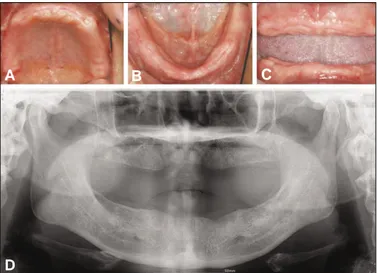
본 증례는 완전 무치악 52세 여환으로 보철치료를 위해 본원 에 내원하였고 전신 병력으로는 고혈압이 있었다. 치과 병력 으로는 10년 전부터 상, 하악 국소의치를 착용하였으며 지대치 동요와 염증으로 잔존치아 발거 하였다. 발치 후 임시의치를 제작하고, 점막조정제(Coe Comfort GC America Inc., Chicago, USA)를 이용하여 의치 내면을 이장하였다. 2 - 3주 간격으로 조 정하고 3개월째 인상채득 및 진단용 납형 제작하고 평가하여 상악에 9개, 하악에 8개의 임플란트를 식립하여 고정성 임플란 트 보철 치료 계획을 수립하였다(Fig. 1).
2. 수술
상악과 하악을 2회로 나누어 수술하였으며 오스템 임플란트 시스템(US II, OSSTEM, Seoul, Korea, ∅3.3 mm × 5개, ∅4 mm × 12개)이 사용되었다. 수술과정에서 모두 35Ncm 이상의 초기 고정력을 얻었고 지속적으로 점막 조정제를 사용하여 임시 의 치를 사용하였다. 식립 5개월째 2차 수술을 시행하였고 임상 및 방사선학적 검사상 모두 성공적인 골 유착을 보였다(Fig. 2).
3. 보철물 제작
2차 수술 후 4주째에 패턴레진(Duralay, Reliance, Dental Mfg. Co., Worth, IL, USA)을 이용하여 인상용 코핑을 연결하고 실리콘 인 상재(ImprintTMII GarantTM, 3M ESPE, Seefeld, Germany)를 이용하여 픽업 인상으로 최종 인상채득 하여 주모형을 제작하였다(Fig.
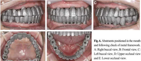
3). 안궁이전 및 악간 관계 기록하여 주모형을 교합기에 부착하 고 전치부에 인공치를 배열하여 중절치의 절단 위치를 결정하 고 배열된 납의치를 구강 내에 시적하고 교합상의 순측 부위 를 제거하여 구순 지지를 평가하였고 납형을 제작하여 구강 내에 시적 후 평가하였다. 하악은 나사-시멘트 유지형 보철형 태(SCRP)로 제작하고 상악은 나사 구멍이 순측을 향하는 우측 중절치와 양측 견치 부위는 시멘트 유지형, 다른 부위는 나사- 시멘트 유지형태의 복합형태 보철물(SCP: screw-cement prosthe- sis)을 제작하기로 계획하였다. 하악 양측 견치부는 ER abut- ment(OSSTEM, Seoul, Korea)를 사용하고 나머지는 부위는 UCLA abutment(OSSTEM, Seoul, Korea)를 사용하여 내관을 제작하였다 (Fig. 4). 내관 위에 납형을 제작하고 cut-back 시행 후 상부 보철물 을 제작하였고 구강 내에 시적하고 적합 및 교합을 평가한 후 도재소성하였다(Figs. 5, 6).
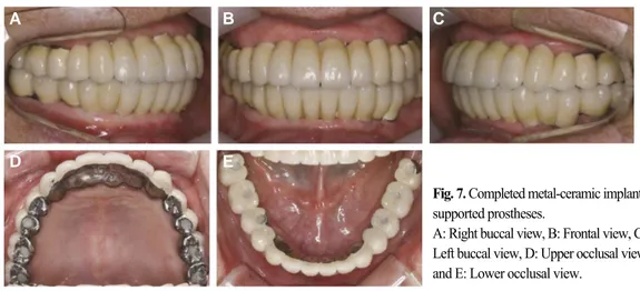
상악 양측 견치와 중절치를 제외하고 내관과 상부 보철물을 자가중합형 레진 시멘트(Resiment�Ready-Mix�, MO, USA)를 이 용하여 구강 내에서 합착한 후, 보철물을 제거하여 잉여 시멘 트를 제거하였다. 시멘트 유지형태 부위는 임시 임플란트 시 멘트(Premier Implant CementTM, Premier Products Company, PA, USA)를 적용하여 장착하였다(Fig. 7).
Fig. 1. Pre-operative intraoral view (A, B and C) and panoramic radiograph (D) after 3-month extraction.
A: Occlusal view of maxilla, B: Occlusal view of mandible, C: Anterior view and
D: Pre-operative panoramic radiograph. Fig. 2. Post-operative panoramic radiograph.
A B C
D
Fig. 3. Impression taking and master model fab- rication.
A, B: Pick-up impression coping of maxilla and mandible connected with Duralay resin, C, D:
Impression of maxilla and mandible, E, F:
Master cast of maxilla and mandible.
Fig. 4. Abutment positioned onto implant replica, Duralay resin modification and abutment milling used to correct implant angulation.
A: Plastic sleeve of UCLA abutments of maxilla modified with Duralay resin and milling, B: Plastic sleeve of UCLA abutments and ER abutments (arrow) of mandible modified with Duralay resin and milling, C: Customized abutments of maxilla fab- ricated with UCLA abutment, D: Customized abutments of mandible fabricated with UCLA and ER abutments (arrow).
Fig. 5. Metal framework fabrication.
A, B: Metal-ceramic waxing of coping on prepared abutment and wax cut- back, C, D: Casted metal framework with access channel of occlusal screw.
Fig. 6. Abutments positioned in the mouth and following check of metal framework.
A: Right buccal view, B: Frontal view, C:
Left buccal view, D: Upper occlusal view and E: Lower occlusal view.
A B C
D E F
A C
B D
A C
B D
A B C
D E
고찰
완전 무치악 환자에서의 임플란트 치료는 크게 고정성 보철 물과 가철성 피개 의치로 나눌 수 있다. 보철물의 형태를 결정 시 구강 위생, 심미성, 악간 공간, 생역학적 측면 등이 고려되어 야 한다. 완전 무치악 환자 중 정상적인 악간 관계를 가지며 구 강 위생이 좋고, 치조제 흡수가 적으며 경제력이 뒷받침되는 환자의 경우 임플란트를 이용한 고정성 금속-도재 임플란트 보철 수복을 고려할 수 있다. 완전 무치악 환자에서 금속-도재 보철물의 상부 구조물이 광범위하게 연결 고정되는 경우 정확 한 보철물의 제작을 위해 치료실과 기공실의 매 과정마다 적 합성에 영향을 미치는 요소를 고려한 술식이 이루어져야 한 다. 임플란트와 지대주간의 적합 기준은 10 ㎛ 또는 그 이하가 요구되어지나 항상 이를 충족시키기에는 한계가 있다.14임플 란트 상부 구조물을 제작할 때의 첫 번째 목적은 보철물과 임 플란트의 금속 계면이 수동적으로 적합하도록 하는 것이다. 그 러나, 모든 과정을 정확히 수행한다 하더라도 현재 사용하는 재료와 보철물 제작 방법에는 많은 부정확성이 잠재되어 있으 며 오차의 원인 요소가 다양하기 때문에 완전한 수동적 적합 을 얻기 어려운 것은 잘 알려진 사실이다.15부정확성으로 인해 야기되는 임플란트 보철물의 오차는 나사 풀림과 임플란트 보 철 구성요소의 파절의 원인이 될 수 있다.6,7,15또한, 지지 골에 유 해한 응력을 유발하여 변연골 흡수와 골 유착의 실패를 유발 할 수도 있다.7,16
보철물의 수동적 적합을 얻기 위한 노력으로 1991년 Voitik13 에 의해 처음 소개된 칼(KAL: Kulzer Abutment Luting) 방식은 스페이서로 작용하는 플라스틱 실린더 이용하여 프레임워크 내면을 더 크게 형성하여 지대주와 상부 보철물 사이 공간을 레진으로 채워 수동적 적합을 얻고 교합면에 나사구멍을 형성 하는 방식이다. 이후 여러 방법들이 소개되었고 국내에서는 나사-시멘트 유지형 보철물(SCRP)로 다양한 지대주를 이용한 방법들이 소개되고 있다.
나사-시멘트 보철물(screw-cement prosthesis)은 나사-시멘트 유지형(SCRP: screw and cement retained implant prosthesis)과 시 멘트 유지형(cement-retained prosthesis)의 복합형태로, 일반적으로 다수 임플란트 중 일부 나사-시멘트 유지형 보철물과 여러 개 의 시멘트 유지형태 보철물을 말하며 시멘트 유지 부위는 보 철물의 수동적 적합을 제공하고 임시 시멘트를 사용하여 탈부 착성을 제공한다.17,18반면 나사유지 부위는 상대적으로 심미적 요구도가 적은 구치부에 적용하여 심미와 교합 안정성을 제공 한다.
본 증례에서 완전 무치악 환자의 식립 각도가 양호하지 못한 일부 임플란트를 부분적으로 시멘트 유지형으로 제작하여 만 족할 만한 기능과 심미적 결과를 얻을 수 있었다(Fig. 8). 이런 보 철물을 적용함으로써 얻는 장점은 나사 유지에 의해 구강 내 Fig. 8. Pre-operative and post-operative facial photographs and panoramic radi- ograph showing improved facial appearance.
A: Pre-operative frontal view, B: Pre-operative lateral view, C: Post-operative frontal view, D: Post-operative lateral view and E: Post-operative panoramic radiograph.
Fig. 7. Completed metal-ceramic implant- supported prostheses.
A: Right buccal view, B: Frontal view, C:
Left buccal view, D: Upper occlusal view and E: Lower occlusal view.
A B C
D E
A B C D
에서 쉽게 탈락하지 않으며 반대로 시멘트 유지 보철물을 임 시 시멘트로 합착하였기에 술자가 원할 때 쉽게 제거가 가능 하다는 것이다. 하지만, 나사-시멘트 보철물은 순수 나사유지 또는 시멘트 유지 보철물과 구강 내 장착 및 하중 부여시 상이 한 거동을 보일 수 있다. 하중에 따른 지대주의 안정성과 시멘 트 사용부의 변연 부적합, 그리고 시멘트 손실에 따른 다양한 합병증이 생길 가능성을 고려해야 한다. 또한, 최종 합착 후 다 시 나사를 조일 때 발생하는 전하중에 의하여 보철물과 임플 란트에 스트레스가 발생 가능하다. 이는 특히 나사 유지부위 에 집중될 수 있다. 따라서 임상적 성공을 위해 기능시 나사 풀 림이나 파절 등 각 부위에 발생 할 수 있는 응력에 대한 관찰이 필요할 것이다. 술자는 임플란트 치료에 앞서 환자의 처한 상 황에 따라 적절한 유지 형태를 선택하고 기능과 심미와 더불 어 환자의 경제성과 술자의 편의성을 고려하고 유지관리 측면 또한 치료 계획에 포함시켜야 할 것이다.
결론
임플란트 보철 제작시 상부 구조물이 광범위하게 연결 고정 되는 경우 순, 협측으로 식립된 부위는 시멘트 유지형태로 제 작하고 다른 부위는 나사-시멘트 유지형태를 복합적으로 사용 하여 나사-시멘트 보철물을 제작할 수 있다. 이러한 방법은 보 철물의 수동적 적합을 얻고 각 임플란트 연결 시스템이 갖고 있는 불리한 점을 극복할 수 있으며 기능과 심미 그리고, 유지 관리 영속성을 통해 다양한 임상 증례에 그 적용 범위를 확대 할 수 있다.
참고문헌
1. Misch CE. Screw-retained versus cement-retained implant- supported prostheses. Pract Periodontics Aesthet Dent 1995;7(9):15- 8.
2. Chee W, Felton DA, Johnson PF, Sullivan DY. Cemented ver- sus screw-retained implant prostheses: which is better? Int J Oral Maxillofac Implants 1999;14:137-41.
3. Misch CE. Contemporary implant dentistry. 2nd ed. St. Louis:
Mosby Inc.; 1999. p. 549-93.
4. Hebel KS, Gajjar RC. Cement-retained versus screw-retained im-
plant restorations: achieving optimal occlusion and esthetics in implant dentistry. J Prosthet Dent 1997;77:28-35.
5. Michalakis KX, Hirayama H, Garefis PD: Cement retained versus screw-retained implant restorations: a critical review.
Int J Oral Maxillofac Implants 2003;18:719-28.
6. Taylor TD. Prosthodontic problems and limitations associated with osseointegration. J Prosthet Dent 1998;79:74-8.
7. Zarb GA, Schmitt A. The longitudinal clinical effectiveness of osseointegrated dental implants: the Toronto study. Part III:
Problems and complications encountered. J Prosthet Dent 1990;64:185-94.
8. Breeding LC, Dixon DL, Bogacki MT, Tietge JD. Use of luting agents with an implant system: Part I. J Prosthet Dent 1992;68:737- 41.
9. Goodacre CJ, Kan JY, Rungcharassaeng K. Clinical complica- tions of osseointegrated implants. J Prosthet Dent 1999;81:537- 52.
10. Pauletto N, Lahiffe BJ, Walton JN. Complications associated with excess cement around crowns on osseointegrated implants: a clin- ical report. Int J Oral Maxillofac Implants 1999;14:865-8.
11. Aparicio C. A new method for achieving passive fit of an interim restoration supported by Bra�nemark implants: a technical note.
Int J Oral Maxillofac Implants 1995;10:614-8.
12. Preiskel HW, Tsolka P. The DIA anatomic abutment system and telescopic prostheses: A clinical report. Int J Oral Maxillofac Implant 1997;12:628-33.
13. Voitik AJ. The Kulzer abutment luting; Kal technique. A direct assembly framework method for osseointegrated implant pros- theses. Implant Soc 1991;2:11-4.
14. Rangert B, Jemt T, Jo¨rneus L. Forces and moments on Branemark implants. Int J Oral Maxillofac Implants 1989;4:241-7.
15. Kan JY, Rungcharassaeng K, Bohsali K, Goodacre CJ, Lang BR.
Clinical methods for evaluating implant framework fit. J Prosthet Dent 1999;81:7-13.
16. Kallus T, Bessing C. Loose gold screws frequently occur in full- arch fixed prostheses supported by osseointegrated implants af- ter 5 years. Int J Oral Maxillofac Implants 1994;9:169-78.
17. Kim SG, Park JU, Jeong JH, Bae C, Bae TS, Chee W. In vitro eval- uation of reverse torque value of abutment screw and marginal opening in a screw- and cement-retained implant fixed partial den- ture design. Int J Oral Maxillofac Implants 2009;24:1061-7.
18. Lindstro¨m H, Preiskel H. The implant-supported telescopic prosthesis: a biomechanical analysis. Int J Oral Maxillofac Implants 2001;16:34-42.
Retrievable SCP (screw-cement prosthesis) implant-supported fixed partial dentures in a fully edentulous patient: a case report
Joo-Hyeun Kim, DDS, MSD, Bo-Hyeok Yun, DDS, MSD, Jung-Eun Jang, DDS, MSD, Jung-Bo Huh, DDS, MSD, PhD, Chang-Mo Jeong*, DDS, MSD, PhD
Department of Prosthodontics, School of Dentistry, Pusan National University, Yangsan, Korea
Implant prostheses were classified into screw-retained prosthesis and cement-retained prosthesis by their method of retaining, and there is screw and cement retained implant prosthesis (SCRP) which has been made reflecting the strengths of these two. The advantages of the SCRP technique are easy retrievability and passive fit of implant prostheses.
However, the occlusal screw holes of implant prostheses can be thought as a disadvantage with respect to esthetics and occlusion. Inappropriately positioned implants also lim- ited the use of the SCRP technique. The present study is reporting about the case where nine implants (US II, OSSTEM, Seoul, Korea) were placed in maxilla and eight in mandible respectively in fully edentulous patients. Then, the cement-retained prosthesis was applied for the part in which the screw hole positioned improperly, and screw-retained pros- thesis for properly positioned implants so that the combined screw-cement prosthesis has been produced where the satisfying result has shown in both function and esthetics.
Three-year follow-up has been done for the patient. (J Korean Acad Prosthodont 2012;50:318-23) Key words: Full edentulous; Implant; Fixed prosthesis; Screw-cement prosthesis; Retrievability
*Corresponding Author: Chang-Mo Jeong
Department of Prosthodontics, School of Dentistry, Pusan National University, Beom-eo li, Mul-geum eup, Yangsan, 626-770, Korea +82 55 360 5130: e-mail, cmjeong@pusan.ac.kr
Article history
Received September 17, 2012 / Last Revision October 8, 2012 / Accepted October 13, 2012
c cc
2012 The Korean Academy of Prosthodontics
This is an Open Access article distributed under the terms of the Creative Commons Attribution Non-Commercial License (http://creativecommons.org/licenses/by-nc/3.0) which permits unrestricted non-commercial use, distribution, and reproduction in any medium, provided the original work is properly cited.