359
J Korean Assoc Maxillofac Plast Reconstr Surg 2011;33(4):359-362
Original Article
원고 접수일 2011년 3월 31일, 게재 확정일 2011년 5월 31일 책임저자 김수관
(501-759) 광주시 동구 서석동 375번지, 조선대학교 치과대학 구강악안면외과학 교실
Tel: 062-220-3815, Fax: 062-228-7316, E-mail: [email protected]
RECEIVED March 31, 2011, ACCEPTED May 31, 2011 Correspondence to Su-Gwan Kim
Departmet of Oral and Maxillofacial Surgery, School of Dentistry, Chosun University, 375, Seosuk-dong, Dong-gu, Gwangju 501-759, Korea Tel: 82-62-220-3815, Fax: 82-62-228-7316, E-mail: [email protected]
CC This is an open access article distributed under the terms of the Creative Commons Attribution Non-Commercial License (http://creativecommons.org/licenses/
by-nc/3.0) which permits unrestricted non-commercial use, distribution, and reproduction in any medium, provided the original work is properly cited.
골유도재생술 시 비탈회 동종골와 우심막유래 차단막의 임상적 활용
임형섭ㆍ김수관ㆍ문성용ㆍ오지수ㆍ정경인ㆍ박진주ㆍ정미애1
조선대학교 치의학대학원 구강악안면외과학교실, 1강원대학교 치위생학과
Abstract
Guided Bone Regeneration Using Mineralized Bone Allograft and Barrier Membrane Derived from Ox Pericardium
Hyoung-Sup Lim, Su-Gwan Kim, Seong-Yong Moon, Ji-Su Oh, Kyung-In Jeong, Jin-Ju Park, Mi-Ae Jeong
1Department of Oral and Maxillofacial Surgery, School of Dentistry, Chosun University,
1
Department of Dental Hygiene, Kangwon National University
Purpose: This study evaluated the clinical applications of implant placement and guided bone regeneration using a mineralized bone allograft and a barrier membrane derived from ox pericardium
Methods: From January 2007 to June 2009, among the patients who received an implant at Chosun University Dental Hospital, patients were selected if they were treated with guided bone regeneration (GBR) with simultaneous implant placement or GBR prior to implant placement. The selected patients were sorted according to the materials and membranes used in GBR, and the implant survival rate was recorded by clinical examination and reviewing the medical records and the radiographs.
Each study list was analyzed by SPSS (version 12.0, SPSS Inc., USA) software and the survival rate was verified by Chi-square tests.
P
values less than 0.05% were deemed significant.Results: 278 implants were placed on a total of 101 patients and 8 implants resulted in failure. Three implants failed among 15 implants with only a mineralized bone allograft. No failure was shown among the 74 implants placed with mineralized bone allograft and a barrier membrane derived from ox pericardium. One group of 4 implant placements showed failure among the 102 implants placed with a mineralized bone allograft and another bone graft material. The group that had a barrier membrane derived from ox pericardium with a mineralized bone allograft or other bone materials showed no implant failure. Three failures were shown among the 21 implants placed with only bone graft and not using a membrane. The group with membranes other than a barrier membrane derived from ox pericardium showed 5 failures among 170 implants.
Conclusion: The implant survival rate of the group with GBR using a mineralized bone allograft was 96.3%, which meant there was little difference compared to the groups of another bone graft materials (98.9%). The implant survival rate of the group without a membrane-was 85.7% and it showed a significant difference compared to the group using a barrier membrane derived from ox pericardium (100%) and the group using another membrane (97.1%).
Key words: Allograft, Bone regeneration, Dental implant, Survival rate
360
임형섭: 골유도재생술 시 비탈회 동종골와 우심막유래 차단막의 임상적 활용J Korean Assoc Maxillofac Plast Reconstr Surg
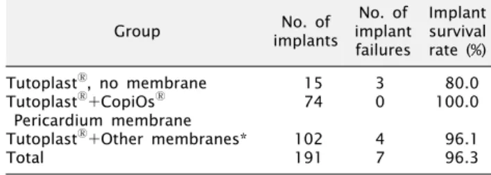
Table 1. Implant survival rates according to membranes with TutoplastⓇ
Group No. of
implants
No. of implant failures
Implant survival rate (%)
TutoplastⓇ, no membrane 15 3 80.0
TutoplastⓇ+CopiOsⓇ 74 0 100.0
Pericardium membrane
TutoplastⓇ+Other membranes* 102 4 96.1
Total 191 7 96.3
*Bio-Gide, Bio-mesh, Gore-tex, Ossix, Tefgen, Osseoguard.
서 론
임프란트의 시술이 증가하고 발전하면서 초기에는 임프란트 식립 시 골질이 좋은 부위에 임프란트를 식립하였으나, 현재는 보철물의 위치와 심미적인 부분을 고려하여 임프란트를 식립하면 서 골유도재생술의 이용이 점점 증가하고 있다. 뿐만 아니라 골유 도재생술은 발치창의 보존, 치조능 증강술, 상악동 골증강술, 임플 란트 주변 골결손부 수복을 위해 행해지는 경우가 많다.
골유도재생술의 성공에 영향을 미치는 요소로서는 적적한 골이 식재의 선택, 차단막의 세포 차단성, 공간 확보 및 유지 및 차단막 의 물리적인 강도 등이 있다.
경조직 결손부의 골유도재생술에 있어 자가골 이식은 가장 이상적으로 알려져 있다. 골형성, 골전도 및 골유도 능력을 모두 가지고 있으며 면역 거부반응이 없고 빠른 치유를 보이는 장점을 가지고 있으나, 채취량이 제한적이고 공여부에 이차결손을 야기 하는 것이 최대의 단점이다[1,2]. 최근 이러한 단점들을 보완할 수 있는 여러 종류의 동종골, 이종골, 합성골 등의 재료들이 소개 되고 사용되고 있다. 이 중 본 연구에서 사용된 Tutoplast
Ⓡ는 비탈회 동종골로서 사체로부터 얻어진 골을 용매로 지방을 제거하 고 탈수되는 과정을 거쳐 가공하고 멸균한 것으로 최근 자가골을 대신하여 골대체재로서 널리 사용 중인 재료이다.
골유도재생술의 술식에서 이식재와 더불어 골이식술의 성공에 많은 영향을 미치는 부분이 차단막이다. 차단막의 요건으로서는 생체 친화성, 공간 확보 및 유지 능력, 조작성 등이 있다. CopiOs
ⓇPericardium membrane은 소의 심막(pericardium)을 처리하여 만들어진 흡수성막으로 장기간의 유지 능력, 조작성 및 연조직 반응이 우수하여 골유도재생술, 상악동 거상술 시 널리 이용되고 있다[3-5].
본 연구에서는 동종골인 Tutoplast
Ⓡ와 우심막 유래 흡수성 막인 CopiOs
ⓇPericardium membrane을 사용하여 시행한 골 유도재생술시 임프란트의 생존율에 대한 평가를 통해 예후를 알아 보고자 한다.
연구방법
1. 연구대상
2007년 1월부터 2009년 6월까지 조선대학교 치과병원에 내원하 여 임프란트를 식립한 환자 중 임프란트 식립과 동시, 혹은 임프란 트 식립 전에 골유도재생술을 시행 받은 환자를 대상으로 하였다 (CDMDIRB-1112-55). 연구 대상 중 흡연을 하는 환자, 임프란트의 생존율에 영향을 줄 수 있는 비조절성 전신질환을 가진 환자는 연구 대상에서 제외되었다. 임상 추적 기간은 보철물 장착 이후 기능을 한 기간이 최소 6개월 이상인 환자로 제한하였다.
2. 연구방법
연구 대상을 골이식 재료, 차단막에 따라 분류하여 임상적 검사, 의무기록지 평가, 방사선 사진을 통한 분석을 통해 임프란 트의 생존율을 기록하였다. 생존 기준은 다음 사항을 포함하였 다; I) 임상적으로 임프란트의 동요도가 없을 것, II) 임프란트 주위에 지속적인 방사선 투과상이 없을 것, III) 임프란트 주위염 이 없을 것.
골이식재의 종류에 따른 분류로는 Tutoplast
Ⓡ를 사용한 그룹 과 사용하지 않은 그룹으로 나누었으며, 차단막의 종류에 따른 분류로서는 차단막을 사용하지 않은 그룹과 CopiOs
ⓇPericar- dium membrane을 사용한 그룹, 다른 차단막을 사용한 그룹으 로 나누었으며, Tutoplast
Ⓡ를 사용한 그룹을 다시 세분화하여 차단막을 사용하지 않고 Tutoplast
Ⓡ를 사용한 그룹, CopiOs
ⓇPericardium membrane과 Tutoplast
Ⓡ를 사용한 그룹, Tutop- last
Ⓡ를 다른 차단막을 사용한 그룹으로 분류하였다.
각 연구항목을 통계분석하기 위하여 SPSS (version 12.0, SPSS Inc., Chicago, IL, USA) software를 이용하였으며, 각 연구항목들은 생존율에 대해 Chi-square test로 유의 수준 0.05%
수준에서 검정하였다.
결 과
총 101명의 환자에서 278개의 임프란트가 식립되었으며, 총 8개의 임프란트가 실패하여, 전체 임프란트 생존율은 97.1%로 조사되었다.
차단막의 사용 없이 Tutoplast
Ⓡ만 단독으로 사용한 그룹은
15개의 임프란트가 식립되었으며, 이 중 3개의 임프란트가 실패
하였다. 3개의 임프란트는 1명의 환자에서 감염을 통해 발생하였
다. Tutoplast
Ⓡ와 CopiOs
ⓇPericardium membrane을 사용한
그룹은 74개의 임프란트가 식립되었으며, 이 중 실패한 경우는
없었다. Tutoplast
Ⓡ와 다른 차단막을 사용한 그룹은 102개의
임프란트가 식립되었고 이 중 4개의 실패가 관찰되었다. 다른
차단막의 경우 Bio-Gide, Bio-mesh, Gore-tex, Ossix, Tefgen,
Hyoung-Sup Lim: Guided Bone Regeneration Using Mineralized Bone Allograft and Barrier Membrane Derived from Ox Pericardium
361
Vol. 33 No. 4, July 2011 Table 2. Implant survival rates according to graft materials
Group No. of
implants
No. of implant failures
Implant survival rate (%)
TutoplastⓇ 191 7 96.3
Other graft materials* 87 1 98.9
Total 278 8 97.1
*Bio-oss etc.
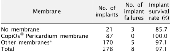
Table 3. Implant survival rates according to membranes
Membrane No. of
implants
No. of implant failures
Implant survival rate (%)
No membrane 21 3 85.7
CopiOsⓇ Pericardium membrane 87 0 100.0
Other membranes* 170 5 97.1
Total 278 8 97.1
*Bio-Gide, Bio-mesh, Gore-tex, Ossix, Tefgen, Osseoguard.
Osseoguard가 사용되었다(Table 1). Tutoplast
Ⓡ를 사용한 그 룹에서 Pericardium 차단막이나 다른 차단막 사이에서의 임프란 트 생존율에 있어 통계학적인 유의성은 없었으나( P >0.05), 차단 막을 사용하지 않은 그룹과는 통계학적으로 유의할 만한 차이를 나타내었다( P =0.004).
골이식재에 대한 분류에서 Tutoplast
Ⓡ를 골이식재로 사용한 그룹에서 191개의 임프란트 중 7개가 실패하였다. 다른 골이식재 를 사용한 그룹에서는 87개의 임프란트 중 1개의 임프란트가 실패하였다(Table 2). Tutoplast
Ⓡ를 골이식재로 사용한 그룹과 다른 이식재를 사용한 그룹 간에는 임프란트 생존율에 있어 통계 학적인 유의성이 나타나지 않았다( P >0.05).
CopiOs
ⓇPericardium membrane을 차단막으로 사용한 경 우 골이식재로 Tutoplast
Ⓡ와 함께 사용한 그룹에서 74개의 임프 란트가 식립되었으며, 다른 골이식재를 사용한 그룹에서 13개의 임프란트가 식립되었고, 두 그룹 모두 임프란트의 실패는 보이지 않았다. 차단막을 사용하지 않은 그룹에서 21개의 임프란트 중 3개의 임프란트 실패가 관찰되었고, 다른 차단막을 사용한 그룹에 서 170개의 임프란트 중 5개가 실패하였다(Table 3). Pericar- dium 차단막을 사용한 그룹과 다른 차단막을 사용한 그룹간에는 임프란트 생존율에 있어 유의할 만한 차이가 없었으나( P >0.05), 차단막을 사용하지 않은 그룹과는 유의할 만한 차이를 나타내었다 ( P =0.002).
고 찰
Tutoplast
Ⓡ는 구강악안면영역의 골결손부에 이식재로 사용되 는 비탈회 동종골로서, 감염성 질환이나 악성 질환이 없는 기부자 의 사후 24시간 후, 무균 상태에서 조직을 채취하여 지방과 골수를 제거하고 저선량의 감마선(gamma irradiation; 17.8 GY)을 시 행한 골이식재로 높은 다공성으로 동결건조골보다 표면적이 넓어 빠른 치유와 완전한 재형성을 촉진한다고 보고되었다[6-8].
Froum 등[9]은 상악동 거상술 시 비탈회 동종골(Puros, Zimmer Dental, Carlsbad, CA)과 이종골(Bio-Oss
Ⓡ, Osteo- health Co., Shirley, NY)를 사용하여 26∼32주 후 골형성 효과 를 비교 분석한 결과, Puros에서 더 많은 양의 신생골과 유골 (osteoid)이 관찰되었다고 보고하였다.
Keith 등[10]은 임프란트 식립 시 블록형 동종골(Puros Block Allograft, Zimmer dental)를 이용하여 골유도 재생술을 시행한 결과 99%의 임프란트 성공률을 보였다. 본 연구에서는 입자형 골을 사용하였으며, 96.3%의 성공률을 보여 입자형과 블록형 모두 높은 성공률을 보여주고 있다.
차폐막의 경우 흡수성과 비흡수성으로 분류되며, 비흡수성 막 의 경우는 생체 적합성이 우수하고 골생성부의 공간 확보가 가능 하다는 장점이 있으나, 연조직이 불충분한 경우나 날카로운 막의 경계부 등에서 주변 점막을 천공시켜 하방의 골 치유에 부정적 영향을 초래할 수 있으며, 막의 제거를 위해 추가적인 수술이 필요하다는 단점을 가지고 있다[11]. 대표적인 비흡수성 막으로는 titanium mesh, Gore-tex 등이 사용되고 있으며, 성공적인 골 증대효과가 보고되고 있다[12-15].
흡수성 막의 경우 막의 제거가 불필요하며 상방 연조직의 치유 가 양호한 장점을 가지고 있으나, 골생성부의 공간 확보에 어려움 이 있고 일단 감염이 되면 하방 골이식재의 손실을 야기하며 분해속도의 차이로 임상적 예견성이 불확실하고 분해 산물이 치유 에 영향을 줄 수 있는 단점을 가지고 있다[16].
우심막유래 차단막은 인체의 여러 분야의 수술에서 널리 사용 되고 있다. 경동맥의 내절제술을 시행한 후 보강을 위하여 아주 많이 사용되고 있으며, 강하고 질긴 섬유소 성분으로 구성되어 있어 인간의 조직과 유사한 강도를 가지고 있으며, 이물 반응 또한 잘 나타나지 않는 것으로 보고되고 있다[17]. 또한 Choi 등[18]은 우심막 패치를 이용한 동물의 담관 복원술에서도 좋은 결과를 나타내었으며, 안과 영역에서는 안구적출술 후 인공 안구 삽입 시 인공 안구의 피복재로서 우수한 효과를 보고하였다[19].
DeBacker 등[20]은 우심막의 장점으로 장력에 견디는 힘이 좋고,
봉합이 용이하면서 봉합을 유지하는 적절한 강도를 가지며, 유연
하고, 원하는 모양으로 변형이 쉬운 기계적인 장점 또한 가지고
있음을 보고하였다. 본 연구에서 사용된 CopiOs
ⓇPericardium
membrane 또한 소의 심막(pericardium)을 처리하여 만들어진
흡수성막으로서 장기간의 유지 능력, 조작성 및 우수한 연조직
반응으로 치과적 영역에서는 골유도재생술, 상악동 거상술 시
널리 이용되고 있다[3-5]. 본 연구에서는 Pericardium 차단막을
사용한 그룹에서 임프란트 성공률이 100%로 다른 차단막을 사용
362
임형섭: 골유도재생술 시 비탈회 동종골와 우심막유래 차단막의 임상적 활용J Korean Assoc Maxillofac Plast Reconstr Surg
한 그룹과는 통계적인 차이는 없었으나, 차단막을 사용하지 않은 그룹에 비해 통계학적으로 높은 생존율을 보였다. 이는 Pericar- dium 차단막이 다른 차단막과 비교하여 충분한 효과를 나타내는 차단막으로 평가할 수 있으며, 또한 향후 지속적으로 골유도 재생 술 및 상악동 거상술 시에도 예지성 있는 차단막으로 사용될 수 있다고 생각된다. 하지만 Pericardium 차단막과 다른 차단막 의 개별적인 비교에 관해서는 다른 차단막들의 개별 숫자가 적어 비교하기 어려워 이에 대해 추가적이고 장기적인 연구가 필요할 것으로 생각된다.
결 론
Tutoplast
Ⓡ와 다른 이식재 간에는 임프란트의 생존율에 있어 통계학적으로 유의할 만한 차이를 보이지 않았다. Pericardium 차단막을 사용한 그룹과 그 외의 다른 차단막을 사용한 그룹 간에는 통계학적인 유의성이 없었으나, 차단막을 사용하지 않은 그룹에 비해서는 통계학적으로 유의할 만한 높은 임프란트 생존율 을 나타내었다.
이를 통해 골이식재의 종류보다는 차단막의 사용 여부에 따라 임프란트 생존율에 영향을 미친다고 생각되며, 차단막 중 CopiOs
ⓇPericardium membrane은 골유도재생술에 있어 임프란트 생존 율을 보장하는 예지성 있는 재료라 생각된다.
References