김성주∙김철환∙김경욱
단국대학교 치과대학 구강악안면외과학교실
타액선 다형성 선종에서 Glycosaminoglycan의 발현
GLYCOSAMINOGLYCAN EXPRESSION IN PLEOMORPHIC ADENOMAS OF THE SALIVARY GLAND
Seong-Joo Kim, Chul-Hwan Kim, Kyung-Wook Kim
Dept. of Oral & Maxillofacial Surgery, School of Dentistry, Dankook University
Pleomorphic adenoma is the most common benign tumor in salivary glands, and occurred in frequency of 60% in parotid gland tumors, and 50% in submandibular gland tumors, and 25% in sublingual gland tumors. Histopathologically, pleomorphic adenoma is composed of epithelial cells and mesenchymal tissues, and called ‘mixed tumor’because of morphological divergency. The cell structures of luminal area are com- posed of polyhedral and cuboidal secretory epithelial cells and modified myoepithelial cells around it, and mesenchymal tissue is composed of some myoepithelial cells and stromal tissue. In stromal tissue, myxoid change, chondroid change, or hyalinization can be seen even if bone tissue. In many studies, tumor cells of pleomorphic adenoma containing modified myoepithelial cell participate in synthesis of glycosaminoglycans .
In this study, tissue sample of pleomorphic adenoma of human salivary gland were obtained from 20 sur- gical specimens, and all specimens were routinely fixed in 10% formalin and embedded. Serial 4-8㎛ thick sections were cut from paraffin blocks. The histopathologic evaluation was done with light microscopy. And, with immunohistochemical staining, characteristics of glycosaminoglycan were observed. And, for biochemi- cal analysis of glycosaminoglycan, isolation of crude glycosaminoglycan from tumor tissue and immuno-blot analysis were carried out. With transmission electromicroscopy, tumor cells and biologic behavior of pleo- morphic adenoma were observed with distribution and expression of glycosaminoglycan in tumor cells,
The results were obtained as follows:
1. In immunohistochemical study, chondroitin 4-sulfate is highly postively stained in myxoid stromal tis- sue, and chondroitin 6-sulfate is highly positively stained in chondroid mesenchymal tissue, both gly- cosaminoglycans are positively stained in non-luminal cell of ductal area.
2. Dermatan sulfate and keratan sulfate is positively stained in periductal non-luminal tumor cells.
3. In immunohistochemical study, heparan sulfate is weakly stained in luminal cells and non-luminal cells around duct, and chondroid mesenchymal tissue.
4. In transmission electromicroscopic view, the tumor cells are composed of modified myoepithelial cells, and contain many microfilaments and well developed rough endoplasmic reticulum.
5. In Immuno-Blot analysis, the expression of glycosaminoglycans is expressed mostly in chondroitin 6- sulfate and chondroitin 4-sulfate.
From the results obtained in this study, tumor cells of pleomorphic adenoma are composed of modified myoepithelial cells, and glycosaminoglycans of chondroitin 4-sulfate and chondroitin 6-sulfate mostly par- ticipate in the development of pleomorphic adenoma, but dermatan sulfate, keratan sulfate and heparan sulfate glycosaminoglycans were expressed variably.
Key words : Glycosaminoglycan expression, Pleomorphic adenoma
Abstract
Ⅰ. 서 론
다형성 선종은 타액선에서 가장 많이 호발하는 양성종양 으로서, 전체 이하선 종양의 60%, 악하선 종양의 50%, 설 하선 종양의 25%를 차지하며, 소타액선 종양의 50%를 차 지하는 가장 많이 빈발하는 타액선 양성 종양이다
1). 임상적 으로 느린 성장속도를 갖고 명확한 경계를 보인다. 모든 연 령층에서 발생할 수 있으나 주로 40대에서 가장 많이 호발 하며 치료는 완전절제술로서, 대타액선에서 호발한 다형성 선종은 소엽절제술이나 타액선 절제술로 치료하며, 피막의 바깥부위에 있는 병소의 경우에는 재발이 흔히 일어나기 때 문에 단순 절제술로는 종양의 재발이 나타날 수 있다. 일반 적으로 재발률은 40-60%로 높은 편이며, 치료 없이 방치 되거나 재발이 된 경우에는 약 3-15%에서 악성 변이가 일 어나며 이를 carcinoma ex pleomorphic adenoma라고 명 명한다
2-4). 조직병리학적으로는 종양 세포는 상피성 세포 조 직과 간엽성 조직으로 구성되어 있으나 그 형태는 매우 다 양하게 나타나 혼합종(mixed tumor)이라고도 명명되어진 다. 종양 세포 중, 선관구조에는 입방형 세포와 방추형의 근 상피세포로 이루어지며, 간엽조직은 근상피세포와 섞여 있 으며, 일부에서는 간질조직에서 초자화가 일어나기도 하고, 혹은 연골성, 지방세포, 심지어는 골조직까지도 형성할 수 있다
5-9).
다형성 선종의 종양세포의 세포외 간질에는 기저막 성분 을 포함하여 다양한 기질을 포함하고 있다. 여러 연구에 의 하면 이러한 근상피세포를 포함하는 다형성 선종 종양 세포 가 proteoglycan의 합성에 관여한다는 사실이 밝혀지게 되 었으며
10), 최근에 들어서는 기질 성분의 분석에 대한 연구는 단클론항체검사를 이용한 면역조직화학적 검사의 발전으로 많은 조직과 장기에서의 proteoglycan의 분석이 가능하게 되었고 혈관이나 근육 등이나 기저막, 모든 연골조직 등에 서의 proteoglycan의 합성의 과정에 대하여 알 수 있게 되 었다
11). 여러 기질 성분 중, chondroitin sulfate와 der- matan sulfate는 근육세포에서 생성되며, heparan sul- fate는 혈관 및 동맥에서, keratan sulfate는 각막 및 연골 에서 주로 생성된다
12-19). Chondroitin sulfate는 정상 타액 선에서 분비되는 분비과립으로도 나타나며, 이러한 기질 성 분은 다형성 선종에서도 나타난다
20).
이전의 연구들에서는 다형성 선종의 형성에 있어서 이 종 양의 점액성 물질들에 기초한 조직화학적 연구에 초점이 이 루어져 왔으며
21), 결체조직에 많이 분포되어 있는 gly- cosaminoglycan이, 이 종양의 상피성 조직 및 결체조직에 모두 나타나는지, 그리고 상피성 조직이 이러한 gly- cosaminoglycan의 합성에 관여하는 지에 대한 면역조직화 학적 연구는 미미한 실정이다.
본 연구에서는 다형성 선종의 종양세포에 존재하는 pro-
teoglycan의 분포, 종양 세포의 간질조직에서의 발현을 통 한 생물학적 양태를 살펴 다형성 선종의 발생 과정에 대한 연구를 시행하여 임상적으로 가장 흔한 타액선 양성 종양인 다형성 선종의 임상적 치료에 도움이 되고자 하였다.
Ⅱ. 연구 재료 및 방법
1. 연구재료
구개에서 발생된 20례의 다형성 선종을 절취하여 10%
중성포르말린에 고정한 후, 4㎛ 파라핀 절편을 제작하고, 통법에 따라 H&E 염색 및 MT 염색을 시행하고 광학현미 경하에서 조직병리학적 특징을 관찰하였다.
2. 면역조직화학적 검사
절취한 조직을 면역조직화학적 염색을 시행하여 조직병리 학적 특성을 관찰하였다. 먼저 절취한 조직을 고정한 후 Poly-L-Lysine으로 처리된 슬라이드에 4-8㎛ 파라핀 절편 을 제작하고 면역조직화학적 염색을 시행하기 위하여 절편 을 0.3% H
2O
2에 5분간 부란시키고 10mM phosphate buffer solution (PBS) 으로 세척한 후, 제 1차 항체로 처 리한 후(Table 1) LSAB(labelled streptavidin biotin)법 으로 처리하고 난 후, 발색제로 Diaminobenzidine을 이용 하였다. 발삼으로 봉입한 다음 Mayer’s Hematoxylin으로 대조염색을 시행하여 광학현미경으로 200배하에서 관찰하 였다(Table 2).
3. Immuno Blot 분석법을 이용한 기질 성분의 추출
1) Glycosaminoglycans의 동정 : 조직을 95% 수용성 에탄올에 넣은 후, acetone으로 건조하고 조직을 분쇄하였 다. Protein-glycosaminoglycan linkage의 결합을 분리하 기 위하여 건조분말 조직을 0.3N NaOH 용액에 넣고 4℃
에서 하루 동안 유지하였다. 혼합물을 4N HCl와 1M Tris- HCl (pH 8.0)을 이용하여 pH가 8.0이 되도록 하고,
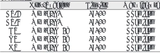
Table 1. Specification of Primary Antibody Used in This Study
Host/Subclass Dilution Manufacture
CS-4 Mouse/IgM 1:100 Chemicom
CS-6 Mouse/IgM 1:100 Chemicom
DS Mouse/IgG2a 1:100 Chemicom
KS Mouse/IgG2b 1:100 Chemicom
HS Mouse/IgG2b 1:100 Chemicom
Pronase-P(5mg/g 건조조직)를 넣고 toluene을 첨가한 후, 40℃에서 20시간 동안 부화시킨 후, 혼합물을 10,000G로 15분간 원심분리하여 상층액을 모으고, 상층부 의 glycosaminoglycan을 1% potassium acetate를 포함 하는 95% 수용성 에탄올에 침전시켰다. 80% 수용성 에탄 올로 씻고 acetone으로 건조시킨 후, 조직 0.1g을 1ml 증 류수에 용해하였다.
2) Glycosaminoglycan의 전기영동
Glycosaminoglycan의 전기영동은 5cm의 cellulose acetate 흡착지에 0.2 M calcium acetate buffer를 이용 하여 1mA/cm 으로 3시간 동안 전극을 걸고, 2㎕를 흡착지 에 점적한 후, 흡착지를 Alcian blue로 염색하고 densito- meter로 관찰하였다.
4. 투과전자현미경(TEM)을 이용한 종양 세포의 관찰
종양 조직을 1㎣의 크기로 잘라 2.5% glutaraldehyde에 넣은 후 고정액은 acetate veronal를 사용하여 pH 7.2로 완충시키고 1% osmium tetraoxide를 사용하여 30-60분 후 고정하였다. 고정액을 증류수로 씻어낸 후 알코올로 탈 수시키고, n-butyl methacrylate에 침윤시킨 후, 젤라틴 캡슐에 포매하고, 60℃에서 중합하고 촉매제는 1% ben- zoyl peroxide를 사용하였다. 0.2㎟ 정도 되는 블록에서 초 박 절편을 제작하고 탄소 격자상에 올려놓고 Uranyl acetate와 Lead citrate로 염색한 후 투과전자현미경 (JEOL JEA CX II, JAPAN)으로 관찰하였다.
Ⅲ. 연구 결과
1. H&E 소견
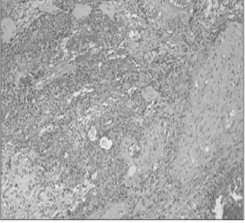
상피세포, 근상피세포 및 간엽조직 성분을 포함하는 매우 다양한 조직소견을 보였으며, 종양은 결합조직의 피낭으로 잘 경계가 되어 있었으며, 상피세포의 양상은 다양하게 나 타나 있어, 분명한 선도관 구조를 보이기도 하였으며 상피 세포의 소규모 엽을 형성하기도 하였으며, 세포 형태는 선 상, 침상, 다각형의 모양을 나타내기도 하였다. 선도관 구조 내에는 호산성 물질을 함유하고 있었으며, 간질조직 부위는 연골성, 초자질성, 점액성 간질조직이 모두 나타났으며, 특 징적으로 소성조직인 점액성 간질조직에 높은 비율로 근상 피세포가 나타났다(Fig. 1, 2).
2. 면역조직화학적 염색 결과 (Table 3)
1) Chondroitin sulfate 염색 소견
Chondroitin 4-sulfate (CS-4)는 점액성 간질 조직 부 위에서 뚜렷한 양성반응을 보였고(Fig. 3), 연골성 조직부 위에서도 양성반응을 나타내었다(Fig. 4). 또 chondroitin 6-sulfate(CS-6)는 종양세포 주위의 점액성 조직에서 뚜렷 한 양성반응을 보였다(Fig. 5).
Table 2. Procedure for Immunohistochemical Staining
Paraffin section preparation
�
Deparaffination & hydration
�
Peroxydase inactivation (0.3% H
2O
2, 5mins.)
�
Serum blocking reagent (20mins.)
�
Primary Ab (4 hrs.)
�
LASB(labelled streptavidin biotin) method (30mins.)
� Deaminobenzidine
�
Counter staining with Mayer’ s hematoxylin
� Balsam mounting
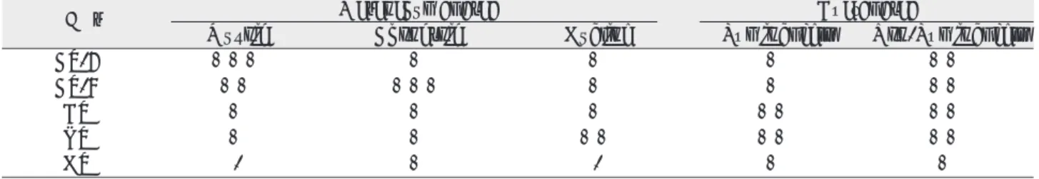
Table 3. Immunostaining Intensity in Various Matrix of Pleomorphic Adenoma
GAGs Mesenchymal area Ductal area
Myxoid Chondroid Hyaline Luminal cell Non-Luminal cell
CS-4 +++ + + + ++
CS-6 ++ +++ + + ++
DS + + + ++ ++
KS + + ++ ++ ++
HS - + - + +
(-: negative, +: mild, ++: moderate, +++: strong stained)
2) Dermatan sulfate (DS) 염색소견
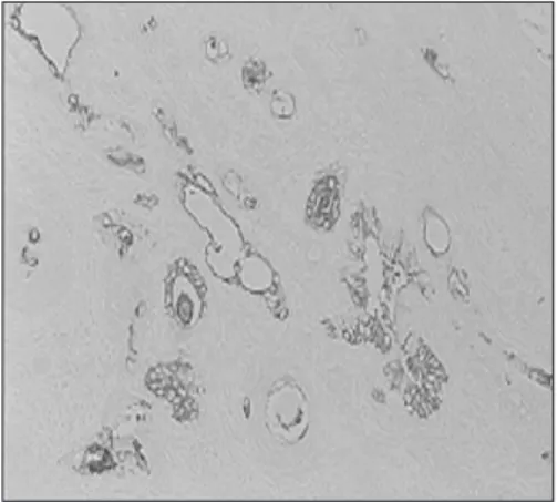
종양세포 사이의 인접한 세포간 간질조직에서 미약한 양 성반응을 보였다(Fig. 6).
3) Keratan sulfate (KS) 염색 소견
연골성 간질 조직의 종양세포에서 국소적인 양성 반응을 보였다(Fig. 7).
4) Heparan sulfate (HS) 염색소견
연골성 조직부위의 종양세포들에서 양성반응을 나타내었 다(Fig. 8).
3. Immuno-blot 분석을 이용한 glyosaminolycan 의 동정
Immuno-blot 분석법을 이용한 기질 성분을 추출한 후, 전기영동을 시행한 결과, chondroitin 4-sulfate와 chon- droitin 6-sulfate에서 강한 뚜렷한 밴드를 형성하는 것으 로 보아 이 종양의 발생 및 성장에 크게 관여하는 것으로 사 료되었다(Fig. 9).
4. 투과전자현미경(TEM)을 이용한 종양 세포의 관찰
선도관 구조 부위의 상피세포는 미세융모와 잘 발달된 rER와 골지체를 보였고, 근상피세포가 여러 겹으로 둥글게 둘러싸고 있었다. 근상피세포의 세포질에는 미세섬유사가 풍부하고 세포사이가 확장되어 있었다(Fig. 10, 11).
Ⅳ. 총괄 및 고찰
다형성 선종은 타액선 종양 중 가장 흔히 빈발하는 종양으 로서
22), 형태조직학적으로 상피세포성분, 근상피세포, 점액 성 및 연골성의 간엽 조직성분 등이 아주 다양하게 나타나 는 것이 특징적이다
6,7). 다형성 선종은 혼합종(mixed tumor)라고도 명명된 양성 종양으로 이 혼합종의 의미는 상피조직과 간엽조직으로 구성된 종양을 의미한다
23). 다형 성 선종의 조직학적인 세포 구성 성분은 크게 상피세포와 근상피세포의 2가지 형태로 세포 구성이 이루어지며
5), 간엽 성 조직은 매우 다양하게 나타난다. 종양조직의 선관구조 부위에서는 선관구조의 내강을 이루는 상피세포는 입방형, 다각형의 세포로 구성되어 있고, 그 주위에 방추형의 근상 피세포로 이루어지며, 간엽조직은 근상피세포와 섞여 있으 며, 일부에서는 간엽조직에서 초자화가 일어나기도 하고, 혹은 연골성, 지방세포, 심지어는 골 조직까지도 나타날 수 가 있다. 다형성 선종에서의 근상피세포는 1898년
Zimonermann에 의해 처음 기술되었으며, 선관 상피층의 기저부위나 개제관 혹은 선조관의 기저부위에 존재한다
8, 9). 정상적인 근상피세포는 세포질에 존재하는 근원섬유의 작 용으로 분비단위의 압력을 높혀 타액의 분비를 촉진하는 기 능을 가지고 있으며, 변화가 많은 분화로 인하여 다양한 간 질조직을 구성하는 주된 세포이다
24).
이 종양의 발생기원은 매우 다양하게 나타나는데 다형성 선종의 세포 분화를 위해 일반적으로 받아들여지고 있는 이 론은 첫째, 상피성분과 간엽성분에서 함께 종양이 발생한다 는 설과, 둘째 상피성분에서 단독으로 종양이 발생하며 간 엽 성분 즉 점액성, 연골성 조직 등은 상피세포에서 직접 만 들어지거나 간엽세포로 변형되어 발생한다는 설이 있으며
25), 셋째, 종양이 상피세포에서 유래되고 자극을 받은 비종양 조직의 간엽성 조직이 이형성(dysplasia)을 이루어 연골성 조직으로의 변성과정을 거친다는 이론이다
26). 현재는 상피 성 기원으로 종양이 발생하는 것이 정설로 받아들여지고 있 다
9,25,27,28).
다형성 선종 종양의 세포 구조의 전자현미경적 소견은 그 구성 세포가 관세포, 선방세포, 근상피세포, 및 미확정세포 로 구성되며, 점액성 및 연골성 기질 부분에는 근상피세포 가 주된 세포로 나타난다고 하였으며
5,9,25,29,30), 본 연구에서 의 투과전자현미경의 결과와 같이, 대부분의 세포로 충만된 부분은 관세포로 구성되어 있었고, 점액성 부분은 근상피세 포가 주로 나타났으며, 그 외에도 미확정세포가 섞여 나타 났다. 근상피세포는 핵이 길게 늘어나 있고, 이 염색질이 핵 막의 변연을 따라 분포하고, 세포소기관은 세포의 변연을 따라 분포하고 세포막으로부터는 세포질 돌기를 내거나 융 모를 형성하여 주위 세포와는 desmosome으로 연결되어 있었다. 이는 다형성선종의 조직학적 발생 기전과 관련된 이전의 미세 구조 연구의 결과와 유사하며
9,25)이는 다형성 선종이 간엽성 기원이라기보다는 상피성 기원에서 유래한 다는 현재의 학설을 뒷받침하여 준다.
다형성 선종은 이와같이 복잡한 조직학적 소견을 보이며,
상피성 세포, 근상피성 세포, 그리고 변형성 근상피세포가
점액성, 초자성, 연골성 형태가 종양 조직 내에 혼재되어 섞
인 형태로 특징지어진다
31). 일반적으로 많은 문헌에서는 근
상피세포가 다형성 선종의 조직학적 다양성을 이루는 가장
주된 역할을 한다고 알려져 왔으나
32-39), 이 종양의 각각 다
른 세포형태학적 차이에 따른 연구는 명백히 밝혀지지 않았
다. 세포외 간질은 다형성 선종의 다양한 조직학적 다양성
에 따른 조절 인자로서의 가능성을 가지고 있다. 정상 분비
선 조직에서의 세포외 간질은 장기형성(organogenesis) 및
형태 발생에 기초적인 역할을 담당하며, 분비선의 분화, 분
화기의 유지 등에도 깊이 관여하는 것으로 알려져 왔다
40, 41).
또한, 세포외 간질 조직 중에서 기저막 부위에 존재하는 단
백은 다형성 선종의 표현형의 중요한 조절 요소로 밝혀져 왔다
42-46).
인체의 결합조직은 많은 세포간 물질과 다양한 종류의 세 포로 구성된 조직으로서, 신체의 거의 모든 장기와 조직을 지지해주고 결합해주는 역할을 한다. 결합조직의 세포간 물 질을 세포외 기질(extracellular matrix)이라고도 하며, 형 태학적으로는 섬유형태의 결합조직 섬유와 정해진 형태가 없는 무정형의 기질로 구성되어 있다. 이 기질은 반용액성 겔의 특징을 갖는 무정형의 투명한 물질에서 유래된 이름이 다. 조직액은 기질과 성글게 유지되어 있어 지지구조들 사 이에서 분자들의 이동과 맥혈관계의 대사산물의 교환을 위 한 매체로 작용하게 된다. 기질은 여러 종류의 길고 선형의 분지하지 않는 긴 다당류 사슬의 혼합형태로 되어 있으며, 각각은 이당류 구조가 반복된 구조이다
47-54). 이당류 구조의 구성은 일반적으로 우론산(uronic acid)과 아미노당으로 구성되어 있으며, 이중 아미노당은 현재 glycosaminogly- can이라고 불리워지며, 이전에는 뮤코다당(mucopolysac- charide)으로 명명되기도 하였다
55,56). 이 glycosaminogly- can은 이당류 구조에 위치한 hydroxyl, carboxyl, sul- phate 기의 존재로 인하여 산성의 성질을 갖는다. 하이알루 론산은 소성결체조직에서 주로 나타나는 glycosaminogly- can이며, chondroitin 4-sulfate, chondroitin 6-sulfate, dermatan sulfate, heparan sulfate, heparin sulfate, 그리고 keratan sulfate 등의 glycosaminoglycan은 하이 알루론산과는 달리, 다양한 단백질 분자와 공유결합이 되어 있어 이른바 proteoglycan을 형성하게 된다. 이러한 pro- teoglycan은 90-95%가 탄수화물로 구성되는 거대한 분자 량을 갖게 된다. Glycosaminoglycan은 특정지지 구조의 투과성을 결정하고 세포의 기저막의 구조에 중요한 역할을 하게 된다. 이러한 glycosaminoglycan은 콜라겐처럼 분비 되기 전에 변화 과정을 거친다. 먼저 단당류나 이당류 구조 의 단계적인 첨가로 다당류가 형성된 후 골지체에서 황산화 와 에피머화 반응에 의해 변화된다
57,58). 마지막으로 gly- cosaminoglycan이 세포를 떠나기 전에, proteoglycan 분 자를 형성하기 위해 core 단백의 serine 잔기에 적합한 gly- cosaminoglycan이 공유결합을 한다
59).
현재의 타액선 종양에 대한 연구들은 면역화학적 검사법 을 통한 proteoglycan의 위치를 밝혀냄으로써, 종양의 각 각 다른 조직학적 형태에 있어서의 proteoglycan의 염색 형태의 비교 연구가 활발하게 진행되고 있다. 특정 단클론 항체 검사법과 효소 전처리법을 이용한 proteoglycan의 동 정 및 분석이 개발된 이후로 각종 조직과 장기에서의 pro- teoglycan에 대한 분석이 가능하게 되어
11)proteoglycan의 조직 분포에 대한 연구가 활발히 이루어졌다. 타액선 종양 에 대한 proteoglycan에 대한 조직화학적, 생화학적 연구
는 Takeuchi 등
20,60)과 Toida 등의 연구
61)에 의해서 많은 발 전이 있어 왔다. 타액선 종양에 존재하는 탄성섬유들은 특 수한 염색방법에 의해서 발견되었으며
62), 이 섬유들은 변형 된 근상피세포들로부터 생성되었음을 밝혀냈으며
63), 다형성 선종에 대한 또 다른 proteoglycan에 관한 면역조직화학적 연구에 의하면, 근상피세포에서 강한 염색반응을 나타내어 특징적인 소견이 관찰되었다
10).
Cutis 등의 연구에
64)의하면 정상 타액선 조직에서는, 선 관주위에서 heparan sulfate의 면역염색에서 강한 반응을 나타났으나 chondroitin sulfate와 dermatan sulfate, 그 리고 keratan sulfate의 면역염색에서는 미약한 반응을 나 타내었다. 또한 선관 내부에는 염색 반응이 나타나지 않았 다. 이의 결과는 정상 타액선에서는 상피세포가 gly- cosaminoglycan의 생성과 분비를 하는 것이 명백하며, 분 비는 선관이 아닌, 세포외 기질로 분비됨을 의미한다. 분비 액 내부에는 glycosaminoglycan이 없는 이유는 생합성된 glycosaminoglycan이 기저막이나 세포 표면으로 이동되기 때문이라고 하였다
65). 반면 타액선 종양인 다형성 선종에서 는 glycosaminoglycan의 분포는 상피 조직과 간질 조직에 서 다양하게 나타난다. 선관 부위의 세포에서는 gly- cosaminoglycan의 분포가 다양하게 나타나지만, 도관 내 부에는 나타나지 않는다. 이는 다형성 선종에서의 gly- cosaminoglycan도 마찬가지로 간질 조직의 구성에 참여한 다는 사실을 증명하여 준다
66).
Sibutani 등의 연구에 의하면
10), 다형성 선종에서의 pro- teoglycan 면역 염색 결과에서, 변형된 근상피세포의 분포 와 변형 연골화(chondroid) 세포의 출현을 보고하였으며, 또한 초자질 조직과 점액성 조직에서의 연골성 변환에 관하 여서도 보고가 된 바 있다
29,37,67,68). 그리고 연골성 조직은 많 은 양의 keratan sulfate proteoglycan을 함유하지만 chondroitin sulfate, dermatan sulfate, heparan sul- fate의 proteoglycan은 소량이 분포한다고 하였다.
근상피세포와 관련한 이전의 연구에 의하면, 타액선 종양 의 다형성 선종에서 proteoglycan의 합성을 하는 세포들은 변형된 근상피세포와 관련이 있음을 보고하였다
37,67). 변형 된 근상피세포는 다형성 선종에서 특이적 형태로 keratin 면 역 염 색 반 응 에 서 양 성 반 응 으 로 나 타 나 며
69-71), vimentin
72), 그리고 S-100 단백
38,70,73-75)에서도 양성반응을 나타낸다.
본 연구에서는 대부분의 glycosaminoglycan은 도관 부
위의 세포보다는 비도관 부위에서 강한 염색반응을 나타냈
으며, 상피세포 주위의 부분적인 점액성 간질 부위에도 강
한 염색반응을 나타냈다. 따라서 glycosaminoglycan은 주
로 비도관 부위의 상피세포에서 생성됨을 알 수 있었으며,
이는 glycosaminoglycan이 세포를 자극하여 세포외 기질
의 합성을 촉진한다는 연구의 결과를 뒷받침 한다
76,77). 이렇 게 생성된 glycosaminoglycan은 비도관 부위의 세포로 하 여금 자가분비(autocrine)기전 등의 피드백 기전을 통해 더 많은 glycosaminoglycan 및 다른 세포외 기질을 분비하도 록 하며 생성된 glycosaminoglycan은 종양 세포의 형태나 운동성, 대사 기능, 세포질 분화 등의 표현형에 중요한 역할 을 하게 된다
78).
본 연구에서 초자질 간질부위에서의 glycosaminoglycan 의 발현이 낮게 나타났는데 이는 초자질 간질부위는 주로 elastin, 제4형 콜라겐, laminin 등의 다른 세포외 간질 물 질들로 구성되어 있기 때문이다
45). 이 연구에서 간엽조직에 서의 점액성 간질 부위와 연골성 간질 부위에서의 gly- cosaminoglycan의 분포의 차이가 많이 나타났는데, 이는 연골성 간질 부위에는 chondroitin 6-sulfate가 강하게 반 응하였고, 점액성 간질 부위에는 chondroitin 4-sulfate가 강하게 반응하였다. 종양세포에서 분비된 glycosaminogly- can에 의해 점액성 간질이 형성되고 이 부위에서 gly- cosaminoglycan의 구성의 변화가 일어나 연골성 간질로의 변성이 일어나고 구성 성분의 변화, 즉 elastin이나 colla- gen등의 다른 세포외간질이 함께 나타나게 되는 것으로 알 려져 있다
79,80).
본 연구에서 heparan sulfate의 염색 반응은 간질 조직 의 연골성 조직에서만 매우 미약하게나마 면역조직화학적 염색반응이 나타나고 다른 간질 조직에서는 나타나지 않았 는데, 일반적으로 세포핵의 heparan sulfate는 세포외 기 질의 다른 분자 및 성장 인자들과 함께 DNA 합성을 억제하 고 세포성장을 조절하는 역할을 하는 것으로 알려져 있어
81-83), 이는 세포 성장과 분화에 있어 물질 대사에서의 연골성 분 화에 따른 것으로 본 연구의 결과에서의 heparan sulfate 는 세포외 간질의 구성성분이라기 보다는 주로 세포성장의 조절과 분화의 결과로 생각되었다.
본 연구의 glycosaminoglycan의 생화학적 분석은 면역 블롯팅(Immuno-Blotting)법을 이용하였다. 전기영동을 시행한 결과, chondroitin 6-sulfate, chondroitin 4-sul- fate 성분이 추출되었으며, 이 결과는 본 연구의 면역조직 화학적 분석에서의 결과를 신뢰할 수 있었으며, Takeuchi 등
23)과 최근의 Zhao 등
84)의 연구 결과와도 유사하게 나타 났다.
본 연구에서는 임상적으로 많이 접할 수 있는 타액선 종양 인 다형성선종의 세포를 조직학적 형태를 비교하여, 변형된 근상피세포를 관찰하고 세포성 성분과 간질성 성분들을 분 석하여 보았으며, 면역조직화학적 검사를 통하여 이 종양의 성장과 근상피세포의 분화와 관련된 세포간 물질에 나타나 는 proteoglycan의 다양한 발현의 연구를 시행하여, 비선 관 상피세포들이 glycosaminoglycan의 주된 분비세포이
며, 주로 chondroitin sulfate가 종양의 성장에 주요 역할 을 하는 것으로 나타나, 다른 타액선 종양과의 차이점을 발 견하였고 이와 같은 연구 결과가 향후 다형성 선종의 임상 적 치료에 도움이 되고자 하였다. 그러나 이 종양의 생성과 형태발생에 관하여 이의 연구로는 부족하며, 많은 proteo- glycan에 대한 성분의 생성과 분비에 대한 더 많은 면역전 자현미경적, 생화학적, 분자유전생물학적 연구가 향후 필요 하리라 사료된다.
Ⅴ. 결 론
본 연구에서는 20례의 다형성 선종을 절취하여 10% 중 성포르말린에 고정한 후 4㎛ 파라핀 절편을 제작하고 염색 을 시행하여 광학현미경하에서 조직병리학적 특징을 관찰 하고, 면역조직화학적 염색을 시행하여 glycosaminogly- can의 특성을 관찰하고, Immuno Blot 분석법을 이용한 glycosaminoglycan의 추출과 투과전자현미경을 이용한 종 양 세포의 관찰을 시행하여, 다형성 선종 세포에 존재하는 glycosaminoglycan의 분포, 종양세포의 간질 조직에서의 발현을 통한 종양세포의 생물학적 양태를 살펴 다음과 같은 결과를 얻었다.
1. 면역조직화학적 검사에서 chondroitin 6-sulfate는 연 골성부위에서, 그리고 chondroitin 4-sulfate는 점액성 부위에서 강한 양성반응을 보였으며, 비도관부위의 종양 세포에서는 양 물질이 모두 발현되었다.
2. Dermatan sulfate와 keratan sulfate는 도관 부위의 종양세포에서 양성 반응을 나타내었다.
3. Heparan sulfate는 연골성 부위와 도관 부위의 종양세 포에서 양성 반응을 나타내었다.
4. 투과전자현미경에서는 종양세포의 대부분은 변형된 근 상피세포의 소견을 보였으며, 방추형세포의 세포질에 많 은 미세 섬유사를 보이고, 한쪽으로 치우친 핵과 잘 발 달된 rER을 보였다.
5. Immunoblot분석에서는 chondroitin 4-sulfate와 chondroitin 6-sulfate가 가장 뚜렷한 양성 반응을 보 였다.
이상의 연구에서 다형성 선종 세포는 변형성 근상피세포
로 구성되며, 종양 세포의 면역조직화학적 검사와
Immuno-blot분석법에서 glycosaminoglycan 중, chon-
droitin 4-sulfate 및 chondroitin 6-sulfate가 주로 종양
발생과 성장에 관여하지만 다양한 발현을 나타낸 der-
matan sulfate, keratan sulfate, heparan sulfate도
다형성 선종의 종양의 발생에 관여하는 것으로 사료되
었다.
참고문헌
1. Attie JN, Sciubba JJ : Tumor of major and minor salivary glands; clinical and pathologic feature. Curr Probl Surg 18 : 65, 1981.
2. Kroll SO, Boyers RC : Mixed tumor of salivary glands:
long-term follow-up. Cancer 30 : 276, 1972.
3. Dardick L, van Nostland AWP, Philips MJ : Histogenesis of salivary gland pleomorphic adenoma(mixed tumor) with evaluation of the role of the myeoepithelial cell. Hum Pathol 13 : 62, 1982.
4. Nishimura T, Furukawa M, Kawahara E et al : Differential diagnosis of pleomorphic adenoma by immuno- histochemical means. J Laryngol Otol 105(12) : 1057, 1991.
5. Mylius EA : The identification and the role of the myoep- ithelial cell in salivary gland tumors. Acta Pathol Microbiol Scand (Suppl 139) 50 : 1, 1960.
6. Seifert G, Langrock I, Donath K : A pathological classifica- tion of pleomorphic adenoma of the salivary glands. HNO 24 : 415, 1976.
7. Naeim F, Frosberg MI, Waisman J et al : Mixed tumors of the salivary glands: growth pattern and Recurrence. Arch Pathol Lab Med 100 : 271, 1976.
8. Fernando MM, Christian M : Histology of the major sali- vary glands Am J Surg Pathol 13 : 879, 1989.
9. Hubner G, Klein HJ, Kleinsasser O et al : Role of myoep- ithelial cells in the development of salivary gland tumors.
Cancer 27 : 1255, 1971.
10. Sibutani T, Iwayama Y, Tsunone M et al : Immunohistochemical localization of glycosaminoglycans with the use of monoclonal antibodies in salivary pleomor- phic adenomas. Histochemical J (submitted)
11. Chounchman JR. Caterson B, Christner JE et al: Mapping by monoclonal antibody detection of glycosaminoglycans in connective tissues. Nature 307 : 650, 1984.
12. Clowes AW, Clowes MM, Wight TN : Localization of pro- teoheparan sulfate in rat aorta. Histochemistry 80 : 379, 1984.
13. Lark MW, Mar H, Lara S et al : Arterial chondroitin sul- fate proteoglycan: Localization with monoclonal antibody.
J Histochem Cytochem 36 : 1211, 1998.
14. Iozzo RV, Clark CC : Chondroitin sulfate proteoglycan is constituent of the basement membrane in rat embryo pari- etal yolk sac. Histochemistry 88 : 23, 1987.
15. Glant T, Leval G : Localization of antigenic components in proteoglycan aggregate of bovine nasal cartilage.
Histochemistry 77 : 217, 1983.
16. Poole AR, Webber C, Pidoux I et al : Localization of a presence of an immunologically related species in other tis- sues. J Histochem Cytochem 34 : 619, 1986.
17. Ratcliffe A, Fryer PR, Hardinghan TE : The distribution of aggregation proteoglycans in articular cartilage.
Comparison of quantitative immunoelectron microscopy with radioimmunoassay and biochemical analysis. J Histochem Cytochem 32 : 193, 1984.
18. Takagi M, Parmiey RT, Denys FR : Ultrastructural cyto- chemistry and immunohistochemistry of proteoglycans associated with epiphyseal cartilage calcification. J Histochem Cytochem 31 : 1089, 1993.
19. Vertel BM, Morrel JJ, Barkman LL : Immunofluorescence studies on cartilage matrix synthesis. The synthesis of link protein, chondroitin sulfate proteoglycan monomer and type II collagen. Exp Cell Res 158 : 423, 1985.
20. Takeuchi J, Sobue M, Yoshida M et al : Pleomorphic ade- noma of the salivary gland with special reference to histo- chemical and electron microscopic studies and biochemical analysis of glycosaminoglycan in vivo and in vitro. Cancer 36 : 1171, 1975.
21. Grishman E : Histochemical analysis of mucopolysaccha- rides occurring in mucus-producing tumors. Cancer 5 : 700, 1952.
22. Spiro RH : Salivary neoplasms: overview of a 35-year experience with 2,807 patients. Head Neck Surg 8 : 177, 1986.
23. Takeuchi J, Sobue M, Yoshida M et al : Pleomorphic ade- noma of the salivary gland. Cancer 36 : 1771, 1975.
24. Harriette J. Kahn, Reuben B, Alexander M et al : Myoepithelial cells in salivary gland tumors. Arch Pathol Lab Med 109 : 190, 1985.
25. Welsh RA, Meyer AT : Mixed tumors of human salivary gland. Histogenesis Arch Pathol 85 : 433, 1968.
26. Quitntarelli G, Robinson L : The glycosaminoglycans of salivary gland tumors. 51 : 19, 1967.
27. Batsakis JG : Salivary gland neoplasia: An outcome of modified morphogenesis and cytodifferentiation. Oral Surg 49 : 229, 1980.
28. Eversole LR : Histogenic classification of salivary tumors.
Arch Pathol 92 : 433, 1971.
29. Doyle LE, JA Lynn, IT Panapiio et al : Ultrastructure of the chondroid regions of benign mixed tumor of salivary gland. Cancer, 22 : 225, 1968.
30. Shirasuna K, Sato M, Miyazaki T : A myoepithelial cell line estabolished from a human pleomorphic adenoma aris- ing in minor salivary gland. Cancer 45 : 287, 1980.
31. Seifert G, Sobin LH : World Health Organization interna- tional histologic classification of tumors. Histologic typing of salivary gland tumors. Springer, Berlin Heidelberg New York, 1991.
32. Araujo VC, Araujo NS : Vimentin as a marker of myoep- ithelial cells in salivary gland tumors. Eur Arch Otorhinolayngol 247 : 252, 1990.
33. Araujo VC, Carvalho YR, Araujo NS : Actin versus vimentin in myoepithelial cells of salivary gland tumors. A comparative study. Oral Surg 77 : 387, 1994.
34. Bataski JG, Kraemer B, Sciubba JJ : The pathology of head and neck tumors. The myoepithelial cells and its par- ticipation in salivary gland neoplasia. Head Neck Surg 5 : 222, 1983.
35. Dardick I, Van Nostrand P : Myoepithelial cells in salivary gland tumors-revisited. Head Neck Surg 7 : 395, 1985.
36. Dardick I, Van Nostrand P, Jeans D et al : Pleomorphic adenoma. I. Ultrastructural organization of “epithelial”
regions. Hum Pathol 14 : 780, 1983.
37. Dardick I, Van Nostrand P, Jeans D et al : Pleomorphic adenoma. II. Ultrastructural organization of “stromal”
regions. Hum Pathol 14 : 798, 1983.
38. Kahn HJ, Baumal R, Marks A et al : Myoepithelial cells in salivary gland tumors. An immunohistochemical study.
Arch Pathol Lab Med 109 : 190, 1985.
39. Morinage S, Nakajima T, Shimosato Y : Normal and neo- plastic myoepithelial cells in salivary glands. Hum Pathol 18 : 1218, 1987.
40. Kibbey MC, Royce LS, Dym M et al : Glandular-like mor- phogenesis of human submandibular tumor cell line A253 on basement membrane components. Exp Cell Res 198 : 343, 1992.
41. Li ML, Aggeler J, Farson DA et al : Influence of a reson-
stituted basement membrane and its components on
casein gene expression and secretion in mouse mammary epithelial cells. Proc Natl Acad Sci USA. 84 : 136, 1987.
42. Caselitz J : Basement membrane antigens as tumor mark- ers. Curr Top Pathol 77 : 223, 1987.
43. Cheng J, Saku T, Okabe H et al : Basement membrane protein adenoid cystic carcinoma. Cancer 69 : 2631, 1992.
44. Lam RMY : An electron microscopic histochemical study of the histogenesis of major salivary gland pleomorphic ade- noma. Ultrastruct Pathol 8 : 207, 1985.
45. Saku T, Cheng J, Okabe H et al : Immunolocalization of basement membrane proteins in the stroma of salivary gland pleomorphic adenoma. J Oral Pathol Med 19 : 208, 1990.
46. Skalova A, Leibo I : Basement membrane proteins in sali- vary gland tumors. Virchows Arch 420 : 425, 1992.
47. Gahmberg CG, Tolvanen M : Why mammalian cell surface proteins are glycoproteins. Trends Biochem Sci 21 : 308, 1996.
48. Kobata A : Structures and functions of the sugar chains of glycoproteins. Eur J Biochem 209 : 483, 1992.
49. Opdenakker G, Rudd P, Ponting C et al : Concepts and principles of glycobiology. FASEB J 7 : 1330, 1993.
50. Varki A : Biological roles of oligosaccharides; all of the theories are correct. Glycobiology 3 : 97, 1993.
51. Weiss WI, Drickamer K : Structural basis of carbohydrate- protein interactions. Biochem 65 : 441, 1996.
52. Caplin MF, Kennedy JF : Carbohydrate Analysis: A prac- tical approach, 1994, 2nd eds IRL press, Oxford.
53. Dwek RA, Edge CJ, Harvey DJ, et al : Analysis of glyco- protein-associated oligosaccharides. Annu Rev Biochem 62 : 65, 1993.
54. McCleary BV, Matheson NK : Enzymic analysis of poly- saccharide structure. Adv Carbohydr Chem Biochem 44 : 147, 1986.
55. Carney SL, Muir H : The structure and function of carti- lage proteoglycans. Physiol Rev 68 : 858, 1988.
56. Izzo RV : Matrix proteoglycans; form molecular design to cellular function. Ann Rev Biochem 67 : 609, 1998.
57. Dwek RA, Edge CJ, Harvey DJ et al : Analysis of glyco- protein-associated oligosaccharides. Annu Rev Biochem 62 : 65, 1993.
58. McCleary BV, Matheson NK : Enzymic analysis of poly- saccharide structure. Adv Carbohydr Chem Biochem 44 : 147, 1986.
59. Jackson RL, Busch SJ, Cardin AD : Glycosaminoglycans;
molecular properties, protein interactions, and role in physiological processes. Physiol Rev 71 : 481, 1991.
60. Takeuchi J, Sobue M, Sato E et al : Variation in gly- cosaminoglycan components of breast tumors. Cancer Res 36 : 2133, 1976.
61. Toida M, Takeuchi J, Hara K : Histochemical studied of intercellular component of salivary gland tumors with spe- cial reference to glycosaminoglycan, laminin, and vascular elements. Virchows Arch[A] 403 : 15, 1984.
62. El-Bardaie AM, Nikai H : Myoepithelial cell staining by levanol fast cyanine 5RN applied to epoxy-embedded semi- thin sections. Acta Histochem Cytochem 19 : 473, 1986.
63. Nikai H, El-Bardaie AM, Takata T et al : Histologic eval- uation of myoepithelial participation in salivary gland tumors. Int J Oral Maxillofac Surg 15 : 597, 1986.
64. Cutis LS, Christian CP, Rendell JK : Gylcosaminoglycan synthesis by adult rat submandibular-gland secretory
units. Arch Oral Biol 32 : 413, 1987.
65. Takeuchi J, Sobue M, Yoshida M, et al : Gylcosaminoglycan-synthetic activity of pleomorphic ade- noma, adenoid cystic carcinoma, and nonneoplastic tubu- loacinar cells of the salivary gland. Cancer 42 : 202, 1978.
66. Harrison JD, Auger DW : Mucosubstance histochemistry of pleomorphic adenoma of parotid and submandibular salivary glands of man: light and electron microscopy.
Histochem J 23 : 293, 1991.
67. Dardick I, Von Nostrand AWP, Phillips MJ : Histogenesis of salivary gland pleomorphic adenoma(mixed tumor) with an evaluation of the role of the myoepithelial cells. Hum Pathol 13 : 62, 1982.
68. Mills SE, Coper PH : An ultrastructural study of cartilagi- nous zones and surrounding epithelium in mixed tumors of salivary glands and skin. Lab Invest 44 : 6, 1981.
69. Dardick I, Rippstrein P, Skimming L, et al : Immunohistochemistry and ultrastructure of myoepitheli- um and modified myoepithelium of ducts of human major salivary glands; histogenic implications for salivary gland tumors. Oral Surg 64 : 703, 1987.
70. Mori M, Tsukitani T, Ninomiya T, et al : Various expres- sion of modified myoepithelial cells in salivary pleomorphic adenomas; Immunohistochemical studies. Path Res Pract 182 : 632, 1987.
71. Palmer RM : The identification of myoepithelial cells in human salivary glands. A review and comparision of light microscopical method. J Oral Pathol 15 : 221, 1986.
72. Shinohara H, Yamada K, Tanaka T et al : Coexpression of keratin and vimentin in salivary pleomorphic adenomas. J Oral Pathol Med 18 : 133, 1989.
73. Mori M, Murase N, Hosaka M et al : Immunohistochemical expression of S-100 protein in reac- tive and neoplastic myoepithelial cells of variant salivary pleomorphic adenoma. Acta Histochem Cytochem 19 : 231, 1986.
74. Naito R, Tsukitani K, Ninomiya T et al : Immunohistochemical localization of S-100 protein subunit αand βin pleomorphic adenoma of the salivary gland.
Virchows Arch(Cell Pathol) 57 : 63, 1989.
75. Nakazato Y, Ishizaki J, Takahashi K et al : Localization of S-100 protein and glial fibrillary acidic protein-related antigen in pleomorphic adenoma of the salivary glands.
Lab Invest 46 : 621, 1982.
76. Meier S, Hay ED : Stimulation of extracellular matrix synthesis in the developing cornea by glycosaminoglycans.
Proc Natl Acad Sci USA 71 : 2310, 1974.
77. Kosher RA, LAsh JW, Minor RR : Environmental enhancement of in vitro chondrogenesis. IV. Stimulation of somite chondrogenesis by exogenous chondromucoprotein.
Dev Biol 35 : 210, 1973.
78. Gospodarowicz D, Greenburg G, Birdwell CR : Determination of cellular shape by the extracellular matrix and its correlation with the control of cellular growth.
Cancer Res 38 : 4155, 1978.
79. Nikai H, Ogawa I, Ijuhin N et al : Ultrastructural cyto- chemical demonstration of elastin in the matrix of salivary gland tumors. Acta Pathol Jpn 33 : 1171, 1983.
80. Toole BP : Proteoglycans and hyaluronnan in morphogene-
sis and differentiation. In Hay ED, Cell Biology of
Extracelluar Matric. New York: Plenum Press, 1991 ;
p305.
81. Furukawa K, Bhavandan VP : Influence of glycosamino- glycan on endogenous DNA synthesis in isolated normal and cancer cell nuclei. Biochem Biophys Acta 697 : 344, 1982.
82. Ishihara M, Gedarko NS, Conrad HE : Involvement of phosphatidylinositol and insulin in coordinated regulation of proteoheparan sulfate metabolism and hepatocyte growth. J Biol Chem 262 : 2708, 1987.
83. Fedarko NS, Ishihara M, Conrad HE : Control of cell divi- sion in hepatoma cells by exogenous heparan sulfate pro- teoglycan. J Cell Physiol 139 : 284, 1989.
84. Zhao M, Takata T, Kudo Y, et al : Biosynthesis of glycosa- mi- noglycans and aggrecan by tumor cells in salivary pleomorphic adenoma: ultrastructural evidence. J Oral Pathol Med 28 : 442, 1999.
저자 연락처
우편번호 330-716
충남 천안시 신부동 산 7-1번지
단국대학교 치과대학 부속 치과병원 구강악안면외과 김 경 욱
원고 접수일 2005년 10월 5일 게재 확정일 2006년 1월 24일
Reprint Requests Kyung-Wook Kim
Dept. of Oral & Maxillofacial Surg., College of Dentistry.
Dankook University, 7-1 Sinbudong, Cheonan, 330-716, Korea Tel: 82-41-550-1994 Fax: 82-41-551-8988
E-mail: [email protected]
Paper received 5 October 2005
Paper accepted 24 January 2006
사진부도 설명
Fig. 1. Compact tumor cells secreting eosinophilic material and spindle or ovoid cells in the loose stroma (×100, H&E).
Fig. 2. Spindle shaped tumor cells in the myxoid stroma and duct formation containing eosinophilic coagu- lum(×200, H&E).
Fig. 3. Strong CS-4 staining in myxoid stroma(×100, CS-4).
Fig. 4. Positive CS-4 staining in cartilaginous area(×400, CS-4).
Fig. 5. Positive CS-6 staining in myxomatous area(×400, CS-6).
Fig. 6. Slight DS positive staining of interstitial area in tumor nest(×200, DS).
Fig. 7. Focal positve KS of tumor cells in cartilaginous area(×100, KS).
Fig. 8. Positive HS of tumor cells in cartilaginous area(×200, HS).
Fig. 9. Immuno-Blot of GAGs in PA: Chondroitin 6-sulfate, Chondroitin 4-sulfate, Heparan sulfate, Dermatan sulfate and Keratan sulfate in descending order.
Fig. 10. Modified epithelial cell showing numerous filaments in ovoid cytoplasm(×5000, TEM).
Fig. 11. Modified epithelial cell showing well-developed rER cisternae and eccentric nucleus(×5000, TEM).
Abbreviations
PA : Pleomorphic adenoma CS-4 : Chondroitin 4-Sulfate CS-6 : Chondroitin 6-Sulfate DS : Dermatan Sulfate KS : Keratan Sulfate HS : Heparan Sulfate
rER : rough Endoplasmic Reticulum
TEM : Transmission Electromicroscopy
GAGs : Glycosaminoglycans
사진부도 ①
Fig. 1 Fig. 2
Fig. 3 Fig. 4
Fig. 5 Fig. 6
사진부도 ②