─ 296 ─ Journal of Trauma and Injury
Vol. 25, No. 4, December, 2012 � Case Report �
흉부 둔상에 의해 발생한 내흉동맥 손상의 카테터경유 혈관색전술 치료 경험
1전북대학교병원 외과, 전북대학교병원 응급의학과 최석진1∙정태오∙이재백∙윤재철
─ Abstract ─
Internal Mammary Artery Injury Caused by Blunt Chest Trauma Treated with Transcatheter Arterial Embolization
Seok Jin Choi, M.D.
1, Tae Oh Jeong, M.D., Jae Baek Lee, M.D., Jae Chol Yoon, M.D.
1Department of Surgery, Chonbuk National University Hospital, Department of Emergency Medicine, Chonbuk National University Hospital
The aorta is the most common major thoracic artery injured by blunt chest trauma. Injuries to major aortic arch branch arteries can also occur but are much less common than aortic injuries in the setting of blunt trauma.
Although internal mammary artery (IMA) injury is uncommon and rarely diagnosed in cases of blunt chest trauma, it is one of the important sources of bleeding in chest trauma. IMA bleeding can cause ongoing blood loss and may lead to serious conditions such as extensive hemothorax, anterior mediastinal hematoma or its catastrophic complication, cardiac tamponade. However such arotic and branch artery injuries are not easily detected by plain radiograph, and are detected indirectly because of associated mediastinal hematoma.
Herein, we report a case of IMA injury caused by blunt chest trauma secondary to pedestrian traffic accident.
The injured patient was successfully treated by transcatheter arterial embolization (TAE). (J Trauma Inj 2012;25:296-299)
Key Words: Trauma, Internal mammary artery, Embolization.
� Address for Correspondence : Seok Jin Choi
Department of Surgery, Chonbuk National University Hospital, 20 Geonji-ro, Deokjin-gu, Jeonju-si, Jeollabuk-do 561-712, Korea
Tel : 82-63-250-1075, Fax : 82-63-250-1075, E-mail : [email protected]
접수일: 2012년 12월 12일, 심사일: 2012년 12월 16일, 수정일: 2012년 12월 16일, 승인일: 2012년 12월 18일
I. 서 론
흉부 둔상에 의한 내흉동맥(internal mammary artery, IMA)의 손상은 흔하지 않다. 그러나 전종격동의 혈종에 의한 심장눌림증(cardiac tamponade)이나 대량의 혈흉 등 을 초래하여 생명이 위태로워질 수 있으므로, 외상 환자
진료 시 항상 염두에 두어야 한다.(1) 최근 전산화 단층촬 영의 발달로 내흉동맥 손상의 진단이 용이해졌으나 대부 분은 혈관조영술이나 수술중에 진단된다. 내흉동맥의 손상 이 의심되는 경우, 개흉술 대신 혈관조영술을 이용한 진단 과 동시에 카테터경유 혈관색전술을 이용하여 좋은 결과 를 기대 할 수도 있다. 저자들은 보행자 교통사고에 의한
─ 297 ─
— 최석진 외: 흉부 둔상에 의해 발생한 내흉동맥 손상의 카테터경유 혈관색전술 치료 경험 —
흉부 둔상 후 드물게 발생하는 내흉동맥의 손상 및 출혈 로 혈역학의 불안정을 보이며 형성된 흉벽의 혈종을 카테 터경유 혈관색전술을 이용하여 성공적으로 치료한 증례를 보고하고자 한다.
II. 증 례
40분 전에 발생한 보행자 교통사고로 우측 안구 주위와 우측 전흉부의 통증 및 우측 발목 주위 통증과 출혈을 호 소하며 75세 여자가 본원 응급실에 내원하였다. 내원 당시 환자의 의식은 명료하였으며, 활력징후는 혈압 120/80 mmHg, 맥박 84회/분, 호흡수 22회/분, 체온 36.4。C으로 안 정적이었다. 이학적 검사상 의식수준은 GCS (Glasgow coma scale) 15로 측정되었고, 우안 주위의 부종과 열상이 있었으며, 양측 흉부의 호흡음 감소는 없었으나, 전흉부 및 우측 흉부의 압통, 그리고 정중쇄골선(midclavicular line)에 서 뼈 마찰음이 있었다. 복부 팽만이나 복부의 압통, 반발 통은 없었다. 응급 초음파 검사(FAST; focused assess- ment with sonography for trauma)상 우측 흉부 연조직의 부종과 소량의 혈종으로 의심되는 저에코 영상이 보였지 만, 심장과 복부에서의 특이 소견은 보이지 않았다. 내원 직후 시행한 혈액 검사에서 백혈구는 6,170 /ml, 혈색소는 10.4 g/dL, 적혈구용적률은 30.3%, 혈소판은 199,000 /mL 였고, 화학 일반검사에서 AST 55 IU/L로 다소 증가된 소 견 외에는 이상 소견을 보이지 않았다. 심장 표지자 검사 에서 TnI 0.648 ng/mL, CK-MB 14.27 ng/mL, Myoglobin 955.7 ng/mL로 증가된 소견을 보였고, 심전도에서 우각차
단(RBBB)과 ST분절의 상승 소견을 보여 시행한 심초음 파 결과 특이 소견은 없었다. 내원 20분 경과 후 경미한 의식 변화와 함께 심박수가 감소(33회/분), 혈압의 감소 (90/50)를 보여 수액소생술을 시행하고, 내경정맥을 통해 중심정맥관을 삽관 하였다. 약 1500 ml 가량의 수액소생술 후 환자의 의식 상태 및 생체 징후가 안정되어 두부 및 흉부, 하지의 전산화 단층촬영 및 단순 방사선 검사를 시 행하였고, 영상 검사에서 우측 흉부의 다발성 늑골 골절과 조영제의 유출을 보이는 소량의 흉벽내 혈종, 소량의 좌측 혈흉이 보였으며(Fig. 1) 우측 족관절과 족부의 다발성 골 절, 그리고 족배동맥 분지의 거짓동맥류(pseudoaneurysm) 도 관찰되었다. 혈색소가 7.1 g/dL로 감소하여 농축 적혈 구 및 혈수판을 수혈 하면서 보존적 치료를 시행하던 중, 수상 후 12시간이 경과한 시점에서 환자의 수축기 혈압이 70 mmHg, 혈색소가 6.2 g/dL로 감소한 소견을 보였다. 수 액 주입 및 수혈을 시행하였고, 생체 징후를 안정화 시킨 후 추적 영상 검사를 시행하였다. 흉부 전산화 단층촬영에 서 우측 흉벽에 있던 작은 혈종의 크기가 약 6×11 cm로 증가하여 우측 폐를 압박하는 소견이 보였고, 혈흉의 증가 또한 관찰할 수 있었다(Fig. 2). 혈종은 우측 내유동맥의 분지 손상에 의한 지속적 출혈로 보여졌다. 즉시 혈관조영 술을 시행하여 손상에 의한 혈액의 유출 부위가 우측 내 흉동맥의 관통 분지(perforating branch)임을 확인하였고, 금속코일과 혈관 내 지혈제를 이용하여 카테터경유 동맥 색전술을 시행하였다(Fig. 3). 색전술 직후 혈관조영술에서 더 이상의 조영제의 유출이 없음이 확인된 후 환자는 중 환자실로 입원되었다. 환자는 시간이 경과하면서 혈역학적
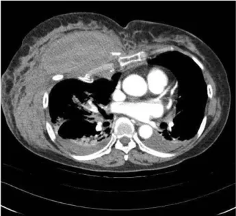
Fig. 2. Follow up contrast enhanced CT revealed a huge hematoma with extravasation and severe right chest wall swelling. The anterior aspect of right lung was markedly compressed by chest wall hematoma.
Fig. 1. Contrast enhanced CT revealed multiple rib fractures on right thoracic cage. Rib fracture and small amout of right chest wall hematoma with extravasation were shown.
으로 안정되었고, 흉벽의 혈종 또한 추적 초음파 및 육안 적으로 감소한 것을 확인할 수 있었다. 환자는 더 이상의 출혈 없이 우측 흉벽의 혈종이 흡수되어 소실되었고, 세균 성 폐렴이 발생하였지만 호전되어 25일간의 중환자 치료 를 마치고 일반병실로 전동되었다.
III. 고 찰
흉부 둔상에 의해 발생하는 가슴 부위 동맥의 흔한 손 상부위는 흉부 대동맥이다. 많은 경우 대동맥 손상은 치명 적 결과를 초래하여 현장에서 사망한다. 대동맥 손상 보다 발생빈도는 낮지만 대동맥의 주요 분지들 또한 손상이 있 을 수 있다.(2) 주요 분지들의 손상은 흔하지 않을 뿐 만 아니라 출혈이 서서히 진행되는 경우 조기 진단이 어려우 며 대개 수술 중 우연히 발견 되는 경우가 많다.(3,4)
내흉동맥은 쇄골하 동맥에서 시작되고 앞갈비사이(ante- rior intercostal branch), 흉골(sternal branch), 그리고 관통 (perforating branch)분지를 갖는다. 내흉동맥은 흉부의 둔 상이나 관통상으로 인한 손상 시 심각한 출혈을 일으킬 수 있는 주요 동맥중의 하나이다.(5,6) 둔상에 의한 내흉동 맥의 손상은 늑골, 쇄골 또는 흉골 같은 주위 뼈조직의 골 절과 동반되어 나타나거나 급격한 가속이나 감속으로 생
긴 전단력이 혈관에 가해지기 때문이다.(7)
손상된 내흉동맥의 진단은 최근 전산화 단층촬영(CT)의 발달과 촬영 빈도의 증가로 CT에 의해 진단되는 경우가 증가하고 있으나 대부분은 혈관조영술로 이루어지거나 수 술 중 발견된다.(8) Multidetector CT (MDCT)의 경우, 둔 상에 의해 발생한 대동맥 손상의 진단에서 높은 민감도 (96%)와 특이도(99%)를 보인 보고가 있으나 내흉동맥 손 상에 의한 혈관외 유출이 보이지 않는 보고도 있다.(8,9) 내유동맥 손상 및 출혈의 정도에 따른 진단의 어려움으로 초기에 출혈을 인지하지 못하는 경우, 지속적인 출혈로 대 량의 혈흉이 발생하거나 종격혈종이 형성될 수 있다. 특히 후자의 경우는 심장의 앞부분에 형성되어 우심방이나 우 심실을 압박하는 심장눌림증(cardiac tamponade)을 초래할 수 있어 적절한 시기에 혈관조영술을 통한 진단 및 색전 술이 이루어져야 할 것으로 보인다. Husted 등은 대동맥의 손상 없이 종격혈종의 크기가 빠르게 증가하는 경우, 내흉 동맥과 늑간동맥의 출혈을 의심하고 혈관조영술과 색전술 을 고려해야 한다고 하였다.(10)
손상된 내흉동맥의 치료는 개흉술이 원칙이며 특히 쇼 크를 보이는 환자에 있어서는 더욱 그러하다. 그러나 쇼크 의 적절한 처치와 함께 카테터경유 동맥색전술을 시행하 여 좋은 결과를 얻은 보고가 있었다.(11) 내흉동맥 손상에 의한 출혈의 경우 색전술을 시행하는 것이 수술에 대한 비용, 시간 및 마취 등으로 인한 위험성을 줄일 수 있을 것으로 보인다.(12) 카테터경유 동맥색전술을 시행하는 경 우, 환자의 혈역학이 불안정하거나 조영제의 유출이 불분 명하여 실패할 가능성이 있으므로 언제든 응급 개흉술을 할 수 있도록 준비해야 할 것이다.
본 증례는 흉부 둔상에 의해 발생한 내흉동맥 손상으로 쇼크와 거대혈종이 발생한 경우로 쇼크에 대한 적절한 처 치와 카테터경유 동맥색전술을 이용해 비수술적인 방법으 로 성공적인 치료한 경우이다. 내흉동맥을 포함한 흉부혈 관 손상이 의심되는 흉부 둔상의 경우 주의 깊은 관찰과 지속적인 감시, FAST, CT 및 혈관조영술 등을 통해 손상 부위를 신속히 확인하고, 적절한 쇼크 처치와 색전술을 시 행하여 효과적으로 비수술적 치료를 할 수 있을 것으로 생각된다.
REFERENCES
01) Irgau I, Fulda GJ, Hailstone D, Tinkoff GH. Internal mammary artery injury, anterior mediastinal hematoma, and cardiac compromise after blunt chest trauma. J Trauma 1995;39:1018-21.
02) Wilkinson J, Jacob TD, Armitage J, Zajko A, Udekwu AO, Peitzman AB. Avulsion of the internal mammary artery caused by blunt trauma. Ann Emerg Med 1993;
─ 298 ─
— Journal of Trauma and Injury Vol. 25, No. 4 —
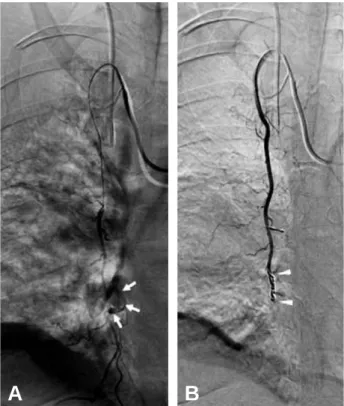
Fig. 3. Selective right internal mammary artery angiogram. (A) Arrows indicate an area of extravasation from perforat- ing branch of internal mammary artery. (B) Arrowheads indicate an area of coil embolization.
A B
─ 299 ─
— 최석진 외: 흉부 둔상에 의해 발생한 내흉동맥 손상의 카테터경유 혈관색전술 치료 경험 —
22:1762-5.
03) Mattox KL, Wall MJ, Jr., Lemaire S. Thoracic great vessel injury. In: Mattox KL, Feliciano DV, Moore EE, eds. Trauma, 7th Edition. New York: McGraw-Hill;
2008:589-606.
04) Mirvis SE, Shanmuganathan K, Buell J, Rodriguez A.
Use of spiral computed tomography for the assessment of blunt trauma patients with potential aortic injury. J Trauma 1998;45:922-30.
05) Henriquez-Pino JA, Gomes WJ, Prates JC, Buffolo E.
Surgical anatomy of the internal thoracic artery. Ann Thorac Surg 1997;64:1041-5.
06) Ritter DC, Chang FC. Delayed hemothorax resulting from stab wounds to the internal mammary artery. J Trauma 1995;39:586-9.
07) Husted JW, Stock JR, Manella WJ. Traumatic anterior mediastinal hemorrhage: control by internal mammary artery embolization. Cadiovasc Intervent Radiol 1982;5:
268-70.
08) Saori Kawamura, Hiroshi Nishimaki, Masakazu
Takigawa, Zong-Bo Lin, Hiroshi Imai, Kazushige Hayakawa, et al. Internal mammary artery injury after blunt chest trauma treated with transcatheter arterial embolization. J Trauma 2006;61:1536-9.
09) Alkadhi H, Wildermuth S, Desbiolles L, Schertler T, Crook D, Marincek B, et al. Vascular emergencies of the thorax after blunt and iatrogenic trauma: multi- detector row CT and three-dimensional imaging.
Radiographics 2004;24:1239-55.
10) Husted JW, Stock JR, Manella WJ. Traumatic anterior mediastinal hemorrhage: Control by internal mammary artery embolization. Cadiovasc Intervent Radiol 1982;5:
268-70.
11) Whigham CJ, Jr., Fisher RG, Goodman CJ, Dodds CA, Trinh CC. Traumatic injury of the internal mammary artery: embolization versus surgical and nonoperative management. Emerg Radilo 2002;9:201-7.
12) Smith DC, Senac MO, Bailey LL. Embolotherapy of a ruptured internal mammary artery secondary to blunt chest trauma. J Trauma 1982;22:333-5.