Abstract (J. Kor. Oral Maxillofac. Surg. 2003;29:272-281)
Ⅰ. 서 론
Cancer chemoprevention은 천연 화합물이나 합성 화합물을 이 용하여 암을 조절, 예방 및 치료하는 의미를 함축한다
1,2). Cancer
chemoprevention이 중요한 이유는 화학요법이나 수술 등의 방법 만으로 충분한 암 치료 효과를 얻을 수 없기 때문이다. 최근에 천 연물로부터 추출한 여러 종류의 물질이 cancer chemoprevention 효과가 우수함이 보고되고 있다. 그 중에서도 Curcuma longa 식 물의 rhizome에서 추출한 curcumin은 항염증작용을 함유하고 있 어서 오래 전부터 전통의약으로 사용되어지고 있다
3-5). Resveratrol 의 구조상 특징은 2개의 phenol 그룹을 함유하며 cis-form 과 trans-form으로 존재하나 포도에는 trans-form만이 존재하며, 일반 적으로 적포도주나 포도쥬스중에는 약 0.1-10 mg/L 정도의 resveratrol를 함유하고 있다. Resveratrol은 구조적인 면에서 phe- nol과 OH기를 함유하여 항산화제의 역할뿐만 아니라 anti-
Curcumin과 resveratrol에 의한 두경부암 유래의 HN-4 세포의 세포주기, 세포사 및 전이관련 단백질의 발현 조절
김사엽∙이상한∙권택규
*경북대학교 치과대학 구강악안면외과학교실, 계명대학교 의과대학 면역학교실
*이 상 한
700-421 대구광역시 중구 삼덕2가 50 경북대학교 치과대학 구강악안면외과학 교실 Sang-Han Lee
Dept. of OMFS, School of Dentistry, Kyungpook National University Samduk 2-ga-50, Jung-gu, Daegu, KOREA, 700-721
Tel : 82-53-420-5911, 5915 Fax : 82-53-426-5365 E mail : [email protected]
EFFECT OF CURCUMIN AND RESVERATROL ON THE CELL CYCLE REGULATION, APOPTOSIS AND INHIBITION OF METASTASIS RELATED PROTEINS IN HN-4 CELLS
Sa-Yub Kim, Sang-Han Lee, Taeg-Kyu Kwon*
Dept. of Oral & Maxillofacial Surgery, School of Dentistry, Kyungpook National University Dept. of Immunology, School of Medicine, Kyemyung University
*Nontraditional or alternative medicine is becoming an increasingly attractive approach for the treatment of various inflammatory dis- orders and cancers. Curcumin is the major constitute of turmoric powder extracted from the rhizomes of the plant Curcuma longa.
Resveratrol is a phytoalexin present in grapes and a variety of medicinal plants. In this report, We investigated the effect of curcumin and resveratrol on regulatory protein of cell cycle, induction of apoptosis and MMP activity.
Treatment with 75 M curcumin for 24 hrs produced morphological changing in HN-4 cells. Curcumin and resveratrol inhibited the cellular growth in HN-4 cells. Inhibition of cell growth was associated with down-regulation of cell cycle regulatory proteins.
Curcumin-induced caspase-3 activation and Bax degradation were dose-dependent with a maximal effect at a concentration of 100 M.
The elevated caspase-3 activity in curcumin treated HN-4 cells are correlated with down-regulation of survivin and cIAP1, but not cIAP2. Curcumin induced a dose-dependent increase of cytochrome c in the cytosol. Curcumin induced-apoptosis was mediated through the release of cytochrome c. In addition, curcumin-induced apoptosis was caused by the generation of reactive oxygen species, which was prevented by antioxidant N-acetyl-cysteine (NAC). Cotreatment with NAC markedly prevented cytochrome c release, Bax cleavage and cell death. Also resveratrol-induced apoptosis was preceded by down-regulation of the anti-apoptotic Bcl-2, cIAP1, and caspase-3 activity. However, resveratrol-induced apoptosis was not prevented by antioxidant NAC. In addition, HN-4 cells release basal levels of MMP2 when cultured in serum-free medium. Treatment of the cells with various concentrations of PMA for 24 hr induced the expression and secretion of latent MMP9 as determined by gelatin zymography. HN-4 cells were treated with various con- centrations of curcumin and resveratrol in the presence of 75 nM PMA, and MMP2 and 9 activities were inhibited by curcumin and resveratrol.
These findings have implications for developing curcumin-based anticancer and anti-inflammation therapies.
Key words: Curcumin, Resveratrol, Cancer chemopreventive agent
platelet aggregation 및 항암등의 생리적 기능 연구가 보고되어 지
고 있다
6-15).
대부분의 cancer chemopreventive agent의 항암 기전은 암세포 에 대하여 생육 억제 및 apoptosis 유도를 야기하므로 이루어지고 있다. 세포주기는 매우 정교하고 복잡한 작용 기전을 통하여 조 절된다. 만약 세포주기 조절에 이상이 생기면 세포는 세포사멸, 성장정지 혹은 암화가 야기된다. 최근 cell cycle의 G1 phase를 조 절하는 하나의 기전은 D type cyclin 이 cyclin dependent kinase(cdk) 4, 5, 6과 결합하여 기질 단백질인 retinoblastoma pro- tein(Rb)의 인산화를 유도함으로서 이루어진다고 보고하고 있다
16-18). 인산화된 Rb는 전사조절 인자인 E2F를 활성화하고, 활성화 된 E2F는 세포주기 조절에 필요한 여러 단백질들을 전사단계에서 조절 한다
19,20). 또 다른 조절 기전은 cdk inhibitor를 통한 cyclin dependent kinase 활성의 조절을 통하여 Rb 단백질의 인산화를 조절 한다
16-18).
세포사멸은 생명체의 발생과 분화에 매우 중요한 과정으로서 정상 세포 및 조직의 유지뿐만 아니라 다양한 질병의 병인에 기 여하고 있다
21,22). 세포사멸 기전은 TNF-α에 의한 세포 사멸 수용 체에 의한 것과 세포내 미토콘드리아에 기인한 것으로 나눌 수 있으며 caspase의 활성, cytochrome c, Bcl-2 등이 관여한다
21,22). 현 재까지 수많은 apoptosis 관련 유전자가 알려 졌는데, 그 중에서 도 Bcl-2는 제일 먼저 발견된 암유발 유전자(proto-oncogene)이다
23,24). Bcl-2는 사람의 림프종의 발생과 밀접하게 관련된 유전자로서 단 백질 분자량은 26 kDa이며 미토콘드리아의 외막, 소포체나 핵막 에 위치해 있고, 다른 암유발 유전자와는 달리 세포증식에 관여 하지 않고 세포의 생존을 조절하는 것으로 밝혀졌다. Bcl-2는 다 양한 기작을 통하여 세포의 사멸을 방지하고 있다. Bcl-2의 세포 사멸 억제 기전은 세포사멸 신호 전달계의 상위 위치를 통제하 기 때문이다. Bcl-2에 의한 조절 기작에는 산화-환원 상태, 막 인 산지질의 비대칭분포, caspase, 미토콘드리아의 막전위, 핵 DNA 의 분절화, 세포사멸 신호 전달계의 활성화 등에 이르기까지 광
범위하다
25-31). 또한 최근에 세포사멸의 실행자로써 caspase의 작
용 기작이 보고 되어지고 있다. 특히 시스테인계의 단백질분해 효소인 caspase가 다양한 apoptosis 자극에 의해서 활성화되며, 활성화된 caspase-3는 세포내의 신호 전달계(PKC δ, PLC-γ1), 구조 단백질(actin), 세포주기조절 단백질(PARP, Rb, p21)등 여러 종류 의 단백질을 특이적으로 분해하므로 일어난다
32-37).
Cancer chemopreventive agent의 작용기전에 apoptosis에 의한 세포 사멸뿐 만 아니라, 암 전이를 억제하는 것 또한 매우 중요하
다
21,22). 진단 및 치료방법의 발달에도 불구하고 암사망의 주요 원
인은 대부분 전이에 기인한다. 종양이 생성 후 주위의 조직으로 부터 영양분의 확산에 의지하나 1-2 mm 이상의 크기로 성장하기 위하여 신생혈관 생성이 필요하다. 이들 종양세포들 중 일부가 종양조직에서 탈리하여 모세혈관이나 venule 을 통하여 순환기 계로 이동하다가 특정 부위에 부착하여 모세혈관을 뚫고 조직 내로 진입한다. 전이과정 중에 종양세포의 탈리와 침윤에 matrix metalloproteinase(MMP)가 관련된다
38-41). 현재까지 16가지의 MMPs
family가 발견되었으며 다음과 같은 특성을 가진다. MMPs는 1) 세포외 기질 성분 중 적어도 한 가지 이상을 분해 시킨다. 따라서 본 연구에서는 MMP 발현조절에 있어서 curcumin의 작용 기전을 중점적으로 규명하고자 하였다. 2) Zn 이온과 결합하는 HEXGH motif를 함유하는 catalytic domain을 함유한다. 3) 비활성형으로 분비하여 활성화 과정을 거쳐 활성화 된다
38-41).
이에 본 연구에서는 curcumin과 resveratrol에 의한 cancer chemopreventive agent 작용 기전을 규명하고자 apoptosis 유도과 정에서의 curcumin의 작용기전과 세포주기조절 단백질의 발현 및 전이에 관련된 MMP 단백질의 활성에 대한 연구를 행하였다.
Ⅱ. 실험재료 및 방법
1. 세포배양과 유지
실험에 사용한 세포는 인간 두경부 암 AMC-Head & Neck- 4(HN-4) 세포주를 KTCC에서 분양 받아서 사용하였다. HN-4 세포 주는 10% bovine calf serum과 1% penicilline-streptomycine이 함유 된 RPMI 배지를 이용하여 세포 성장 최적조건인 37℃, 5% CO
2로 평형된 CO
2배양기에서 배양하였다. 배양된 세포는 약 48시간 간 격으로 계대 배양하여 6-well plate 또는 12-well plate에서 동일한 수의 세포를 split하여 실험하였다.
2. 세포 성장율 측정
세포 성장율을 측정하기 위하여 살아있는 세포를 trypan blue 로 1:1 염색하여 hemo-cytometer를 사용하여 측정하였다. 즉 1×
10
6/ml의 세포를 6-well plate에 도포하고, curcumin 및 resveratrol 을 처리한 다음 계속하여 24시간 동안 배양한 후, trypan blue 염 색하여 세포의 수를 측정하였다. 대조군은 curcumin 및 resvera- trol을 처리하지 않은 군의 세포수를 측정하여 실험군과 비교 검 토하였다.
3. 총 단백질 추출 및 단백질 전기영동
세포를 6-well plate에 동일한 수를 도포하고 curcumin 및 resver- atrol을 처리한 후 24 시간동안 배양하였다. 24 시간후 세포를 cold-phosphate buffered saline (PBS) buffer로 세척하고 trypsin 처 리한 후 3000 rpm에서 5 분간 원심분리하여 세포를 수확하였다.
수확한 세포는 각종 단백질 분해효소 저해제가 함유된 lysis buffer (50 mM Tris-HCl, pH 8.0, 5 mM EDTA, 150 mM NaCl, 0.5%
Nonidet-40)로 용해한 다음, 10,000 x g에서 20분간 원심분리하여
상층액을 취하여 총 단백질 분획을 얻었다. 총 단백질은 Bradford
시약을 사용하여 정량하였다. 정량된 총 단백질 50 μg 을 10 %
reducing SDS-polyacrylamide gel에서 전기영동을 통하여 분리하
였다.
4. Western Blot 분석
시료를 처리한 세포에서 총 단백질을 얻고 전기영동을 하여 분 리한 단백질은 nitrocellulose membrane으로 20% methanol, 25 mM Tris, 및 192 mM glycine buffer 하에서 transfer tank를 이용하여 이 동하였다. 단백질이 이동된 membrane은 Ponceau solution으로 transfer의 유무를 확인한 후, 5% non-fat dry milk solution으로 30 분간 실온에서 반응하여 blocking 하였다. Blocking 후 blocking buffer로 1000배 희석한 특이한 antibody와 membrane을 4시간 이 상 반응하였다. 반응이 끝난 후 Tris-Tween buffered saline(TTBS) 으로 5분 간격으로 6회 세척하였다. 계속하여 Horse radish perox- idase가 부착된 이차항체인 goat anti-IgG antibody와 2시간 반응시 키고 다시 한번 TTBS로 6회 세척하였다. 세척이 끝나면 증류수 로 세척하고 ECL solution으로 1분간 반응하고 코닥 필름에 감광 하여 나타난 band의 두께를 비교하여 단백질 발현 유무 및 그 정 도를 확인하였다.
5. Curcumin 및 resveratrol에 의한 apoptosis 조절 단 백질의 발현조사
Curcumin 및 resveratrol에 의한 apoptosis 유도시 apoptosis 조절 단백질의 발현을 조사하기 위하여 HN-4 세포에 curcumin를 처리 한 후 수확한 세포를 PBS로 세척하고 lysis buffer로 용해한다.
Apoptosis 를 조절하는 단백질의 발현 정도를 조사하기 위하여 동일한 량의 단백질 SDS-gel에 전기영동하여 nitrocellulose mem- brane에 전달하여 Bcl-2, Bcl-xL, Bax, caspase-3와 PLC-γ1 antibody 로 hybridization 시킨 후 second antibody를 다시 hybridization하여 ECL 방법으로 측정하였다..
6. Cytochrome c 유리
HN-4 세포에 다양한 농도의 curcumin을 처리하여 24시간 배양 하여 수확한 세포를 PBS buffer로 1회 washing 한 후 0.16 μM PMSF가 함유된 50 ㎕의 Hypotonic buffer (20 mM HEPES, pH 7.4 ; 10 mM KCl ; 2 mM MgCl
2; 1 mM EDTA) 를 첨가하여 voltexing을 통해 4℃에서 20 분간 반복하여 용해시켰다. 15,000 rpm에서 원 심하여 세포질 단백질을 회수한 후 spectrophotometer를 이용하 여 상기의 단백질 정량법에 의해 정량한 후 40 μg의 단백질을 12%
reducing SDS-PAGE 상에서 전기영동하여 Western Blot 분석을 수 행하였다.
7. Caspase-3 활성 측정
6-well plate에 동일한 수의 세포를 도포하고 serum이 첨가되지 않은 RPMI 배지를 이용하여 세포를 24 시간 결식시켰다. 다양한 농도로 curcumin을 처리한 후 24시간 동안 배양시킨 후 상기의 방법으로 단백질을 추출하여 정량하였다. Sample 30 μg의 단백질 을 100 μM의 caspase-3 substrate I이 첨가된 200 ㎕의 PBS buffer와
혼합하여 시간별로 405 nm에서 caspase-3 활성을 측정하였다.
8. Zymography 방법
6-well plate에 동일한 수의 세포를 도포하고 세포가 부착된 것 을 확인하고 serum이 첨가되지 않은 RPMI 배지를 이용하여 세포 를 24시간 결식시켰다. 그 후 다양한 농도의 curcumin을 처리한 후 24시간 동안 배양시켜 배양 상등액을 회수하여 이를 non reducing dye와 혼합하여 0.1% gelatin이 포함된 10%-PAGE에서 전 기 영동하였다. 전기영동 후 gel을 증류수로 2회 washing 한 후 10 분 간격으로 1시간 동안 2.5% Triton X-100으로 세척하였다. 세척 한 gel을 developer buffer(50 mM Tris-HCl, pH 7.6; 10 mM CaCl
2)를 이용하여 37℃에서 24시간 반응시킨 후 단백질 염색용액(0.5%
coomassie brilliant blue R250, 30% isopropyl alcohol, 10% acetic acid)으로 30분 동안 염색시킨 후 탈색 용액 (10% methanol, 10%
acetic acid)으로 탈색시켜 나타난 band의 두께를 비교하여 단백 질 발현정도를 비교하였다.
Ⅲ. 성 적
1. Curcumin과 resveratrol 처리 시 세포주기 조절인자 들의 발현양상
다양한 농도의 curcumin과 resveratrol을 각각 AMC-Head and Neck-4(HN-4) 세포에 처리한 후 세포주기 조절인자들의 발현양 상을 조사하였다. 다양한 농도의 curcumin을 24시간 처리한 후 단백질을 추출하여 Western blot 분석을 시행하였다. 세포주기 중 G1/S 기의 전이 과정에 작용하는 cdk2 단백질은 curcumin 농도에 관계없이 발현 정도가 일정하였으나, 초기 G1 기에 작용하는 cdk4와 G2/M 기에 작용하는 cdk1의 단백질 발현은 농도 의존적 으로 발현이 감소하였다. 세포주기 조절의 주요 조절인자인 cyclin B, D3, E의 발현은 curcumin 처리에 따라 변화가 미약하였 다. Cdks 저해제인 p21과 p27 단백질 발현은 고농도의 curcumin 에 의하여 억제됨을 확인하였다. Resveratrol에 의한 HN-4 세포에 서 세포주기 조절 단백질 발현의 양상을 조사하였다(Fig. 1).
Resveratrol에 의한 세포주기 조절 단백질의 발현정도는 curcumin 처리와 유사하였으나, cyclin A와 cdk4 단백질의 발현이 resvera- trol 처리 농도 의존적으로 급격한 감소 현상을 보였다(Fig. 2).
2. Curcumin과 resveratrol 처리에 의한 Bcl-2 family 단백질의 발현
Curcumin과 resveratrol에 의한 Bcl-2 family 단백질의 발현에 미
치는 영향을 조사하기 위하여 HN-4 세포에 다양한 농도의 cur-
cumin과 resveratrol을 24 시간 처리한 후 단백질을 추출하여
Western blot 분석을 시행하였다. 일반적으로 anti-apoptosis 기능
이 알려진 Bcl-2와 Bcl-xL 단백질의 발현은 curcumin 처리에 의하
여 변화가 없었다. 그러나 resveratrol 처리 시 Bcl-2 단백질 발현은
농도 의존적으로 단백질의 발현이 감소함을 보였다. Curcumin과 resveratrol 처리 시 pro-apoptosis 기능을 함유한 Bax 단백질 발현 의 증가 현상은 보이지 않았으나 Bax 단백질의 절단 현상을 확인 하였다(Fig. 3).
3. Curcumin과 resveratrol 처리에 의한 IAP family 단 백질의 발현
Curcumin과 resveratrol에 의한 caspases 저해제로 밝혀진 IAP family 단백질의 발현에 미치는 영향을 조사하기 위하여 HN-4 세 포에 다양한 농도의 curcumin을 24시간 처리한 후 cIAP1, cIAP2, XIAP 및 survivin 단백질의 발현을 Western blot 방법으로 확인하 였다. cIAP2 단백질은 curcumin 처리에 의하여 영향을 받지 않았 으나, cIAP1, XIAP 및 survivin 단백질은 curcumin 농도 의존적으로 감소 현상을 보였다(Fig. 4). Resveratrol에 의한 IAP family 단백질 의 발현 양상을 조사한 결과 survivin 의 발현 감소는 낮은 농도에 서 확인 되었으며, cIAP1과 XIAP는 resveratrol 처리에 의하여 영향 을 받지 않았다.
4. Curcumin에 의한 caspase-3 활성에 대한 영향
Curcumin에 의한 apoptosis 과정중의 caspase-3의 관련성을 조 사하기 위하여 다양한 농도의 curcumin을 처리한 후 caspase-3에 특이적인 기질을 사용하여 caspase-3 활성을 측정하였다.
Caspase-3의 활성은 50 μM curcumin 시 급격한 증가 현상을 보였 다(Fig. 5). 또한 pro-caspase-3의 단백질 발현현상을 조사한 결과 50 μM curcumin 처리부터 감소 현상을 보였다. Resveratrol에 의한 pro-caspase-3의 단백질 발현 현상을 조사한 결과 고농도에서 pro-caspase-3 감소 현상을 보였다 (Fig. 6).
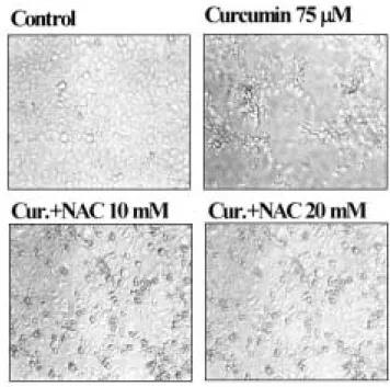
5. Curcumin과 N-acetylcysteine (NAC) 처리에 의한 HN-4 세포의 형태적 변화
Curcumin 처리에 의한 HN-4 세포의 형태적 변화를 관찰하기 위하여 75 μM 농도의 curcumin 을 24 시간 처리한 후 형태적 변화 를 관찰하였다. Curcumin 을 처리하지 않은 세포는 fibroblast 와 같은 건강한 상태의 형태를 보였다. HN-4 세포는 75 μM curcumin 을 처리 시 plate 에 골고루 분포하지 않으며, 사멸한 세포와 둥근 형태의 세포로 변하였으며 일부 세포는 부유 상태로 존재하였 다. Curcumin 처리에 의한 세포의 형태 변화에 항산화제인 NAC 를 처리한 후 세포 형태를 관찰하였다. 20 mM NAC 과 curcumin 을 동시에 처리한 경우 많은 세포들이 건강한 상태의 세포로 전 환됨을 확인하였다(Fig. 12).
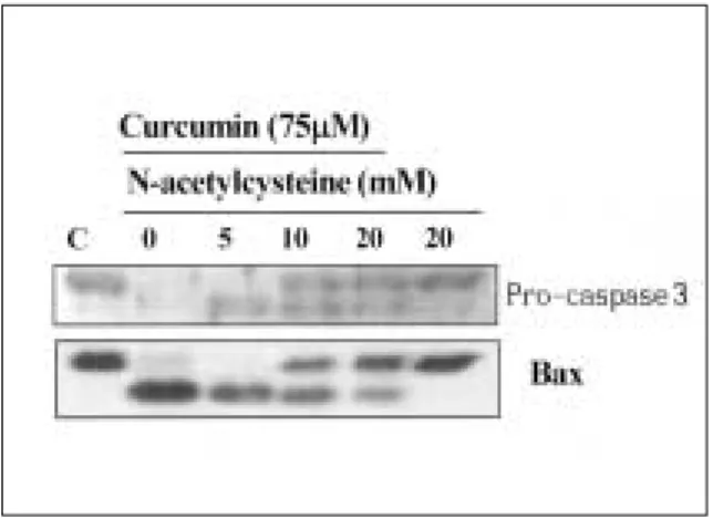
6. Curcumin 과 NAC 처리에 의한 apoptosis 관련 단 백질 발현 양상
Curcumin 처리에 의한 apoptosis 관련 단백질 발현 변화에 항산
화제인 NAC 의 영향을 조사 하기 위하여 curcumin과 NAC를 동 시에 처리한 후 pro-caspase-3와 Bax 단백질 발현을 조사하였다.
Pro-caspase-3는 75 μM curcumin에 완전히 활성화되었으나, NAC 과 curcumin을 동시에 처리한 세포에서는 일부 pro-caspase-3가 존재함을 확인하였다. Caspase의 기질 단백질로 알려진 Bax 단백 질의 발현 변화를 측정한 결과 Bax 단백질 발현 정도는 차이가 없었으나 분절되는 현상은 NAC 에 의하여 저해됨을 확인하였다 (Fig. 8).
7. Resveratrol과 NAC 처리에 의한 apoptosis 관련 단 백질 발현 양상
Resveratrol 처리에 의한 apoptosis 관련 단백질 발현 변화에 항 산화제인 NAC 의 영향을 조사하기 위하여 resveratrol과 NAC를 동시에 처리한 후 pro-caspase-3와 PLC-γ1 단백질 분절 현상을 조 사하였다. 100 μM resveratrol 처리에 의해 pro-caspase-3 량은 감소 하였다. 100 μM resveratrol과 다양한 농도의 NAC 첨가한 후 pro- caspase-3 량은 변화가 없었다. Caspase의 기질 단백질로 알려진 PLC-γ1 단백질 분절 현상을 측정한 결과 NAC 처리에 영향을 미 치지 않았다(Fig. 8).
8. Curcumin과 NAC 처리에 의한 세포질로 cy- tochrome c의 유리 현상
Curcumin에 의한 apoptosis 유도과정에서의 미토콘드리아의 cytochrome c가 세포질로 유리되는지를 조사하기 위하여 75 μM curcumin을 처리한 후 세포질에 유리된 cytochrome c를 확인하였 다. 75 μM curcumin 처리 시 세포질로의 cytochrome c 유리 현상 이 NAC에 의해 저해 여부를 확인하였다. 5 mM NAC과 75 μM cur- cumin 처리 시 세포질로 cytochrome c 유리가 급격히 감소하였 다. 세포질 단백질의 정량을 위하여 Hsp70 단백질을 Western blot 으로 확인한 결과 동일량의 단백질 loading을 확인 할 수 있었다 (Fig. 9).
9. Curcumin 처리에 의한 MMP 단백질의 발현 및 활성 에 대한 영향
Curcumin에 의한 암전이 억제 능력을 조사하기 위하여 MMP2
와 9의 활성에 미치는 영향을 조사하였다. 다양한 농도의 cur-
cumin을 HN-4 세포에 24 시간 처리 한 후 배양액을 취하여 MMP
활성을 조사하였다. MMP 활성은 gelatin gel을 이용한 zymogra-
phy 방법으로 측정하였다. MMP9 활성은 HN-4 세포에서 확인 할
수 없었다. MMP2의 활성은 75 μM curcumin 처리부터 급격히 감
소하였다. 암을 유발할 수 있는 protein kinase C(PKC)의 activator
로 작용하는 phorbol myristate acetate(PMA)를 HN-4 세포에 처리
한 후 MMP 활성을 조사한 결과 PMA는 특이적으로 MMP9의 활
성 증가를 유도하였다. Curcumin에 의한 PMA 유도 MMP9의 저해
능력을 조사하기 위하여, 75 nM PMA와 curcumin을 동시에 처리
한 후 MMP 활성을 측정한 결과 MMP9 및 MMP2의 활성을 50 μM curcumin 부터 급격히 저해 현상을 보였다(Fig. 10).
10. Resveratrol 처리에 의한 MMP 단백질의 발현 및 활성에 대한 영향
Resveratrol에 의한 암전이 억제 능력을 조사하기 위하여 MMP2 와 9의 활성에 미치는 영향을 조사하였다. 다양한 농도의 resvera- trol을 HN-4 세포에 24시간 처리 한 후 배양액을 취하여 MMP 활 성을 조사하였다. MMP 활성은 gelatin gel을 이용한 zymography 방법으로 측정하였다. MMP9 활성은 HN-4 세포에서 확인 할 수 없었으나, MMP2의 활성은 고농도의 resveratrol에 의하여 미약하 게 저해되었다. PMA를 HN-4 세포에 처리한 후 MMP 활성을 조사 한 결과 PMA는 특이적으로 MMP9의 활성 증가를 유도하였다.
Resveratrol에 의한 PMA 유도 MMP2와 9의 저해능력을 조사하기 위하여, 75 nM PMA와 resveratrol을 동시에 처리한 후 MMP2와 9 의 활성을 측정한 결과 MMP2와 MMP9의 활성이 resveratrol에 의 해 미약하게 저해됨을 확인하였다(Fig. 11).
Ⅳ. 고 찰
Cancer chemoprevention은 천연 화합물이나 합성 화합물을 이 용하여 암을 조절, 예방 및 치료하는 의미를 함축한다
1,2). Cancer chemo-prevention이 중요한 이유는 화학요법이나 수술 등의 방 법만으로 충분한 암 치료 효과를 증대할 수 없기 때문이다. 최근 에 천연물로부터 추출한 여러 종류의 물질이 cancer chemo-pre- vention 효과가 우수함이 보고 되고 있다. Curcumin은 Curcuma longa의 rhizome으로부터 추출한 노란색의 pigment로 오래 전부 터 임상적 가치를 인정받아 오고 있다
3-5). 또한 포도에서 추출한 resveratrol의 효과는 적포도주를 많이 음용한 사람의 경우 낮은 심장관련 질환 발병률과의 상관관계를 보이므로 흥미를 유발하 였다
8).
본 연구에서는 curcumin과 resveratrol에 의한 두경부암 세포주 인 HN-4 세포에서의 세포주기 조절 및 apoptosis 유도과정과 암 전이 과정에 주요한 기능을 하는 MMP 활성에 미치는 영향을 규 명하고, 더 나아가 apoptosis 과정에 관련된 여러 단백질의 발현 조절을 분자 생물학적 기전을 이해하고자 하였다.
두경부암 세포주에 다양한 농도의 curcumin과 resveratrol을 24 시간 처리한 후 세포주기 조절 단백질 발현을 비교 분석한 결과 curcumin과 resveratrol은 비슷한 세포주기 조절 단백질의 발현 경 향을 보였다. Resveratrol에 의하여 cyclin A의 발현이 급격한 감소 를 보였다. 이 결과는 백혈병세포주인 U937 세포에 resveratrol이 S기의 arrest를 야기하는 결과와 일치한다
42). Curcumin과 resvera- trol에 의한 세포 생육 억제의 원인은 세포주기 조절 단백질 중 cdk4의 발현 감소가 하나의 원인으로 작용하는 것으로 생각된 다.
녹차의 주성분인 epigallocatechin gallate(EGCG) 등 여러 종류의 cancer chemo-preventive agent는 암세포에 대한 특이적인 apopto-
sis 유도 기능을 가지고 있다
43,44). 본 연구에서는 curcumin과 resveratrol 처리에 의한 두경부암 세포의 세포사멸이 apoptosis와 관련 있음을 조사하기 위하여 Bcl-2 family 단백질과 caspase 효소 저해제인 IAP family 단백질의 발현정도를 조사하였다. 두경부암 세포에 curcumin을 처치한 결과 anti-apoptotic 기능을 함유한 Bcl- xL 및 Bcl-2 단백질 발현의 변화는 확인할 수 없었다. 이는 cur- cumin에 의한 두경부암 세포의 apoptosis 유도 기전은 Bcl-2 단백 질의 down-regulation과는 무관함을 제시한다. 그러나 resveratrol 에 의한 apoptosis 과정에서는 Bcl-2 단백질의 발현 감소를 확인 하였다. Curcumin과 resveratrol에 의한 IAP family 단백질의 발현 양상을 조사한 결과 cIAP1과 survivin 단백질의 발현 감소가 농도 의존적으로 야기됨을 확인하였다. Curcumin과 resveratrol에 의한 apoptosis과정 중에 주요한 작용을 하는 caspase-3 활성을 조사하 였다. 32 kDa의 pro-caspase-3는 비활성화 형태이며 caspase-3가 활성화되면 상위의 caspase에 의해 절단되어 활성화되어야 한다
45,46). 비활성화 형태의 pro-caspase-3 단백질의 양적 감소는 활성화 형 태의 caspase-3가 증가함을 의미한다
45,46). 두경부암 세포에 다양한 농도의 curcumin을 처리한 결과 caspase-3의 비활성 형태의 pro- caspase-3 단백질의 양적 감소와 caspase-3 활성의 증대를 확인하 였다. 세포주기 조절의 주요한 기능을 하는 Rb 단백질, 세포의 신 호 전달계의 주요한 PKC delta와 PLC- γ1 단백질 및 Bax 단백질 등이 caspase-3의 기질로 작용한다
32,33). 본 실험에서도 curcumin 처 리에 의하여 Bax 단백질의 분절을 확인하였으며 이 결과는 cur- cumin에 의한 apoptosis 유도 과정에 caspase-3가 관여된 것으로 생각된다.
Caspase의 활성은 IAP family 단백질 외에도 cytochrome c의 관 련성이 보고되었다
30,31). Caspase-3 활성의 신호전달의 과정에서의 upstream의 조절인자로 cytochrome c가 미토콘드리아 막에서 해 리하여 cytosol로 분리되면 caspase-9과 Apaf-1 단백질과 결합하여 cas-pase-9를 활성화시킨다. 활성화된 caspase-9은 다시 caspase-3 를 활성화시킨다
47,48).
Curcumin 처리에 의한 cytosol로의 cytochrome c 유리정도를 측 정한 결과 두경부 암 세포에서 cytochrome c 의 유리를 확인하였 다.
두경부 암 세포에서 curcumin에 의한 apoptosis 유도과정이 활 성산소에 의하여 야기되는 것을 확인하기 위하여 항산화제 기능 이 있는 NAC을 처리한 결과 curcumin에 의한 apoptosis 유도를 억 제함을 확인하였다. Curcumin과 NAC의 동시 처리는 caspase-3 활 성의 억제 뿐 만아니라 Bax 단백질의 분절 및 cytochrome c 유도 현상도 억제하였다. 그러나 resveratrol에 의한 apoptosis 기전은 NAC 처리에 의하여 caspase-3 활성 및 PLC-γ1 단백질 분절 억제 현상을 관찰 할 수 없었다. 이상의 결과는 curcumin에 의한 apop- tosis 유도 기전과 resveratrol에 의한 apoptosis 유도 기전이 상이함 을 암시한다.
Curcumin과 resveratrol에 의한 암전이 과정에 주요한 역할을 하
는 MMP2와 MMP9 활성에 미치는 영향을 조사한 결과 MMP2는
고농도의 curcumin 및 resveratrol 처리에 의하여 활성이 억제되었
다. 이상의 결과들을 근간으로 하여 curcumin과 resveratrol 처리
에 의한 두경부암 세포의 생육억제, 세포주기 조절 단백질의 발 현 변화 및 apoptosis 유도와 암 전이 관련 단백질인 MMP 활성 억 제 등의 결과는 curcumin과 resveratrol이 cancer chemopreventive agent로 작용하는 핵심 기전의 일부가 규명되리라 생각된다.
Ⅴ. 요 약
본 연구에서는 두경부암 HN-4 세포에서 cancer chemopreven- tive agent인 curcumin과 resveratrol를 처리하여 HN-4 세포의 생육 억제의 원인이 apoptotic cell death에 의하여 일어나며 세포 주기 조절 단백질의 발현 및 암 전이에 관련된 MMP 활성 저해 기전에 대하여 이해하고자 하였다.
HN-4 세포에 다양한 농도(10-100 μM) curcumin을 처리하여 세 포주기 조절 단백질의 발현을 측정한 결과 cdk1과 cdk4 단백질 이 농도 의존적으로 발현이 감소하였으며, resveratrol 처리에서는 cyclin A 단백질의 특이적인 감소 현상을 확인하였다. Curcumin에 의한 apoptosis 유도 기전을 조사한 결과 anti-apoptotic 기능이 있 는 Bcl-2 및 Bcl-xL 단백질 발현 감소 현상은 없었으나, caspase 저 해 IAP family 단백질중 cIAP1과 survivin 단백질 발현 현상이 처리 농도 의존적으로 감소하였다. Resveratrol을 처리한 경우 Bcl-2 및 survivin 단백질 발현 감소현상을 확인하였다. Curcumin과 resver- atrol에 의한 apoptosis 과정은 caspase-3 의존적인 apoptosis 유도 기전을 보였다. Curcumin에 의한 apoptosis 과정은 항산화제인 NAC 처리에 의해서 저해되었다. Curcumin과 NAC 동시 처리는 cytochrome c 유리, caspase-3 활성화 및 Bax 단백질 분절 현상을 억제하였다. 그러나 resveratrol에 의한 apoptosis 과정은 NAC 처리 에 의하여 억제되지 않았다. Curcumin과 resveratrol에 의한 암 전 이 관련 단백질인 MMP2와 9의 활성 저해효과도 확인하였다.
결론적으로, curcumin에 의한 항암효과는 세포주기 조절 및 apoptosis 유도 및 전이 관련 단백질의 활성 억제를 통하여 야기 되는 것으로 생각되어 진다.
참고문헌
1. Kelloff GJ, Johnson JR, Crowell JA, Boone CW, DeGeorge JJ, Steele VE, Mehta MU, Temeck JW, Schmidt WJ, Burke G, and Sigman CC : Approaches to the development and marketing approval of drugs that prevent cancer. Cancer Epidemiol. Biomarkers Prev., 1995 ; 4 :1-10.
2. Ames BN, Gold LS, and Willett WC : The causes and prevention of cancer. Proc. Natl. Acad. Sci. U. S. A., 1995 ; 92 : 5258-5265.
3. Ammon HP, and Wahl MA : Pharmacology of Curcuma longa.
Planta. Med., 1991 ; 57 :1-7.
4. Huang MT, Smart RC, Wong CQ, and Conney AH : Inhibitory effect of curcumin, chlorogenic acid, caffeicacid, and ferulic acid on tumor promotion in mouse skin by\12-O-tetradecanoylphor bol-13-acetate.
Cancer Res., 1998 ; 48 : 5941-5946.
5. Huang MT, Newmark HL, and Frenkel K : Inhibitory effects of cur- cumin on tumorigenesis in mice. J. Cell Biochem. Suppl., 1997 ; 27 : 26-34.
6. Soleas, GJ., Diamandis, EP., and Goldberg, DM. : Wine as a biologi- cal fluid:history, production, and role in disease prevention. J. Clin.
Lab. Anal., 1997 ; 11: 287-313.
7. Soleas, GJ., Diamandis, EP., and Goldberg, DM. : Resveratrol: a mol-
ecule whose time has come? And gone? Clin. Biochem., 1997 ; 30 : 91-113.
8. Fremont, L. : Biochemical effect of resveratrol. Life Sci., 2000 ; 66 : 6663-6673.
9. Jang, M., Cai, L., Udeani, GO., Slowing, KV., Thomas, CF., Beecher, CW. Fong, HH., Farnsworth, NR., Kinghorn, AD., Mehta, RG., Moon, RC., and Pezzuto, JM. : Cancer chemopreventive activity of resvera- trol, a natural product derived from grapes. Science, 1997 ; 275 : 218-220.
10. Fauconneau, B., Waffo-Teguo, P., Huguet, F. Barrier, L., Decendit, A., and Merillon, JM. : Comparative study of radical scavenger and antioxidant properties of phenolic compounds from Vitis vinifera cell cultures using in vitro tests. Life Sci., 1997 ; 61 : 2103-2110.
11. Pace-Asciak, CR., Rounova, O., Hahn, SE., Diamandis, EP., and Goldberg, DM. : The red wine phenolics trans resveratrol and quercetin block human platelet aggregation and eicosanoid synthe- sis :Implication for protection against coronary heart disease. Clin.
Chim. Acta., 1996 ; 246 :163-182.
12. Mgbonyebi, OP., Russo, J., and Russo, IH. : Antiproliferative effect of synthetic resveratrol on human breast epithelial cells. Int. J.
Oncol., 1998 ; 12 : 865-869.
13. Clement, MV., Hirpara, JL., Vhawdhury, SH., and Pervaiz, S. : Chemopreventive agent resveratrol, a natural product derived from grapes, triggers CD95 signaling-dependent apoptosis in human tumor cells. Blood, 1998 ; 92: 996-1002.
14. Surh, YJ., Hurh, YJ., Kang, JY., Lee, E., Kong, G., and Lee, SJ. : Resveratrol, an antioxidant present in red wine, induces apoptosis in human promyelocytic leukemia (HL60) cells. Cancer letters, 1999
; 140:1-10.
15. Ragione, FD., Cucciolla, V., Borriello, A., Pietra, VD., Racioppi, L., Soldati, G., Manna, C., Galletti, P., and Zappia, V. : Resveratrol arrests the cell division cycle at S/G2 phase transition. Biochem.
Biophys. Res. Commun., 1998 ; 250: 53-58.
16. Hunter, T. and Pines, J. : Cyclins and cancer. II: Cyclin D and CDK inhibitors come of age. Cell, 1994 ; 79: 573-82.
17. Morgan, DO. : Principles of CDK regulation. Nature, 1995 ; 374:
131-134.
18. Sherr, CJ. and Roberts, JM. : Inhibitors of mammalian G1 cyclin- dependent kinases. Genes Dev. 1995 ; 9: 1149-1163.
19. Ikeda, MA., Jakoi, L. and Nevins, JR. : A unique role for the Rb pro- tein in controlling E2F accumulation during cell growth and differ- entiation. Proc. Natl. Acad. Sci. U. S. A. 1996 ; 93: 3215-3220.
20. Moberg, K., Starz, MA. and Lees, JA. : E2F-4 switches from p130 to p107 and pRB in response to cell cycle reentry. Mol. Cell. Biol. 1996
; 16: 1436-1449.
21. Wyllie, AH. : Apoptosis. Br. J. Cancer, 1993 ; 67: 205-208.
22. Thompson, CB. : Apoptosis in the pathogenesis and treatment of disease. Science, 1995 ; 267: 1456-1462.
23. Tsujimoto, Y., Jaffe, E., Cossman, J., Gorham, J., Nowell, PC. and Croce, CM. : Clustering of breakpoints on chromosome 11 in human B-cell neoplasms with the t(11;14) chromosome translocation.
Nature, 1985 ; 315: 340-343.
24. Tsujimoto, Y. and Croce, CM. : Analysis of the structure, transcripts, and protein products of bcl-2, the gene involved in human follicular lymphoma. Proc. Natl. Acad. Sci. U. S. A. 1986 ; 83: 5214-5218.
25. Lawrence, MS., Ho, DY., Sun, GH.,Steinberg, GK. and Sapolsky, RM.
: Over expression of Bcl-2 with herpes simplex virus vectors pro- tects CNS neurons against neurological insults in vitro and in vivo. J.
Neurosci., 1996 ; 16: 486-496.
26. Shimizu, S., Eguchi, Y., Kosaka, H., Kamiike, W., Matsuda, H. and Tsujimoto, Y. : Prevention of hypoxia-induced cell death by Bcl-2 and Bcl-xL. Nature, 1995 ; 374: 811-813.
27. Tudor, G., Aguilera, A,. Halverson, DO. ,Laing, ND. and Sausville, EA. : Susceptibility to drug-induced apoptosis correlates with differ- ential modulation of Bad, Bcl-2 and Bcl-xL protein levels. Cell Death Differ., 2000 ; 7: 574-586.
28. illardon, F., Moll, I., Meyer, M. and Michaelidis, TM. : Alterations in cell death and cell cycle progression in the UV-irradiated epidermis
of bcl-2-deficient mice. Cell Death Differ., 1999 ; 6: 55-60.
29. Kyprianou, N., King, ED., Bradbury, D. and Rhee, JG. : bcl-2 over- expression delays radiation-induced apoptosis without affecting the clonogenic survival of human prostate cancer cells. Int. J. Cancer, 1997 ; 70: 341-348.
30. Yang, J., Liu, X., Bhalla, K., Kim, CN., Ibrado, AM., Cai, J., Peng, TI., Jones, DP. and Wang, X. : Prevention of apoptosis by Bcl-2: release of cytochrome c from mitochondria blocked. Science, 1997 ; 275:
1129-1132.
31. Kluck, RM., Bossy-Wetzel, E., Green, DR. and Newmeyer, DD. : The release of cytochrome c from mitochondria: a primary site for Bcl-2 regulation of apoptosis. Science, 1997 ; 275: 1132-1136.
32. Koriyama, H., Kouchi, Z., Umeda, T., Saido, TC., Momoi, T., Ishiura, S. and Suzuki, K. : Proteolytic activation of protein kinase C delta and epsilon by caspase-3 in U937 cells during chemotherapeutic agent-induced apoptosis. Cell Signal., 1999 ; 11: 831-838.
33. Park, JW., Choi, YJ., Suh, SI., Baek, WK., Suh, MH., Jin, IN., Min, DS., Woo, JH., Chang, JS., Passaniti, A., Lee, YH. and Kwon, TK. : Bcl-2 overexpression attenuates resveratrol-induced apoptosis in U937 cells by inhibition of caspase-3 activity. Carcinogenesis, 2001
; 22: 1633-1639.
34. Mashima, T., Naito, M., Noguchi, K., Miller, DK., Nicholson, DW.
and Tsuruo, T.: Actin cleavage byCPP-32/apopain during the devel- opment of apoptosis. Oncogene, 1997 ; 14: 1007-1012.
35. Decker, P., Isenberg, D. and Muller, S. : Inhibition of caspase-3- mediated poly(ADP-ribose) polymerase (PARP) apoptotic cleavage by human PARP autoantibodies and effect on cells undergoing apoptosis. J. Biol. Chem., 2000 ; 275: 9043-9046.
36. An, B. and Dou, QP. : Cleavage of retinoblastoma protein during apoptosis: an interleukin 1 beta-converting enzyme-like protease as candidate. Cancer Res., 1996 ; 56: 438-442.
37. Zhang, Y., Fujita, N. and Tsuruo, T. : Caspase-mediated cleavage of p21Waf1/Cip1 converts cancer cells from growth arrest to undergo- ing apoptosis. Oncogene, 1999 ; 18: 1131-1138.
38. John, A. and Tuszynski, G. : The role of matrix metalloproteinases in tumor angiogenesis and tumor metastasis. Pathol. Oncol. Res.,
2001 ; 7: 14-23.
39. Stamenkovic, I. : Matrix metalloproteinases in tumor invasion and metastasis. Semin. Cancer Biol., 2000 ; 10: 415-433.
40. Curran, S. and Murray, G. I. : Matrix metalloproteinases: molecular aspects of their roles in tumour invasion and metastasis. Eur. J.
Cancer, 2000 ; 36: 1621-1630.
41. Nelson, AR., Fingleton, B., Rothenberg, ML. and Matrisian, LM. : Matrix metalloproteinases: biologic activity and clinical implications.
J. Clin. Oncol., 2000 ; 18: 1135-4119.
42. Park, JW., Choi, YJ., Jang, MA., Lee, YS., Jun, DY., Suh, SI., Baek, WK., Suh, MH., Jin, IN. and Kwon, TK. : Chemopreventive agent resveratrol, a natural product derived from grapes, reversibly inhibits progression through S and G2 phases of the cell cycle in U937 cells. Cancer Lett., 2001 ; 163: 43-49.
43. Chung, LY., Cheung, TC., Kong, SK., Fung, KP., Choy, YM., Chan, ZY. and Kwok, TT. : Induction of apoptosis by green tea catechins in human prostate cancer DU145 cells. Life Sci., 2001 ; 68:
1207-1214.
44. Islam, S., Islam, N., Kermode, T., Johnstone, B., Mukhtar, H., Moskowitz, RW., Goldberg, VM., Malemud, CJ. and Haqqi, TM. : Involvement of caspase-3 in epigallocatechin-3-gallate-mediated apoptosis of human chondrosarcoma cells Biochem. Biophys. Res.
Commun., 2000 ; 270:793-797.
45. Grutter, MG. : Caspases: key players in programmed cell death.
Curr. Opin. Struct. Biol., 2000 ; 10: 649-655.
46. Nunez, G., Benedict, MA., Hu, Y. and Inohara. N. : Caspases: the proteases of the apoptotic pathway. Oncogene, 1998 ; 17: 3237- 3245.
47. Srinivasula, SM., Ahmad, M., Fernandes-Alnemri, T. and Alnemri, ES.
:Autoactivation of procaspase-9 by Apaf-1-mediated oligomerization.
Mol. Cell, 1998 ; 1: 949-957.
48. Li, P., Nijhawan, D., Budihardjo, I., Srinivasula, SM., Ahmad, M., Alnemri, ES. and Wang, X. : Cytochrome c and dATP-dependent formation of Apaf-1/caspase-9 complex initiates an apoptotic protease cascade. Cell, 1997 ; 91: 479-489.
사진부도 ①
Fig. 1. The expression levels of cell cycle regulatory proteins by curcumin. HN-4 cells were treated with indicated concentrations of curcumin. Equal amounts of cell lysates (40 μg) were resolved by SDS-PAGE, transferred to membrane and probed with specific antibodies. A representative study is shown; two additional experiments yielded similar results.
Fig. 2. The expression levels of cell cycle regulatory proteins by resveratrol. HN-4 cells were treated with indicated concentrations of resveratrol. Equal amounts of cell lysates (40 μg) were resolved by SDS-PAGE, transferred to membrane and probed with specific antibodies.
Fig. 3.The expression levels of Bcl-2 family proteins in HN-4 cells after treatment with curcumin and resveratrol. HN-4 cells were treated with indicated concentrations of curcumin and resveratrol. Equal amounts of cell lysates (40 μg) were resolved by SDS-PAGE, transferred to membrane and probed with specific antibodies(anti-Bcl-2, anti-Bcl-xL and anti-Bax). A representative study is shown; two additional experiments yielded similar results.
Fig. 4. The expression levels of IAP family proteins in HN-4 cells after treatment with curcumin and resveratrol. HN-4 cells were treated with indicated concentrations of curcumin and resveratrol. Equal amounts of cell lysates (40 μg) were resolved by SDS- PAGE, transferred to membrane and probed with specific antibodies(anti-cIAP1, anti-cIAP2, anti-XIAP and anti-survivin). A representative study is shown; two additional experiments yielded similar results.
사진부도 ②
Fig. 5.Effect of curcumin on caspase-3 activity. HN-4 cells were treated with the indicated concentrations of curcumin for 24 h and harvested in lysis buffer.
Enzymatic activities of caspase-3 were determined by incubation of 20 μg of total protein with 200 μM chromogenic substrate (DEVD-pN) in a 100 μl assay buffer for 2 h at 37℃. The release of chromophore p-nitroanilide (pNA) was monitored spectrophot- ometrically (405 nm). Data are mean values from three independent experiments and bars represent standard deviations.
Fig. 6. Effect of curcumin and resveratrol on pro- caspase-3 expression levels. HN-4 cells were treated with indicated concentrations of curcumin and resveratrol. Equal amounts of cell lysates (40 μ g) were subjected to electrophoresis and analyzed by Western blot for caspase-3.
Fig. 7. Effect of curcumin and NAC on expression levels of pro-caspase-3 and Bax. HN-4 cells were treated with indicated concentrations of curcumin and curcumin plus NAC. Equal amounts of cell lysates (40 μg) were subjected to electrophoresis and analyzed by Western blot for caspase-3 and Bax.
Fig. 8. Effect of resveratrol and NAC on expression levels of pro-caspase-3 and proteolytic cleavage of PLC-γ1. HN-4 cells were treated with indicated concentrations of resveratrol and curcumin plus NAC. Equal amounts of cell lysates (40 μg) were subjected to electrophoresis and analyzed by Western blot for caspase-3 and PLC-γ1. The proteolytic cleavage of PLC-γ1 was indicated by arrow.
사진부도 ③
Fig. 9. Release of cytochrome c in curcumin-treated HN-4 cells. HN-4 cells were treated with indicated concentrations of curcumin and curcumin plus NAC.
Cytosolic extracts were prepared as described under Material and Methods. Cytosolic protein (30 μg) was resolved on 12% SDS-PAGE and then transferred to membrane and probed with specific anti-cytochrome c antibody, or with anti-Hsp70 to serve as control for the level of protein loaded.
Fig. 11. Effect of resveratrol on MMP2 and 9 activity.
HN-4 cells were cultured in serum free medium with indicated concentrations of curcumin and PMA plus curcumin. After 24 h, media samples were collected and analyzed for gelatinase activity by zymography.
Fig. 10. Effect of curcumin on MMP2 and 9 activity.
HN-4 cells were cultured in serum free medium with indicated concentrations of curcumin and PMA plus curcumin. After 24 h, media samples were collected and analyzed for gelatinase activity by zymography.
Fig. 12. Effect of curcumin and NAC on the morphology of HN-4 cells. HN-4 cells were treated with 75 μM curcumin, 75 μM curcumin plus 10 mM NAC and 75 μM curcumin plus 20 mM NAC for 24h.