Genetic Testing of Korean Familial
Hypercholesterolemia Using Whole-Exome Sequencing
Soo Min Han1, Byungjin Hwang2, Tae-gun Park2, Do-Il Kim3, Moo-Yong Rhee4, Byoung- Kwon Lee5, Young Keun Ahn6, Byung Ryul Cho7, Jeongtaek Woo8, Seung-Ho Hur9, Jin- Ok Jeong10, Sungha Park11,12, Yangsoo Jang11,12, Min Goo Lee1, Duhee Bang2*, Ji Hyun Lee13*, Sang-Hak Lee11,12*
1 Department of Pharmacology, Brain Korea 21 PLUS Project for Medical Sciences, Severance Biomedical Science Institute, Yonsei University College of Medicine, Seoul, Korea, 2 Department of Chemistry, Yonsei University, Seoul, Korea, 3 Cardiology Division, Department of Internal Medicine, Haeundae Paik Hospital, Inje University College of Medicine, Busan, Korea, 4 Cardiovascular Center, Dongguk University Ilsan Hospital, Goyang, Korea, 5 Cardiology Division, Department of Internal Medicine, Gangnam Severance Hospital, Yonsei University College of Medicine, Seoul, Korea, 6 Heart Center of Chonnam National University Hospital, Gwangju, Korea, 7 Cardiology Division, Department of Internal Medicine, Kangwon National University Hospital, Kangwon National University College of Medicine, Chunchon, Korea,
8 Endocrinology Division, Department of Internal Medicine, Kyunghee University School of Medicine, Seoul, Korea, 9 Cardiology Division, Department of Internal Medicine, Keimyung University Dongsan Medical Center, Daegu, Korea, 10 Cardiology Division, Department of Internal Medicine, School of Medicine, Chungnam National University, Chungnam National University Hospital, Daejeon, Korea, 11 Cardiology Division, Department of Internal Medicine, Severance Cardiovascular Hospital, Seoul, Korea,
12 Cardiovascular Research Institute and Cardiovascular Genome Center, Yonsei University Health System, Seoul, Korea, 13 Department of Oral Biology, College of Dentistry, Yonsei University, Seoul, Korea
*duheebang@yonsei.ac.kr(DB);jihyni@yuhs.ac(JHL);shl1106@yuhs.ac(SHL)
Abstract
Familial hypercholesterolemia (FH) is a genetic disorder with an increased risk of early- onset coronary artery disease. Although some clinically diagnosed FH cases are caused by mutations inLDLR, APOB, or PCSK9, mutation detection rates and profiles can vary across ethnic groups. In this study, we aimed to provide insight into the spectrum of FH-causing mutations in Koreans. Among 136 patients referred for FH, 69 who met Simon Broome crite- ria with definite family history were enrolled. By whole-exome sequencing (WES) analysis, we confirmed that the 3 known FH-related genes accounted for genetic causes in 23 pa- tients (33.3%). A substantial portion of the mutations (19 of 23 patients, 82.6%) resulted from 17 mutations and 2 copy number deletions inLDLR gene. Two mutations each in the APOB and PCSK9 genes were verified. Of these anomalies, two frameshift deletions in LDLR and one mutation in PCSK9were identified as novel causative mutations. In particu- lar, one novel mutation and copy number deletion were validated by co-segregation in their relatives. This study confirmed the utility of genetic diagnosis of FH through WES.
a11111
OPEN ACCESS
Citation: Han SM, Hwang B, Park T-g, Kim D-I, Rhee M-Y, Lee B-K, et al. (2015) Genetic Testing of Korean Familial Hypercholesterolemia Using Whole-Exome Sequencing. PLoS ONE 10(5): e0126706.
doi:10.1371/journal.pone.0126706 Academic Editor: Klaus Brusgaard, Odense University hospital, DENMARK
Received: December 24, 2014 Accepted: April 7, 2015 Published: May 11, 2015
Copyright: © 2015 Han et al. This is an open access article distributed under the terms of theCreative Commons Attribution License, which permits unrestricted use, distribution, and reproduction in any medium, provided the original author and source are credited.
Data Availability Statement: Sequencing data are accessible at Sequence Read Archive (http://trace.
ncbi.nlm.nih.gov/Traces/sra/, accession number SRA:
SRA245003).
Funding: Financial support was provided by the Basic Science Research Program through the National Research Foundation of Korea, which was funded by the Ministry of Education, Science, and Technology (2012R1A1A2039828,
2012R1A4A1029061, SH Lee). Funding was also provided by the Creative Allied Project (Korean Research Council of Fundamental Science and Technology, SH Lee) and Korea Health Technology
Introduction
Familial hypercholesterolemia (FH) is a genetic disorder characterized by high levels of serum low-density lipoprotein cholesterol (LDL-C) and an increased risk of premature coronary ar- tery disease. It is commonly caused by loss-of-function mutations inLDLR, mutations in APOB, or less-frequent gain-of-function mutations within PCSK9. FH is clinically diagnosed based on serum cholesterol levels, physical examinations, and family history. Clinical diagnos- tic criteria and guidelines have been developed by experts in this field, though controversies re- main. [1] The confirmation of mutations through DNA testing can allow for targeted family screening. It has also been proven to be highly efficient for patient identification. [2]
To this date, several genetic techniques are available for the diagnosis of FH. Assay systems that are designed for the most common mutations have typically been used. [3] However, ge- netic screening through conventional molecular diagnostic techniques has limitations for effec- tive FH diagnosis due to the wide variety of types and locations of mutations in known genes [4], as well as the existence of undiscovered or potential FH-causing genes. [5–7] Next-genera- tion sequencing (NGS) is a powerful tool for discovering genetic mutations in large genomic regions and novel disease-related genes. Several studies have demonstrated the utility of NGS in the diagnosis of FH. [8–10]
In the present study, we utilized whole-exome sequencing (WES) in Korean FH patients to provide insight into the spectrum of mutations causing FH. We also investigated clinical phe- notypes in FH patients with and without mutations.
Materials and Methods Patient enrollment
Patients with FH and their family members were recruited at nine sites in Korea. Each center’s Institutional Review Board (IRB) approved the protocol. Initially 136 suspicious patients were referred for FH, and among them, 66 were excluded due to uncertain family history. One pa- tient was a family member of a formerly enrolled proband and was also excluded. Ultimately, the exomes of 69 FH patients were analyzed. All subjects or their representatives gave written informed consent. All clinical investigations were conducted in accordance with the principles of the Declaration of Helsinki. This study was approved by the IRB of Severance Hospital at Yonsei University College of Medicine in Korea (IRB No.: 4-2008-0267).
Clinical and laboratory Assessment
The clinical diagnosis of FH is based on the Simon Broome criteria of the UK. [11] Specifically, the criteria for definite FH were defined as total cholesterol (TC)> 290 mg/dL or
LDL-C> 190 mg/dL the presence of tendon xanthomas in either the patient or relatives, or ge- netic evidence of mutations inLDLR, APOB or PCSK9. The criteria for possible FH were de- fined as the cholesterol levels described above, with the addition of a family history of
premature myocardial infarction or raised cholesterol> 290 mg/dL. At the time of enrollment, each patient underwent medical history interviews, comprehensive physical examinations, and laboratory assessments.
TC, triglycerides (TGs), high-density lipoprotein cholesterol (HDL-C), and LDL-C were measured in all subjects. Patients fasted and avoided alcohol and smoking for at least 12 hours prior to blood sampling. Samples were analyzed within 4 hours by laboratories that were certi- fied by the Korean Society of Laboratory Medicine.
R&D Project (Korea Health Industry Development Institute, Ministry of Health & Welfare; HI14C0070, JH Lee; HI13C2163, D Bang).
Competing Interests: The authors have declared that no competing interests exist in this study.
Control Group
Exome sequencing data of Koreans without the FH phenotype were used as controls (n = 390).
We collected 95 raw exome data sets from the National Biobank of Korea. The other 295 exome data sets were generated at Yonsei University. All control data sets were processed under the same analysis pipeline that processed the FH patients’ exomes. The control group had no conditions that were known to affect plasma lipid levels.
WES and data analysis
Exome sequencing was performed by the Agilent SureSelect Enrichment System, according to the manufacturer's protocol. Due to differences in collection times, the SureSelect All Exon 50Mb kit was used in 44 patients for the first set of exome analysis, whereas the SureSelect All Exon V4+UTRs kit was used in 25 patients for the second set of exome analysis. Sequencing of exome was performed on Illumina HiSeq2000/2500 platforms with 101-bp and 150-bp paired- end sequencing for each set. The sequencing paired reads were mapped to the reference ge- nome, NCBI Build 37 (hg19), by Novoalign (v2.07.18). Primarily aligned reads were re-aligned locally near indels and were further processed with recalibrated quality scores, using the Ge- nome Analysis Toolkit (v2.3.6). [12,13] PCR duplicates were removed by Picard (v1.6.7). Se- quencing data are accessible at Sequence Read Archive (http://trace.ncbi.nlm.nih.gov/Traces/
sra/; accession number SRA: SRA245003). Variant calls were conducted using the Unified GATK Genotyper (v2.3.6) and retained only if being called with a minimum of 8× coverage.
The summary of the overall exome sequencing data for each set is shown inS1 Table. Addi- tionally, annotations and functional effect predictions for single nucleotide variants (SNVs) were performed by PolyPhen-2 (v2.2.2), [14] SIFT, [15] and ANNOVAR. [16] Small indels were annotated by ANNOVAR. Additionally, we rescreened total variants for splicing sites and variable promoter regions, which were likely to be missed by conventional annotation pro- grams. For splicing site variants, we screened ±50bp of exons from all transcript isoforms that were downloaded from the UCSC Table Browser. We screened for known pathogenic variants in the promoters ofLDLR, APOB, and PCSK9 by referring to the public database (The Human Gene Mutation Database, HGMD). Finally, we inferred changes in copy number from exome- sequenced reads by the CoNIFER (COpy Number Inference From Exome Reads) algorithm, [17] which utilizes singular value decomposition normalization. Copy number variants were validated by the TaqMan Copy Number Assay (S2 Table).
Pathogenicity prediction and mutation validation
Pathogenic mutations inLDLR, APOB, and PCSK9 were classified based on previously re- ported causal variants from public databases (LOVD-LDLR, LSBD-UMD-LDLR,
LOVD2-LDLR, and HGMD). The pathogenicity of unreported variants inLDLR, APOB, and PCSK9 were predicted based on the 1) deleterious effects of amino acid changes and evolution- ary conservation, as predicted by Polyphen-2 and SIFT algorithms, 2) frequency in 390 Korean controls and public databases (1000 Genomes Project, dbSNP135, and NHLBI GO Exome Se- quencing Project [ESP]), and 3) co-segregation of identical variants in family members, if avail- able. The predicted deleterious mutations that were absent in controls yet present in affected family members were classified as novel pathogenic mutations. All variants classified as known or novel pathogenic mutations in the three FH genes were confirmed by Sanger sequencing.
Results
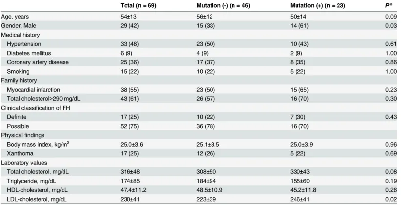
Clinical characteristics of study subjects
The characteristics of enrolled patients are shown inTable 1. We found that 23 of 69 patients harbored FH-linked mutations inLDLR, APOB, or PCSK9. We defined mutation-positive pa- tients by confirming known and novel genetic aberrations in these three genes. All other pa- tients were classified as mutation negative. Compared to mutation-negative patients,
mutation-positive patients were more frequently males with higher LDL-C levels (223±39 mg/
dL vs. 246±41 mg/dL;p = 0.02). In addition, mutation-positive patients tended to be younger than mutation-negative patients. However, there was no difference in history of coronary ar- tery disease between the two groups.
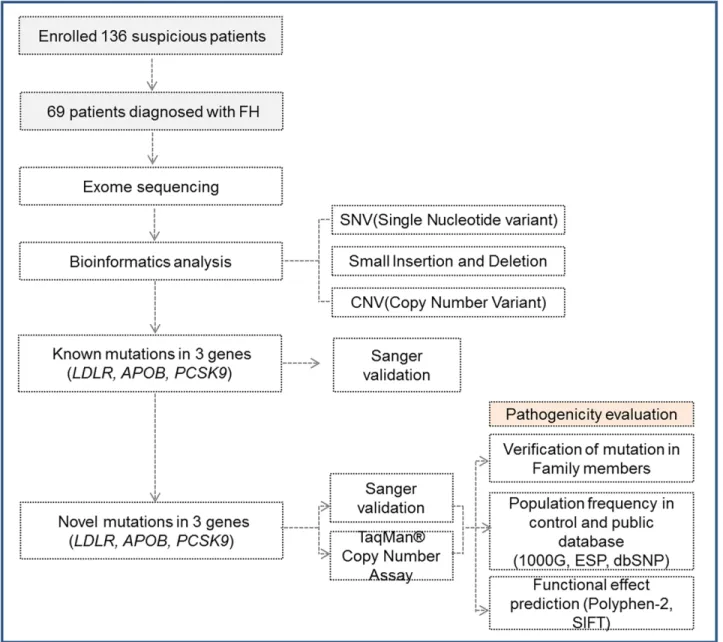
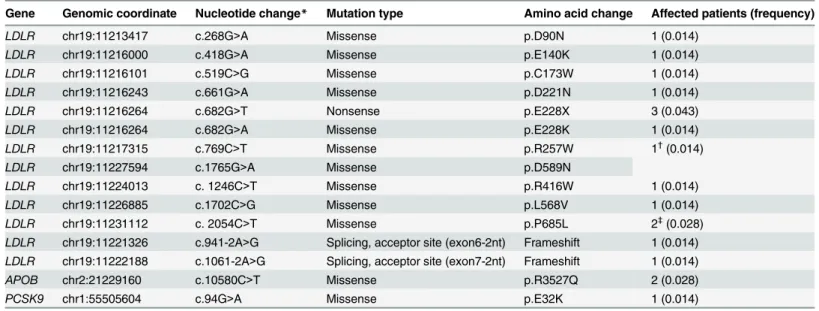
Detection and validation of known mutations in three FH-linked genes
The exomes of 69 FH patients were analyzed, as described (Fig 1). Of the 23 patients with mu- tations in 3 FH-linked genes, known causal mutations were found in 18 patients (Table 2).Among them, 15 patients were identified as having pathogenic mutations inLDLR. Specifically, two patients had the p.P685L mutation and four patients had either the p.E228X or p.E228K mutation. During preliminary screening by Sanger sequencing prior to WES, we found three more patients with the p.P685L mutation, which suggests the presence of potential mutational
Table 1. Clinical characteristics of enrolled familial hypercholesterolemia patients.
Total (n = 69) Mutation (-) (n = 46) Mutation (+) (n = 23) P*
Age, years 54±13 56±12 50±14 0.09
Gender, Male 29 (42) 15 (33) 14 (61) 0.03
Medical history
Hypertension 33 (48) 23 (50) 10 (43) 0.61
Diabetes mellitus 6 (9) 4 (9) 2 (9) 1.00
Coronary artery disease 25 (36) 17 (37) 8 (35) 0.86
Smoking 15 (22) 10 (22) 5 (22) 1.00
Family history
Myocardial infarction 38 (55) 23 (50) 15 (65) 0.23
Total cholesterol>290 mg/dL 43 (61) 26 (57) 16 (70) 0.30
Clinical classification of FH
Definite 17 (25) 10 (22) 7 (30) 0.43
Possible 52 (75) 36 (78) 16 (70)
Physicalfindings
Body mass index, kg/m2 25.0±3.6 25.1±3.5 25.0±3.9 0.96
Xanthoma 17 (25) 12 (26) 5 (22) 0.69
Laboratory values
Total cholesterol, mg/dL 316±48 308±50 330±43 0.08
Triglyceride, mg/dL 174±85 184±94 155±60 0.19
HDL-cholesterol, mg/dL 47.4±11.2 48.5±10.9 45.2±11.8 0.26
LDL-cholesterol, mg/dL 230±41 223±39 246±41 0.02
*Chi-square test or t-test was used where appropriate.
Values are mean± standard deviation or n (%).
HDL: high-density lipoprotein; LDL: low-density lipoprotein; Mutation (-): No known or novel pathogenic mutations in three FH-linked genes (LDLR, APOB, PCSK9); Mutation (+): Known or novel pathogenic mutations in LDLR, APOB, or PCSK9.
doi:10.1371/journal.pone.0126706.t001
hotspots in Korean FH cases. In addition, both the p.R257W and p.D589N homozygous muta- tions were found in one patient. This patient had an LDL-C level of 340 mg/dL, which was rela- tively higher than those of patients with only one heterozygous mutation. Two patients harbored the p.R3527Q mutation inAPOB, the most common causal variation of APOB in other populations. [18] Lastly, one patient had the p.E32K mutation inPCSK9. [19,20]
Discovery and validation of novel mutations in the three FH-linked genes
In addition to known mutations, novel disruptive mutations were detected in three FH pa- tients. Two novel mutations were predicted to disrupt theLDLR gene due to a frame shift (Table 3). Neither mutation was observed in any control exome data or public databases. The 13-nt deletion mutation (c.320_332delGACGTGCTCCCAG) was thought to be a pathogenicFig 1. Exome sequencing analysis of familial hypercholesterolemia (FH). The steps for identifying FH-causing variants in three genes are shown, in addition to the subsequent genetic analyses of whole-exome sequencing data that led to the identification of pathogenicity.
doi:10.1371/journal.pone.0126706.g001
Table 2. Known pathogenic mutations in three FH-linked genes (n = 69).
Gene Genomic coordinate Nucleotide change* Mutation type Amino acid change Affected patients (frequency)
LDLR chr19:11213417 c.268G>A Missense p.D90N 1 (0.014)
LDLR chr19:11216000 c.418G>A Missense p.E140K 1 (0.014)
LDLR chr19:11216101 c.519C>G Missense p.C173W 1 (0.014)
LDLR chr19:11216243 c.661G>A Missense p.D221N 1 (0.014)
LDLR chr19:11216264 c.682G>T Nonsense p.E228X 3 (0.043)
LDLR chr19:11216264 c.682G>A Missense p.E228K 1 (0.014)
LDLR chr19:11217315 c.769C>T Missense p.R257W 1†(0.014)
LDLR chr19:11227594 c.1765G>A Missense p.D589N
LDLR chr19:11224013 c. 1246C>T Missense p.R416W 1 (0.014)
LDLR chr19:11226885 c.1702C>G Missense p.L568V 1 (0.014)
LDLR chr19:11231112 c. 2054C>T Missense p.P685L 2‡(0.028)
LDLR chr19:11221326 c.941-2A>G Splicing, acceptor site (exon6-2nt) Frameshift 1 (0.014) LDLR chr19:11222188 c.1061-2A>G Splicing, acceptor site (exon7-2nt) Frameshift 1 (0.014)
APOB chr2:21229160 c.10580C>T Missense p.R3527Q 2 (0.028)
PCSK9 chr1:55505604 c.94G>A Missense p.E32K 1 (0.014)
*Nucleotide location number was assigned according to the low-density lipoprotein receptor (LDLR; NM_000527), apolipoprotein B (APOB; NM_000384), and proprotein convertase subtilisin/kexin type 9 (PCSK9; NM_174936) mRNA sequences.
†A patient (P49) with p.R257W (homozygote) and p.D589N (homozygote).
‡Screening the remaining cohort by Sanger sequencing identified three more patients with p.P685L.
Variants were characterized in published studies and validated in the present study by Sanger sequencing.
doi:10.1371/journal.pone.0126706.t002
Table 3. Novel pathogenic mutations in three FH-linked genes (n = 69).
Detailed information of novel mutations Pathogenicity†
Gene Genomic
coordinate
Nucleotide change* Mutation type
Amino acid change
Affected patients (frequency)
MAF in Korean controls (n = 390)
Frequency in public databases‡
Polyphen-2 prediction (probability)
SIFT prediction (score)
LDLR chr19:11215902– 11215914
c.321_333del- GACGTGCTCCCAG
Frameshift deletion
p.
C109Sfs§
1 (0.014) 0 Novel NA NA
LDLR chr19:11240299 c.2500_2502del- GATinsC||
Frameshift deletion/
insertion p.
D834Rfs
1 (0.014) 0 Novel NA NA
PCSK9 chr1:55518070 c.643C>T Missense p.R215C 1 (0.014) 0 Novel# Probably
damaging (1)
Damaging (0.008)
*Nucleotide location number was assigned according to the low-density lipoprotein receptor (LDLR; NM_000527) and proprotein convertase subtilisin/
kexin type 9 (PCSK9; NM_174936) mRNA sequences.
†Prediction for frameshift mutations ofLDLR is not available from the Polyphen-2 and SIFT algorithms and is not marked.
‡Public databases include the 1000 Genomes Project, dbSNP135, and NHLBI GO Exome Sequencing Project.
§The frameshift mutation changes the cysteine at position 109, as four nucleotides after the deletion compensate for the frameshift effect until threonine (108).
||The replacement of nucleotides 2500 to 2502 (GAT) occurred by‘C’ at the cis position.
#The p.R215H (c.644G>A) is a gain-of-function mutation in the catalytic domain of PCSK9. [22,23] Variants were validated by Sanger sequencing.
NA: Not available.
doi:10.1371/journal.pone.0126706.t003
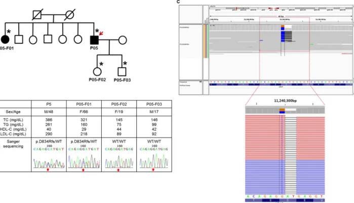
due to frame shift disruptions that introduced a premature stop codon. The p.D834Rfs muta- tion resulted from two concurrent mutations (c.2500_2502delGAT and c.2500insC) consecu- tively at the cis-position and was confirmed as a D834Rfs/- heterozygous mutation, using the Integrative Genomics Viewer (Fig 2). Though it occurred at a relatively posterior position among the 860 coding region ofLDLR, we could still confirm p.D834Rfs as causal variant that co-segregated within the corresponding family (P05;Fig 2). One variant inPCSK9, p.R215C, was identified within an evolutionarily conserved loop within the catalytic domain. [21] Con- sidering that gain-of-function mutations within this loop have been confirmed, [22,23] p.
R215C is likely to be a gain-of-function variant.
Copy number analysis and validation
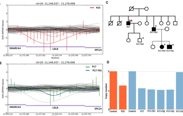
In the CNV analysis, novel copy number deletions were detected in two patients. A fragment spanning from exon 1 to exon 12 ofLDLR was inferred as being lost in one patient (P25) by the CoNIFER algorithm (Fig 3A). [17] The copy number was measured to be half of that of the control by Taqman copy number assay, which detected intron 5 (Fig 3D). Another copy num- ber deletion ranging from exon 8 to exon 12 ofLDLR was detected and validated by Taqman assay (Fig3Band3D). Notably, the copy number loss co-segregated in other affected family members as well, indicating the deletion to be the causal alteration for FH in the corresponding family (P17; Fig3Cand3D).
Fig 2. Pedigree analysis of a patient withLDLR p.D834Rfs/- mutation. (A) A simplified pedigree of the P05 family. The upper right arrow indicates the proband; squares indicate males, and circles indicate females. Open and filled symbols indicate unaffected and affected individuals, respectively. Asterisks indicate family members who underwent clinical examinations and molecular analyses. WT refers wild-type. (B) Clinical examination data and sequencing chromatograms. Vertical arrows indicate the mutation site. (C) Integrative Genomics Viewer screenshot of p.D834Rfs/-. Sequencing reads show that a single nucleotide substitution (G>C) and frameshift deletion (AT/-) occurred at the cis position.
doi:10.1371/journal.pone.0126706.g002
Discussion
In recent years, NGS has been applied in studies on FH and demonstrated as an efficient tool to identify causing mutations in known and novel genes. [10,24] The current study utilized WES to identify FH-causing mutations in Korean FH cases and presented the values of WES in the diagnosis of FH. The exome-based diagnosis was comprehensive, as it confirmed genetic variations that were not merely limited to SNVs and included small insertions or deletions and copy number variations. In fact, we confirmed 23 causative mutations in 3 known FH genes, including 2 copy number deletions, through WES-based genetic testing. Importantly, this sug- gests that newly generated DNA sequences can be used for the discovery and genetic diagnosis of novel FH-causing genes without the need for additional costs. The WES data in genetically undiagnosed patients from this study will be used for further analyses.
A DNA test, along with family screening, is confirmatory and clinically relevant, especial- ly for individuals with borderline-high serum LDL-C levels. Furthermore, the co-segregation analysis of variants in families provides strong evidence of the pathogenicity of undefined
Fig 3. Copy number variation (CNV) detection inLDLR. SVD-ZRPKM values were used to detect CNVs by the CoNIFER algorithm and were calculated by transforming reads per kilobase per million values into standardized z-scores, based on the mean and standard deviation across all analyzed exomes. (A) The SVD-ZRPKM regional plot of the P25 patient with a large copy number deletion inLDLR. (B) The SVD-ZRPKM regional plot of the P17 patient and family member (P17-F01) with an inherited copy number deletion inLDLR. Green and blue indicate SVD-ZRPKM values of P17 and P17-F01, respectively. Values are plotted based on P17. (C) Pedigree of the P17 patient with CNV. The upper right arrow indicates the proband; squares indicate males, and circles indicate females. Open and filled symbols indicate unaffected and affected individuals, respectively. Asterisks indicate family members who underwent clinical examinations and CNV analyses. (D) TaqMan Copy Number Assay for P25, P17, and family members of P17. Red indicates the assay for P25 by probe #1 within intron 5; blue indicates the assay for P17 and other members by probe #2 (overlapped from intron 10 to exon 11). The assay was performed in duplicate and repeated. Results were plotted by CopyCaller software v.2.0.
doi:10.1371/journal.pone.0126706.g003
variants in genetic testing. During initial exploratory screening through WES analysis, we found a total of four novel variants inLDLR with uncertain pathogenicity. Among them, only two variants were found to be causative, based onin silico and family co-segregation analyses. In the case ofAPOB, we found several novel variants with uncertain pathogenicity located outside of LDLR-binding regions. All variants were ruled out by family co-segrega- tion analysis.
Of the FH cases with confirmed mutations, one patient was homozygous for both p.R257W and p.D589N mutations inLDLR and had relatively higher LDL-C levels than those of patients with only one heterozygous mutation. The exact same double homozygous mutation has been previously described in one Taiwanese patient with FH. [25] Both patients exhibited tendon xanthomas without presenting typical homozygous phenotypes. Therefore, we hypothesize that this mutation does not fully abolish LDLR activity. However, further functional studies are needed.
Cholesterol levels in Asian FH patients are lower than those of Western patients. [26] The mean LDL-C of patients in this study was 230 mg/dL, which was similar to that of Japanese pa- tients [27] but lower than those of Chinese or Taiwanese patients. [25,28] In our study, the mutation carriers had significantly higher LDL-C levels. This finding is in accordance with prior reports and indicates that a higher level of LDL-C is an important characteristic of muta- tion-positive patients. [29,30] The proportion of male was higher in mutation carriers. The mean age of females was 57 years and higher than that of males. Post-menopausal females can be subject to nonspecific elevation of blood cholesterol, which may have increased the number of false-positive cases in our study. [31] The prevalence of coronary artery disease in our cohort was 36%, and this can vary widely in Asian populations. [26] There was no difference in the prevalence between patients with and without mutations.
The mutation detection rate in the 3 known FH genes was 33% in our study, which is lower than those of recent reports. [25,26,32,33] One possible reason is that the majority of subjects were initially classified as possible FH patients whose mutation rates were lower than those of definite FH patients. In addition, the low-sequencing coverage of known FH-causing genes, es- peciallyPCSK9, may have lowered the mutation detection rate (S1 Fig). Therefore, we cannot rule out the possibility of undetected mutations in these regions. For the use of WES in the ge- netic diagnosis of FH, further efforts are needed to improve coverage for regions with insuffi- cient coverage in FH-causing genes. As sequencing costs are dropping, the cost for WES becomes quite low. As a result, high-depth WES can achieve high coverage in FH-targeted genes and display better diagnostic performance.
There are several points that haven’t fully covered for the genetic diagnosis in FH cases without defined mutations. Originally, we set the values of WES in an attempt to identify novel FH-causing genes, going beyond the confirmatory exome-based diagnosis of FH. However, when applying existing statistical methods to reveal novel FH-causing genes, we found that the tests were underpowered and could not fully verify the causality of putative genes. These were mainly due to the small sample size. Furthermore, we could not define polygenic FH [34,35]
through WES by calculating LDL-C gene scores, as most of these scores were calculated accord- ing to the number of SNPs that occurred mostly in introns. Further research is needed to evalu- ate polygenic cause in Korean FH cases without defined mutations.
In summary, we identified 23 mutations in known FH genes (19 inLDLR, two in APOB, and two inPCSK9) using WES in 69 FH patients who met Simon Broome criteria with definite family history. We also identified three new causative mutations: two frame shift deletions in LDLR and one mutation in PCSK9.
Supporting Information
S1 Fig. Summary statistics of 3 FH genes.Variant callable portion was defined as locus cov- ered at least of 8× fold coverage by sequencing.
(TIF)
S1 Table. Summary statistics of whole-exome sequencing data.
(DOCX)
S2 Table. Detailed information of TaqMan Copy Number Assay.
(DOCX)
Acknowledgments
Biospecimens and data were provided by the Korean Genome Analysis Project (4845–301), the Korean Genome and Epidemiology Study (4851–302), and the Korea Biobank Project (4851–
307, KBP-2013-42), which were in turn supported by the Korea Center for Disease Control.
Support was also provided by the Korean Society of Lipidology and Atherosclerosis and the Cardiovascular Research Center. We are grateful to Jiyeong Jeong, RN, for her excellent assis- tance with clinical data collection and patient care.
Author Contributions
Conceived and designed the experiments: DB JHL SHL. Performed the experiments: SMH. An- alyzed the data: SMH BH TP. Contributed reagents/materials/analysis tools: DK MR BL YKA BRC JW SH JJ SP YJ SHL. Wrote the paper: SMH DB JHL SHL. Reviewed the manuscript and gave suggestions: MGL.
References
1. Brice P, Burton H, Edwards CW, Humphries SE, Aitman TJ. Familial hypercholesterolaemia: a pressing issue for European health care. Atherosclerosis. 2013; 231(2):223–6. Epub 2013/11/26. doi:10.1016/j.
atherosclerosis.2013.09.019PMID:24267231.
2. Umans-Eckenhausen MA, Defesche JC, Sijbrands EJ, Scheerder RL, Kastelein JJ. Review of first 5 years of screening for familial hypercholesterolaemia in the Netherlands. Lancet. 2001; 357 (9251):165–8. Epub 2001/02/24. doi:10.1016/s0140-6736(00)03587-xPMID:11213091.
3. Sharma P, Boyers D, Boachie C, Stewart F, Miedzybrodzka Z, Simpson W, et al. Elucigene FH20 and LIPOchip for the diagnosis of familial hypercholesterolaemia: a systematic review and economic evalu- ation. Health technology assessment (Winchester, England). 2012; 16(17):1–266. Epub 2012/04/04.
doi:10.3310/hta16170PMID:22469073.
4. Usifo E, Leigh SE, Whittall RA, Lench N, Taylor A, Yeats C, et al. Low-density lipoprotein receptor gene familial hypercholesterolemia variant database: update and pathological assessment. Annals of human genetics. 2012; 76(5):387–401. Epub 2012/08/14. doi:10.1111/j.1469-1809.2012.00724.xPMID:
22881376.
5. Awan Z, Choi HY, Stitziel N, Ruel I, Bamimore MA, Husa R, et al. APOE p.Leu167del mutation in famil- ial hypercholesterolemia. Atherosclerosis. 2013; 231(2):218–22. Epub 2013/11/26. doi:10.1016/j.
atherosclerosis.2013.09.007PMID:24267230.
6. Garcia-Rios A, Perez-Martinez P, Fuentes F, Mata P, Lopez-Miranda J, Alonso R, et al. Genetic varia- tions at ABCG5/G8 genes modulate plasma lipids concentrations in patients with familial hypercholes- terolemia. Atherosclerosis. 2010; 210(2):486–92. Epub 2010/02/23. doi:10.1016/j.atherosclerosis.
2010.01.010PMID:20172523; PubMed Central PMCID: PMCPmc2905734.
7. Fouchier SW, Dallinga-Thie GM, Meijers JC, Zelcer N, Kastelein JJ, Defesche JC, et al. Mutations in STAP1 are associated with autosomal dominant hypercholesterolemia. Circulation research. 2014;
115(6):552–5. Epub 2014/07/19. doi:10.1161/circresaha.115.304660PMID:25035151.
8. Maglio C, Mancina RM, Motta BM, Stef M, Pirazzi C, Palacios L, et al. Genetic diagnosis of familial hypercholesterolaemia by targeted next-generation sequencing. Journal of internal medicine. 2014;
276(4):396–403. Epub 2014/05/03. doi:10.1111/joim.12263PMID:24785115.
9. Vandrovcova J, Thomas ER, Atanur SS, Norsworthy PJ, Neuwirth C, Tan Y, et al. The use of next-gen- eration sequencing in clinical diagnosis of familial hypercholesterolemia. Genetics in medicine: official journal of the American College of Medical Genetics. 2013; 15(12):948–57. Epub 2013/05/18. doi:10.
1038/gim.2013.55PMID:23680767.
10. Futema M, Plagnol V, Whittall RA, Neil HA, Humphries SE. Use of targeted exome sequencing as a di- agnostic tool for Familial Hypercholesterolaemia. Journal of medical genetics. 2012; 49(10):644–9.
Epub 2012/10/12. doi:10.1136/jmedgenet-2012-101189PMID:23054246; PubMed Central PMCID:
PMCPmc3475071.
11. Marks D, Thorogood M, Neil HA, Humphries SE. A review on the diagnosis, natural history, and treat- ment of familial hypercholesterolaemia. Atherosclerosis. 2003; 168(1):1–14. Epub 2003/05/07. PMID:
12732381.
12. DePristo MA, Banks E, Poplin R, Garimella KV, Maguire JR, Hartl C, et al. A framework for variation dis- covery and genotyping using next-generation DNA sequencing data. Nature genetics. 2011; 43 (5):491–8. Epub 2011/04/12. doi:10.1038/ng.806PMID:21478889; PubMed Central PMCID:
PMCPmc3083463.
13. McKenna A, Hanna M, Banks E, Sivachenko A, Cibulskis K, Kernytsky A, et al. The Genome Analysis Toolkit: a MapReduce framework for analyzing next-generation DNA sequencing data. Genome re- search. 2010; 20(9):1297–303. Epub 2010/07/21. doi:10.1101/gr.107524.110PMID:20644199;
PubMed Central PMCID: PMCPmc2928508.
14. Adzhubei IA, Schmidt S, Peshkin L, Ramensky VE, Gerasimova A, Bork P, et al. A method and server for predicting damaging missense mutations. Nature methods. 2010; 7(4):248–9. Epub 2010/04/01.
doi:10.1038/nmeth0410-248PMID:20354512; PubMed Central PMCID: PMCPmc2855889.
15. Kumar P, Henikoff S, Ng PC. Predicting the effects of coding non-synonymous variants on protein func- tion using the SIFT algorithm. Nature protocols. 2009; 4(7):1073–81. Epub 2009/06/30. doi:10.1038/
nprot.2009.86PMID:19561590.
16. Wang K, Li M, Hakonarson H. ANNOVAR: functional annotation of genetic variants from high-through- put sequencing data. Nucleic acids research. 2010; 38(16):e164. Epub 2010/07/06. doi:10.1093/nar/
gkq603PMID:20601685; PubMed Central PMCID: PMCPmc2938201.
17. Krumm N, Sudmant PH, Ko A, O'Roak BJ, Malig M, Coe BP, et al. Copy number variation detection and genotyping from exome sequence data. Genome research. 2012; 22(8):1525–32. Epub 2012/05/16.
doi:10.1101/gr.138115.112PMID:22585873; PubMed Central PMCID: PMCPmc3409265.
18. Futema M, Whittall RA, Kiley A, Steel LK, Cooper JA, Badmus E, et al. Analysis of the frequency and spectrum of mutations recognised to cause familial hypercholesterolaemia in routine clinical practice in a UK specialist hospital lipid clinic. Atherosclerosis. 2013; 229(1):161–8. Epub 2013/05/15. doi:10.
1016/j.atherosclerosis.2013.04.011PMID:23669246; PubMed Central PMCID: PMCPmc3701838.
19. Noguchi T, Katsuda S, Kawashiri MA, Tada H, Nohara A, Inazu A, et al. The E32K variant of PCSK9 ex- acerbates the phenotype of familial hypercholesterolaemia by increasing PCSK9 function and concen- tration in the circulation. Atherosclerosis. 2010; 210(1):166–72. Epub 2009/12/17. doi:10.1016/j.
atherosclerosis.2009.11.018PMID:20006333.
20. Mabuchi H, Nohara A, Noguchi T, Kobayashi J, Kawashiri MA, Inoue T, et al. Genotypic and phenotypic features in homozygous familial hypercholesterolemia caused by proprotein convertase subtilisin/kexin type 9 (PCSK9) gain-of-function mutation. Atherosclerosis. 2014; 236(1):54–61. Epub 2014/07/12. doi:
10.1016/j.atherosclerosis.2014.06.005PMID:25014035.
21. Rader DJ, Cohen J, Hobbs HH. Monogenic hypercholesterolemia: new insights in pathogenesis and treatment. The Journal of clinical investigation. 2003; 111(12):1795–803. Epub 2003/06/19. doi:10.
1172/jci18925PMID:12813012; PubMed Central PMCID: PMCPmc161432.
22. Cameron J, Holla OL, Laerdahl JK, Kulseth MA, Ranheim T, Rognes T, et al. Characterization of novel mutations in the catalytic domain of the PCSK9 gene. Journal of internal medicine. 2008; 263(4):420– 31. Epub 2008/02/13. doi:10.1111/j.1365-2796.2007.01915.xPMID:18266662.
23. Cunningham D, Danley DE, Geoghegan KF, Griffor MC, Hawkins JL, Subashi TA, et al. Structural and biophysical studies of PCSK9 and its mutants linked to familial hypercholesterolemia. Nature structural
& molecular biology. 2007; 14(5):413–9. Epub 2007/04/17. doi:10.1038/nsmb1235PMID:17435765.
24. Motazacker MM, Pirruccello J, Huijgen R, Do R, Gabriel S, Peter J, et al. Advances in genetics show the need for extending screening strategies for autosomal dominant hypercholesterolaemia. European heart journal. 2012; 33(11):1360–6. Epub 2012/03/13. doi:10.1093/eurheartj/ehs010PMID:22408029.
25. Chiou KR, Charng MJ. Detection of mutations and large rearrangements of the low-density lipoprotein receptor gene in Taiwanese patients with familial hypercholesterolemia. The American journal of cardi- ology. 2010; 105(12):1752–8. Epub 2010/06/12. doi:10.1016/j.amjcard.2010.01.356PMID:20538126.
26. Miyake Y, Yamamura T, Sakai N, Miyata T, Kokubo Y, Yamamoto A. Update of Japanese common LDLR gene mutations and their phenotypes: Mild type mutation L547V might predominate in the
Japanese population. Atherosclerosis. 2009; 203(1):153–60. Epub 2008/08/23. doi:10.1016/j.
atherosclerosis.2008.07.005PMID:18718593.
27. Bujo H, Takahashi K, Saito Y, Maruyama T, Yamashita S, Matsuzawa Y, et al. Clinical features of famil- ial hypercholesterolemia in Japan in a database from 1996–1998 by the research committee of the min- istry of health, labour and welfare of Japan. Journal of atherosclerosis and thrombosis. 2004; 11 (3):146–51. Epub 2004/07/17. PMID:15256765.
28. Hu M, Lan W, Lam CW, Mak YT, Pang CP, Tomlinson B. Heterozygous familial hypercholesterolemia in Hong Kong Chinese. Study of 252 cases. International journal of cardiology. 2013; 167(3):762–7.
Epub 2012/04/03. doi:10.1016/j.ijcard.2012.03.048PMID:22464486.
29. Williams RR, Hunt SC, Schumacher MC, Hegele RA, Leppert MF, Ludwig EH, et al. Diagnosing hetero- zygous familial hypercholesterolemia using new practical criteria validated by molecular genetics. The American journal of cardiology. 1993; 72(2):171–6. Epub 1993/07/15. PMID:8328379.
30. Civeira F, Ros E, Jarauta E, Plana N, Zambon D, Puzo J, et al. Comparison of genetic versus clinical di- agnosis in familial hypercholesterolemia. The American journal of cardiology. 2008; 102(9):1187–93, 93.e1. Epub 2008/10/23. doi:10.1016/j.amjcard.2008.06.056PMID:18940289.
31. Lee YH, Lee SG, Lee MH, Kim JH, Lee BW, Kang ES, et al. Serum cholesterol concentration and prev- alence, awareness, treatment, and control of high low-density lipoprotein cholesterol in the Korea Na- tional Health and Nutrition Examination Surveys 2008–2010: Beyond the Tip of the Iceberg. Journal of the American Heart Association. 2014; 3(1):446. Epub 2014/02/28. doi:10.1161/jaha.113.000650 PMID:24572249; PubMed Central PMCID: PMCPmc3959713.
32. Humphries SE, Whittall RA, Hubbart CS, Maplebeck S, Cooper JA, Soutar AK, et al. Genetic causes of familial hypercholesterolaemia in patients in the UK: relation to plasma lipid levels and coronary heart disease risk. Journal of medical genetics. 2006; 43(12):943–9. Epub 2006/12/05. doi:10.1136/jmg.
2006.038356PMID:17142622; PubMed Central PMCID: PMCPmc2563208.
33. Taylor A, Wang D, Patel K, Whittall R, Wood G, Farrer M, et al. Mutation detection rate and spectrum in familial hypercholesterolaemia patients in the UK pilot cascade project. Clinical genetics. 2010; 77 (6):572–80. Epub 2010/03/20. doi:10.1111/j.1399-0004.2009.01356.xPMID:20236128.
34. Talmud PJ, Shah S, Whittall R, Futema M, Howard P, Cooper JA, et al. Use of low-density lipoprotein cholesterol gene score to distinguish patients with polygenic and monogenic familial hypercholestero- laemia: a case-control study. Lancet. 2013; 381(9874):1293–301. Epub 2013/02/26. doi:10.1016/
s0140-6736(12)62127-8PMID:23433573.
35. Futema M, Plagnol V, Li K, Whittall RA, Neil HA, Seed M, et al. Whole exome sequencing of familial hypercholesterolaemia patients negative for LDLR/APOB/PCSK9 mutations. Journal of medical genet- ics. 2014; 51(8):537–44. Epub 2014/07/06. doi:10.1136/jmedgenet-2014-102405PMID:24987033;
PubMed Central PMCID: PMCPmc4112429.