Infarct Core Expansion on Computed Tomography before and after Intravenous Thrombolysis
Dongbeom Song1, Joonsang Yoo1, Jang-Hyun Baek1, Jinkwon Kim1,2, Hye Sun Lee3, Young Dae Kim1, Hyo Suk Nam1, and Ji Hoe Heo1
1Department of Neurology, Yonsei University College of Medicine, Seoul;
2Department of Neurology, CHA Bundang Medical Center, CHA University, Seongnam;
3Department of Biostatistics, Yonsei University College of Medicine, Seoul, Korea.
Purpose: Infarct core can expand rapidly in acute stroke patients receiving intravenous tissue plasminogen activator (IV t-PA). We investigated changes in the extent of infarct core during IV t-PA treatment, and explored the associative factors of this infarct core expansion in patients with proximal artery occlusion.
Materials and Methods: We included patients who were considered for sequential intra-arterial therapy (IAT) due to occlusion of intracranial proximal artery after IV t-PA. Patients who had a baseline Alberta Stroke Program Early Computed Tomography (CT) Score (ASPECTS) ≥6 and who underwent two consecutive CT scans before and shortly after IV t-PA infusion were enrolled. Pa- tients were classified into no, moderate, and marked expansion groups based on decreases in ASPECTS (0-1, 2-3, and ≥4, respec- tively) on follow-up CT. Collateral status was graded using CT angiography.
Results: Of the 104 patients, 16 (15.4%) patients showed moderate and 13 (12.5%) patients showed marked infarct core expansion on follow-up CT scans obtained at 71.1±19.1 min after baseline CT scan. Sixteen (15.4%) patients had an ASPECTS value <6 on the follow-up CT. None of the patients with marked expansion were independent at 3 months. Univariate analysis and ordinal lo- gistic regression analysis demonstrated that the infarct core expansion was significantly associated with collateral status (p<0.001).
Conclusion: Among patients who were considered for IAT after IV t-PA treatment, one out of every seven patients exhibited marked expansion of infarct core on follow-up CT before IAT. These patients tend to have poor collaterals and poor outcomes de- spite rescue IAT.
Key Words: Acute stroke therapy, ischemic stroke, CT scan, collateral circulation, tissue plasminogen activator
INTRODUCTION
Intra-arterial therapy (IAT) using a stent retriever is safe and effective in acute stroke with intracranial proximal artery oc- clusion.1-8 However, despite successful reperfusion by IAT,
some patients do not show clinical improvement. One of the plausible explanations for this futile reperfusion is rapid con- version of ischemic penumbra into irreversible infarct core.9,10 While patients who already have extensive areas of irrevers- ible damage are usually excluded from reperfusion therapy,8,11 some salvageable areas can be converted to irreversible in- farct core during the reperfusion treatment. However, there are few reports on infarct core progression in the hyperacute stage in patients with proximal artery occlusion, and its preva- lence, related factors, and clinical significance are largely un- known.
Before the efficacy of IAT was proven, IAT was sometimes performed as a rescue treatment to patients who do not re- spond to intravenous tissue plasminogen activator (IV t-PA) after follow-up imaging study. By analyzing two consecutive Received: August 14, 2017 Revised: December 11, 2017
Accepted: December 22, 2017
Corresponding author: Dr. Ji Hoe Heo, Department of Neurology, Yonsei Univer- sity College of Medicine, 50-1 Yonsei-ro, Seodaemun-gu, Seoul 03722, Korea.
Tel: 82-2-2228-1605, Fax: 82-2-393-0705, E-mail: [email protected]
•The authors have no financial conflicts of interest.
© Copyright: Yonsei University College of Medicine 2018
This is an Open Access article distributed under the terms of the Creative Com- mons Attribution Non-Commercial License (http://creativecommons.org/licenses/
by-nc/4.0) which permits unrestricted non-commercial use, distribution, and repro- duction in any medium, provided the original work is properly cited.
pISSN: 0513-5796 · eISSN: 1976-2437 Yonsei Med J 2018 Mar;59(2):310-316
https://doi.org/10.3349/ymj.2018.59.2.310
computed tomography (CT) scans acquired before and shortly after IV t-PA, we investigated the change in the extent of the in- farct core during IV t-PA treatment, its associative factors, and clinical significance.
MATERIALS AND METHODS
Study population
We included patients who were potential candidate for IAT due to persistent occlusion of intracranial proximal artery after IV t-PA (Actilyse, Boehringer-Ingelheim, Ingelheim, Germany) and who had two consecutive CT scans before and shortly af- ter IV t-PA. This group was derived from a cohort that was de- veloped to investigate the factors associated with thrombus res- olution after IV t-PA.12,13 In the cohort, two consecutive non- contrast CT (NCCT) scans were acquired before and shortly after IV t-PA infusion in the same scanner (LightSpeed Plus, GE Healthcare, Milwaukee, WI, USA or SOMATOM Sensation 64, Siemens Healthcare, Erlangen, Germany) (Supplementary Material, only online), and CT angiography (CTA) was taken with follow-up NCCT. For this study, we included patients who had unilateral intracranial proximal artery [internal carotid ar- tery (ICA), middle cerebral artery (MCA) M1, or M2] occlusion on CTA between January 2009 and December 2014. We exclud- ed patients who already had a large infarct core in the initial NCCT, indicated by an Alberta Stroke Program Early CT Score (ASPECTS) <6. Patients received IV infusion of t-PA within 3 hours of symptom onset until December 2012 and within 4.5 hours thereafter. Additional IAT was considered if patients did not show a satisfactory clinical response [<50% improvement as measured by the National Institutes of Health Stroke Scale (NIHSS) score] to IV t-PA infusion.
Image analysis
In this study, infarct core was defined as low-density area on NCCT and calculated based on the ASPECTS scoring system.
ASPECTS was measured with NCCT using revised methodol- ogy, which does not account for isolated cortical swelling.14 Patients were classified into three groups: no, moderate, and marked expansion groups, defined based on a decrease of 0–1, 2–3, and ≥4, respectively, in ASPECTS between the two scans.
The CTA-collateral score (CTA-CS) was measured with recon- structed maximum intensity projection CTA images as fol- lows: 0=absence of collateral supply to the occluded vascular territory; 1=collateral supply filling <50%, but >0% of the oc- cluded vascular territory; 2=collateral supply filling >50%, but
<100% of the occluded vascular territory; and 3=collateral supply filling 100% of the occluded vascular territory.15 Two stroke neurologists, who were blinded to all clinical informa- tion, other than the side of the infarction, independently re- viewed the CT scans and measured ASPECTS and CTA-CS.
Disagreements between the two readers were resolved by con-
sensus. Good collateral status was defined as CTA-CS ≥2.
Clinical variables and outcomes
Stroke mechanisms were determined based on the Trial of Org 10172 in Acute Stroke Treatment (TOAST) classification. The initial stroke severity was assessed using the NIHSS, and the functional outcome was measured using the modified Rankin Scale (mRS) score at 90 days. The degree of reperfusion was graded by the Thrombolysis in Cerebral Infarction (TICI) scale in the final run of IAT. TICI scales of the patients who did not undergo IAT were graded with 24-hour follow-up MR angiog- raphy. We obtained outcome data and clinical variables, such as vascular risk factors, laboratory results, and time metrics, from the aforementioned prospective cohort. Successful reper- fusion was defined as TICI ≥2b, and a favorable outcome was defined as mRS ≤2. Symptomatic intracranial hemorrhage (ICH) was defined as any hemorrhage associated with neuro- logical deterioration, as indicated by a decrease of 4 points on the NIHSS within 7 days (European Cooperative Acute Stroke Study III definition).16 This study was approved by the Institu- tional Review Board of Severance Hospital (IRB No. 4-2013- 0828), Yonsei University Health System, with a waiver of writ- ten informed consent from the patients or their qualified next- of-kin because of the retrospective nature of the study.
Statistical analysis
Values are presented as a number (%), mean±standard devia- tion (SD), or median [interquartile range (IQR)], as appropriate.
We compared the baseline characteristics, treatment modali- ties, time parameters, and imaging characteristics between the mild, moderate, and severe infarct expansion groups. Anal- ysis of variance or Kruskal-Wallis test, χ2 test, and Fisher’s exact test were used, as appropriate. The Spearman’s rank correlation coefficient was computed between the ASPECTS difference and the CTA-CS. Variables achieving p-values less than 0.1 in the univariate analyses with an ordinal association and clini- cally important time variables were adjusted for using multi- variate analyses (ordinal logistic regression analysis). Univari- ate analyses (independent sample t-test or Wilcoxon rank sum test for continuous variables, and χ2 test or Fisher’s exact test for categorical variables) were also performed to compare the baseline characteristics, treatment modalities, time parame- ters, and imaging characteristics between the favorable and unfavorable outcome groups. Variables achieving p-values less than 0.1 in the univariate analyses for favorable outcomes were entered for multivariate analyses (binomial logistic regres- sion analysis). Inter-rater agreements were assessed using lin- ear weighted κ statistics. Statistical analyses were performed using the R Statistical Software. Results with a two-sided p-val- ue <0.05 were considered statistically significant.
RESULTS
Baseline characteristics
During the study period, 176 patients in the CT-based throm- bus imaging cohort received IV t-PA treatment for an anterior circulation stroke with serial CT scans acquired before and af- ter the IV t-PA treatment. After excluding 65 patients without proximal artery occlusion on CTA and seven patients with AS- PECTS ≤6 on initial CT, 104 patients were included for this study. The mean age of the study patients was 67.3±10.4 years, and 60 (57.7%) of the patients were men. The median NIHSS score at admission was 16 (IQR, 13−19). Ninety (86.5%) patients were treated with combined IV t-PA and IAT, while 14 (13.5%) patients were treated with IV t-PA alone. The reasons for not performing IAT despite the presence of occlusion on CTA were rapid improvement of clinical symptom in eight patients, ear- ly ischemic change in more than one third of MCA territory in four patients, presence of hemorrhage on follow-up CT in one patient, and active tuberculosis in one patient. The primary modality used in the IAT was the stent retriever in 52 (57.8%) patients, intra-arterial urokinase infusion in 31 (34.4%) patients, modified thrombus suction using a Penumbra catheter in four (4.4%) patients, and carotid stent placement in three (3.3%) patients. Baseline characteristics of the study group appear in Table 1.
Infarct core expansion
Follow-up CT scans were obtained at 71.1±19.1 minutes after baseline CT scan. On the follow-up CT scan, 75 (72.1%) pa- tients showed almost no infarct core expansion. However, 16 (15.4%) patients showed moderate and 13 (12.5%) patients showed marked infarct core expansion (Table 1). Although we excluded patients with a baseline ASPECTS <6, there were 16 (15.4%) patients who had ASPECTS <6 in the follow-up scan (Fig. 1). Inter-rater agreements for the expansion of the infarct core [linear weighted κ, 0.641; 95% confidence interval (CI), 0.485−0.797], ASPECTS (linear weighted κ, 0.666; 95% CI, 0.609−0.723), and CTA-CS (linear weighted κ, 0.625; 95% CI, 0.530−0.721) were all good.
Factors associated with infarct core expansion
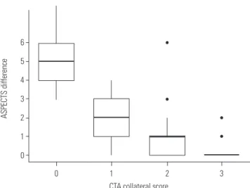
Univariate analyses revealed an association of the infarct core expansion with ICA occlusion, poor collateral status, and se- vere initial neurological deficits, but not with the time interval between the two consecutive CT scans. The proportion of pa- tients with hypercholesterolemia was significantly higher in the moderate expansion group; however, there was no ordinal association between the history of hypercholesterolemia and infarct core expansion. In the ordinal logistic regression analy- sis, the infarct core expansion was significantly associated with collateral status (odds ratio, 9.232; 95% CI, 4.484−22.209; p<
0.001) (Table 2). A significant correlation between the differ- ence in ASPECTS on the consecutive CT scans, and CTA-CS
(Fig. 2) (Spearman’s rank correlation coefficient ρ=-0.733; p<
0.001) was also noted.
Infarct core expansion and clinical outcomes
Fifty eight (55.8%) patients showed a favorable clinical out- come at 3 months. Infarct core expansion was associated with unfavorable clinical outcomes in the univariate (Supplementary Table 1, only online) and multivariate analyses after adjusting for confounding variables, such as sex, age, prothrombin time, time from onset to IV t-PA, initial NIHSS score, initial occlu- sion site, and successful reperfusion. None of the 13 patients with a marked expansion (≥4 point decrease in ASPECTS) of the ischemic core were independent at 3 months. In addition, only two out of the 16 patients with ASPECTS <6 on the follow- up CT showed a favorable clinical outcome. Although we could not perform multivariate analyses given the low numbers of symptomatic ICH and death events within 3 months, a signifi- cant association between infarct core expansion and symp- tomatic ICH and death within 3 months was observed in the univariate analyses (Table 1).
DISCUSSION
This study investigated the expansion of infarct core during IV t-PA treatment in patients with proximal artery occlusion. The study showed that 1) infarct core markedly expanded in about 12% of the patients with proximal artery occlusion at the end of IV t-PA infusion, 2) these patients had poor functional out- comes when they were treated with IAT after the infusion of IV t-PA, and 3) poor collaterals were predictive of a marked expan- sion of the infarct core.
IAT using a stent retriever is the treatment of choice in pa- tients with intracranial proximal artery occlusion even if they already received IV t-PA treatment based on the recent results from randomized controlled trials.2-8 This study simulated the situation in which additional IAT is required after IV t-PA treat- ment, as we included patients who had proximal artery occlu- sion after IV t-PA. Indeed, the majority of the patients in this study population was treated with IAT. In patients who receive IAT on top of IV t-PA, IAT is sometimes performed after the in- fusion of IV t-PA is completed. However, our study demonstrat- ed that infarct core could expand significantly during this time interval in some patients. In this study, 12% of the patients who did not have a large infarct core on the initial scan showed marked infarct core progression (decrease of ASPECTS ≥4).
Despite a rescue IAT, none of these patients recovered to an in- dependent life at 3 months. Although we do not know what the prognosis of these patients would be if they were to receive IAT as a combined therapy without waiting for completion of IV t-PA infusion or follow-up imaging, our results indirectly suggest that combined IAT with infusion of IV t-PA should be initiated as soon as possible and that patients who are expected
Table 1. Baseline Characteristics and Outcomes According to Infarct Core Expansion
Variables Total (n=104) No expansion
(n=75)
Moderate expansion (n=16)
Marked expansion
(n=13) p value
Sex, male 60 (57.7) 44 (58.7) 7 (43.8) 9 (69.2) 0.366
Age (yr) 67.3±10.4 66.1±11.1 71.4±4.62 69.2±10.2 0.137
Hypertension 70 (67.3) 47 (62.7) 13 (81.2) 10 (76.9) 0.314
Diabetes mellitus 26 (25.0) 18 (23.1) 6 (35.3) 3 (23.1) 0.822
Hypercholesterolemia* 9 (8.7) 4 (5.3) 4 (25.0) 1 (7.7) 0.036
Current smoker 19 (18.3) 15 (20.0) 1 (6.3) 3 (23.1) 0.361
Old cerebrovascular accident 32 (30.8) 23 (30.7) 5 (31.2) 4 (30.8) 1.000
Atrial fibrillation 53 (51.0) 34 (45.3) 10 (62.5) 9 (69.2) 0.170
TOAST classification 0.137
Negative evaluation 11 (10.7) 4 (5.41) 4 (25.0) 3 (23.1)
Large-artery atherosclerosis 21 (20.4) 19 (25.7) 1 (6.25) 1 (7.69)
Cardiac embolism 54 (52.4) 37 (50.0) 9 (56.2) 8 (61.5)
More than two causes 14 (13.6) 11 (14.9) 2 (12.5) 1 (7.69)
Other determined 3 (2.91) 3 (4.05) 0 (0.00) 0 (0.00)
Occlusion site 0.004
ICA 37 (35.6) 19 (25.3) 8 (50.0) 10 (76.9)
M1 46 (44.2) 37 (49.3) 7 (43.8) 2 (15.4)
M2 21 (20.2) 19 (25.3) 1 (6.25) 1 (7.69)
Systolic blood pressure (mm Hg) 148.0±27.4 148.5±27.4 148.1±32.1 144.5±22.7 0.891
Hemoglobin (g/dL) 13.8±1.5 13.8±1.6 13.4±1.6 14.3±1.4 0.313
Platelet count (×109/L) 225.8±61.1 228.4±61.4 209.8±73.5 230.9±40.5 0.522
Prothrombin time, international
normalized ratio 0.99±0.10 0.98±0.10 1.00±0.12 1.02±0.10 0.359
Partial thrombin time (sec) 30.1 (6.0) 29.6 (5.2) 32.4 (8.1) 30.3 (6.7) 0.240
Blood sugar level (mg/dL) 137.3±50.8 138.8±54.1 134.4±48.9 131.9±33.5 0.877
Total cholesterol (mg/dL) 167.5±36.8 168±36.5 162±41.3 169±35.7 0.830
Low density lipoprotein (mg/dL) 99.4±32.8 101.2±35.1 91.7±28.5 97.8±24.0 0.622
NIHSS 15.5 [13.0−19.0] 15.0 [11.0−19.0] 15.0 [14.0−20.0] 18.0 [17.0−22.0] 0.015 Initial ASPECTS 9.0 [8.0−10.0] 9.0 [8.0−10.0] 9.0 [8.75−9.25] 9.0 [7.0−10.0] 0.626
Collateral score 2.0 [1.0−3.0] 2.0 [2.0−3.0] 1.0 [1.0−2.0] 0.0 [0.0−0.0] <0.001
Time from onset to the initial CT (min) 72.2±38.7 73.9±39.2 67.5±37.9 68.0±39.2 0.770
Time from onset to IV t-PA (min) 98.5±38.6 101.0±38.6 91.3±37.8 94.2±41.3 0.619
Time interval between the CTs (min) 71.1±19.1 71.1±19.8 71.1±20.4 71.4±14.1 0.999
Groups based on tPA-eligible time
window 0.769
3-hour group 26 (25.0) 19 (25.3) 3 (18.8) 4 (30.8)
4.5-hour group 78 (75.0) 56 (74.7) 13 (81.2) 9 (69.2)
IAT 90 (86.5) 65 (86.7) 15 (93.8) 10 (76.9) 0.462
Stent retriever 52 (57.8) 35 (53.8) 11 (73.3) 6 (60.0) 0.403
Successful reperfusion 75 (72.1) 59 (78.7) 11 (68.8) 5 (38.5) 0.014
Symptomatic ICH 5 (4.81) 2 (2.67) 3 (18.8) 0 (0.0) 0.046
mRS at 3 months 2.0 [1.0−5.0] 2.0 [1.0−3.5] 2.0 [0.75−5.0] 5.0 [4.0−6.0] <0.001
Favorable outcome at 3 months 58 (55.8) 49 (65.3) 9 (56.2) 0 (0.0) <0.001
Death within 3 months 15 (14.4) 8 (10.7) 2 (12.5) 5 (38.5) 0.048
TOAST, Trial of Org 10172 in Acute Stroke Treatment; ICA, internal carotid artery; M1, first segment of the middle cerebral artery; M2, second segment of the middle cerebral artery; NIHSS, National Institutes of Health Stroke Scale; ASPECTS, Alberta Stroke Program Early CT score; CT, computed tomography; IV t-PA, intravenous tissue plasminogen activator; ICH, intracranial hemorrhage; mRS, modified Rankin Score; IAT, intra-arterial therapy.
Values represent a number (%), mean±standard deviation, or median [interquartile range].
*Hypercholesterolemia was defined as 1) fasting cholesterol level >240 mg/dL, 2) fasting low density lipoprotein level >160 mg/dL, or 3) history of taking lipid lowering agent due to hypercholesterolemia.
to have infarct core expansion within an hour may not be an ideal candidates for combined IAT. While the poor outcomes in patients with infarct core expansion could be due to the higher failure of reperfusion in this group, the prognostic ef- fect of infarct core progression was still significant after adjust- ing the successful reperfusion in the multivariate analysis.
We sought to identify factors associated with infarct core ex- pansion and found that collateral status was one of its main associative factors. Collaterals are virtually the only source of blood supply in the presence of a proximal artery occlusion.17,18 Collaterals help the tissues at risk to maintain their viability until final reperfusion is achieved. Previous studies have shown that the rate of infarct growth is determined by collateral sta- tus19-21 and that better collateral status in the hyper-acute stage is associated with more favorable clinical outcomes.22-24 The results of this study are in line with those of previous studies.
Although the elapsed time would be another factor determin- ing the infarct core expansion,19,25,26 no definite association was observed between time between the two CT scans and the infarct core expansion in our study. This might be account- ed for, in part, by the relatively constant time interval between the CTs, with a mean of ~70 minutes and an SD of ~14 minutes
in this study. Nevertheless, our findings suggest that collateral blood supply might be more important than mere time.
We excluded patients who already have considerable infarct core in the initial imaging study (ASPECTS<6) based on recent clinical trial results8,11 because the primary aim of this study was to evaluate the infarct core expansion after the initial imaging.
However, it should be acknowledged that the lack of evidence for clinical efficacy of IAT in patients with low ASPECTS does not necessarily mean that IAT should not be indicated in this group of patients. It is also difficult to provide specific sugges- tions regarding the use of collateral score in selecting an IAT candidate with this study because the patients in this study received IAT as a rescue therapy after waiting for IV t-PA re- sponse and that about 40% of the patients were not treated with a stent retriever, a currently standard modality of choice for IAT.8
There are several limitations to this study. First, occlusion sites and collateral scores were evaluated not in the initial im- aging studies but in the follow-up studies after t-PA infusion because this study population was derived from the cohort for CT-based thrombus imaging. Since there is a slight chance of clot resolution and dynamic change of collateral status over Fig. 1. Distribution of ASPECTS on initial and follow-up CT images. AS-
PECTS, Alberta Stroke Program Early Computed Tomography Score; CT, computed tomography.
45 40 35 30 25 20 15 10 5 0 10
ASPECTS
9 8 7 6 5 4 3 2 1 0
Initial CT Follow-up CT
Table 2. Ordinal Regression Analysis for Infarct Core Expansion
Variables Odds ratio (95% confidence interval) p value
Collateral score 9.232 (4.484−22.209) <0.001
Occlusion site
M2 Reference
M1 1.730 (0.259−16.666) 0.595
ICA 5.642 (0.891−55.459) 0.091
Time from onset to IV t-PA (min) 0.995 (0.980−1.009) 0.462
Time interval between the CT scans (min) 0.992 (0.963−1.022) 0.610
Initial NIHSS 1.108 (0.964−1.296) 0.167
ICA, internal carotid artery; M1, first segment of middle cerebral artery; M2, second segment of middle cerebral artery; IV t-PA, intravenous tissue plasminogen activator; NIHSS, National Institutes of Health Stroke Scale; CT, computed tomography.
6 5 4 3 2 1 0
ASPECTS difference
Number of patients
CTA collateral score
0 1 2 3
Fig. 2. Difference in ASPECTS on consecutive CT scans according to collateral status. ASPECTS, Alberta Stroke Program Early Computed Tomography Score; CT, computed tomography; CTA, CT angiography.
the period of t-PA infusion, this should be considered as a limi- tation of our study. Second, patients who did not have proxi- mal artery occlusion after IV t-PA treatment were not included in this study. Thus, the findings of this study cannot be applied to patients with successful recanalization after IV t-PA or those with distal artery occlusions. Third, while about 30% of patients had a history of previous stroke, data on pre-stroke mRS were not available. Therefore, this should be considered in the in- terpretation of outcome results. Finally, this study has a mod- erate sample size and a retrospective single-center design.
Therefore, generalization of our results must be performed with caution.
In conclusion, our study revealed that some patients who received IV t-PA showed a rapid expansion of the infarct core during IV t-PA infusion, and they showed poor functional out- comes after rescue IAT. These patients with a rapid expansion of infarct core had poor collateral circulations. Our findings support the important role of collateral status for maintaining tissue viability and suggest that IAT should be started as soon as possible without waiting for completion of t-PA infusion.
ACKNOWLEDGEMENTS
This study was supported by a grant by Korea Healthcare Tech- nology Research and Development Project, funded by the Ministry for Health and Welfare, Republic of Korea (HI15C2814, HI15C1056).
ORCID
Dongbeom Song https://orcid.org/0000-0002-7175-4948 Ji Hoe Heo https://orcid.org/0000-0001-9898-3321
REFERENCES
1. Badhiwala JH, Nassiri F, Alhazzani W, Selim MH, Farrokhyar F, Spears J, et al. Endovascular thrombectomy for acute ischemic stroke: a meta-analysis. JAMA 2015;314:1832-43.
2. Berkhemer OA, Fransen PS, Beumer D, van den Berg LA, Lingsma HF, Yoo AJ, et al. A randomized trial of intraarterial treatment for acute ischemic stroke. N Engl J Med 2015;372:11-20.
3. Campbell BC, Mitchell PJ, Kleinig TJ, Dewey HM, Churilov L, Yassi N, et al. Endovascular therapy for ischemic stroke with perfusion- imaging selection. N Engl J Med 2015;372:1009-18.
4. Goyal M, Demchuk AM, Menon BK, Eesa M, Rempel JL, Thorn- ton J, et al. Randomized assessment of rapid endovascular treat- ment of ischemic stroke. N Engl J Med 2015;372:1019-30.
5. Jovin TG, Chamorro A, Cobo E, de Miquel MA, Molina CA, Rovira A, et al. Thrombectomy within 8 hours after symptom onset in ischemic stroke. N Engl J Med 2015;372:2296-306.
6. Saver JL, Goyal M, Bonafe A, Diener HC, Levy EI, Pereira VM, et al. Stent-retriever thrombectomy after intravenous t-PA vs. t-PA alone in stroke. N Engl J Med 2015;372:2285-95.
7. Goyal M, Menon BK, van Zwam WH, Dippel DW, Mitchell PJ, Demchuk AM, et al. Endovascular thrombectomy after large-ves- sel ischaemic stroke: a meta-analysis of individual patient data
from five randomised trials. Lancet 2016;387:1723-31.
8. Powers WJ, Derdeyn CP, Biller J, Coffey CS, Hoh BL, Jauch EC, et al. 2015 American Heart Association/American Stroke Associa- tion focused update of the 2013 guidelines for the early manage- ment of patients with acute ischemic stroke regarding endovas- cular treatment: a guideline for healthcare [rofessionals from the American Heart Association/American Stroke Association. Stroke 2015;46:3020-35.
9. Song D, Lee K, Kim EH, Kim YD, Kim J, Song TJ, et al. Value of uti- lizing both ASPECTS and CT angiography collateral score for out- come prediction in acute ischemic stroke. Int J Stroke 2015;10:
1018-23.
10. Saver JL, Goyal M, van der Lugt A, Menon BK, Majoie CB, Dippel DW, et al. Time to treatment with endovascular thrombectomy and outcomes from ischemic stroke: a meta-analysis. JAMA 2016;
316:1279-88.
11. Menon BK, Campbell BC, Levi C, Goyal M. Role of imaging in current acute ischemic stroke workflow for endovascular therapy.
Stroke 2015;46:1453-61.
12. Kim YD, Nam HS, Kim SH, Kim EY, Song D, Kwon I, et al. Time- dependent thrombus resolution after tissue-type plasminogen ac- tivator in patients with stroke and mice. Stroke 2015;46:1877-82.
13. Nam HS, Kim EY, Kim SH, Kim YD, Kim J, Lee HS, et al. Prediction of thrombus resolution after intravenous thrombolysis assessed by CT-based thrombus imaging. Thromb Haemost 2012;107:786-94.
14. Puetz V, Dzialowski I, Hill MD, Demchuk AM. The Alberta Stroke Program Early CT Score in clinical practice: what have we learned?
Int J Stroke 2009;4:354-64.
15. Tan JC, Dillon WP, Liu S, Adler F, Smith WS, Wintermark M. Sys- tematic comparison of perfusion-CT and CT-angiography in acute stroke patients. Ann Neurol 2007;61:533-43.
16. Hacke W, Kaste M, Bluhmki E, Brozman M, Dávalos A, Guidetti D, et al. Thrombolysis with alteplase 3 to 4.5 hours after acute isch- emic stroke. N Engl J Med 2008;359:1317-29.
17. Liebeskind DS. Collateral circulation. Stroke 2003;34:2279-84.
18. Shuaib A, Butcher K, Mohammad AA, Saqqur M, Liebeskind DS.
Collateral blood vessels in acute ischaemic stroke: a potential therapeutic target. Lancet Neurol 2011;10:909-21.
19. Jung S, Gilgen M, Slotboom J, El-Koussy M, Zubler C, Kiefer C, et al.
Factors that determine penumbral tissue loss in acute ischaemic stroke. Brain 2013;136(Pt 12):3554-60.
20. Miteff F, Levi CR, Bateman GA, Spratt N, McElduff P, Parsons MW.
The independent predictive utility of computed tomography an- giographic collateral status in acute ischaemic stroke. Brain 2009;
132(Pt 8):2231-8.
21. Campbell BC, Christensen S, Tress BM, Churilov L, Desmond PM, Parsons MW, et al. Failure of collateral blood flow is associated with infarct growth in ischemic stroke. J Cereb Blood Flow Metab 2013;33:1168-72.
22. Bang OY, Saver JL, Kim SJ, Kim GM, Chung CS, Ovbiagele B, et al.
Collateral flow predicts response to endovascular therapy for acute ischemic stroke. Stroke 2011;42:693-9.
23. Leng X, Fang H, Leung TW, Mao C, Miao Z, Liu L, et al. Impact of collaterals on the efficacy and safety of endovascular treatment in acute ischaemic stroke: a systematic review and meta-analysis. J Neurol Neurosurg Psychiatry 2016;87:537-44.
24. Menon BK, Qazi E, Nambiar V, Foster LD, Yeatts SD, Liebeskind D, et al. Differential effect of baseline computed tomographic an- giography collaterals on clinical outcome in patients enrolled in the Interventional Management of Stroke III Trial. Stroke 2015;46:
1239-44.
25. Khatri P, Abruzzo T, Yeatts SD, Nichols C, Broderick JP, Tomsick TA; IMS I and II Investigators. Good clinical outcome after ischemic
stroke with successful revascularization is time-dependent. Neu- rology 2009;73:1066-72.
26. Khatri P, Yeatts SD, Mazighi M, Broderick JP, Liebeskind DS, Dem- chuk AM, et al. Time to angiographic reperfusion and clinical
outcome after acute ischaemic stroke: an analysis of data from the Interventional Management of Stroke (IMS III) phase 3 trial.
Lancet Neurol 2014;13:567-74.