Digital subtraction angiography vs. real-time fluoroscopy for detection of intravascular injection during
transforaminal epidural block
Kibeom Park 1 , Saeyoung Kim 2
1
Department of Anesthesiology and Pain Medicine, Keimyung University School of Medicine, Daegu, Korea
2
Department of Anesthesiology and Pain Medicine, Kyungpook National University School of Medicine, Daegu, Korea
Background: Transforaminal epidural block (TFEB) is an effective treatment option for radicular pain. To reduce complications from intravascular injection during TFEB, use of imaging modalities such as real-time fluoroscopy (RTF) or digital subtraction angiography (DSA) has been recom- mended. In this study, we investigated whether DSA improved the detection of intravascular in- jection during TFEB at the whole spine level compared to RTF.
Methods: We prospectively examined 316 patients who underwent TFEB. After confirmation of final needle position using biplanar fluoroscopy, 2 mL of nonionic contrast medium was injected at a rate of 0.5 mL/s under RTF; 30 s later, 2 mL of nonionic contrast medium was injected at a rate of 0.5 mL/s under DSA.
Results: Thirty-six intravascular injections were detected for an overall rate of 11.4% using RTF, with 45 detected for a rate of 14.2% using DSA. The detection rate using DSA was statistically dif- ferent from that using RTF (p=0.004). DSA detected a significantly higher proportion of intravas- cular injections at the cervical level than at the thoracic (p=0.009) and lumbar (p=0.011) levels.
Conclusion: During TFEB at the whole spine level, DSA was better than RTF for the detection of intravascular injection. Special attention is advised for cervical TFEB, because of a significantly higher intravascular injection rate at this level than at other levels.
Keywords: Analgesia; Complications; Epidural; Radiculopathy; Spine
Yeungnam Univ J Med 2019;36(2):109-114 https://doi.org/10.12701/yujm.2019.00122
Received: October 29, 2018 Revised: January 18, 2019 Accepted: January 21, 2019 Corresponding author:
Saeyoung Kim
Department of Anesthesiology and Pain Medicine, Kyungpook National University School of Medicine, 130, Dongdeok-ro, Jung-gu, Daegu 41944, Korea Tel: +82-53-200-5873 Fax: +82-53-426-2760
E-mail: [email protected]
Introduction
Transforaminal epidural block (TFEB) is an effective diagnostic and treatment option for spinal radicular pain [1]. The transforaminal approach is target-specific, compared with other approaches for epidural blocks [2]. Potential risks associated with TFEB include infection [3,4], dural puncture [5,6], bleeding, and intravascular injection [5]. To reduce complications resulting from intravascular injection of drugs, several methods have
been proposed, including use of short-beveled or blunt-type needles, large-diameter needles, non-particulate steroids, or imaging modalities such as real-time fluoroscopy (RTF) or digital subtraction angiography (DSA) [7]. There are no case reports or studies about fatal neurologic events resulting from intravascular injection of non-particulate steroids, but administration of local anesthetics may cause rare complications in the central nervous system during a cervical TFEB. Local anesthetics depress respiration and consciousness during a cervical root block [8].
Copyright © 2019 Yeungnam University College of Medicine
This is an Open Access article distributed under the terms of the Creative Commons Attribution Non-Commercial License (http://creativecommons.org/licenses/by-nc/4.0/)
which permits unrestricted non-commercial use, distribution, and reproduction in any medium, provided the original work is properly cited.
Injection of a local anesthetic through a vertebral artery can cause loss of consciousness and seizures [9].
RTF reportedly failed to detect 29.0% of intravascular injections compared to DSA during lumbosacral TFEB [10]. However, Kim et al. [11] found no benefit with use of DSA compared to RTF during lumbosacral TFEB. DSA has disadvantages such as increased radiation exposure to the physician and patient and high cost of equipment compared to RTF [12]. DSA for TFEB was reported to increase the effective radiation dose by 2.3- to 4.3-fold compared to conventional fluoroscopy [13].
To our knowledge, no report has prospectively compared DSA and RTF during TFEB at the whole spine level, including cervical, thoracic, lumbar, and sacral levels, in the same patient.
The present study investigated whether DSA improved the detection rate for intravascular injection during TFEB at the whole spine level, compared to that using RTF.
Materials and methods
1. Patients and exclusion criteria
The present study was approved by the Institutional Review Board of our hospital (DSMC 2015-09-042), and informed written consent was obtained from all participants.
We prospectively examined 316 TFEB procedures. Inclusion criteria were age over 18 years and radicular pain from herniated nucleus pulposus, spinal stenosis, post-spinal surgery syndrome;
zoster-induced pain; or pain owing to other conditions such as complex regional pain syndrome. Exclusion criteria were pregnancy, allergy to contrast medium and local anesthetics, participant refusal, and persistent contraindication to epidural block such as coagulopathy and infection at the injection site.
2. Intervention and data collection
Two pain-management physicians were involved in this study.
Both physicians were board-certified in the department of pain medicine, and had more than 8 years of working experience.
TFEB was performed by one physician and simultaneously observed by the other physician.
Before the procedure, all participants were monitored with an electrocardiogram, pulse oximetry, and noninvasive blood pressure measurement. A 20- or 22-G cannula was inserted in the hand. The participants did not receive sedation. Under fluoroscopic guidance, TFEB was performed using a Quincke type, 25-G, 9-cm spinal needle (Taechang Industrial Co., Kongju, Korea). For cervical level injection, the participant was placed in a supine position on a table with the head slightly extended.
The fluoroscope (Ziehm Vision, Ziehm Imaging, Nuremberg,
Germany) was rotated obliquely 45-55° to the ipsilateral side to provide the best view of the selected neural foramen. The needle was advanced to the superior articular process, at the division between the caudal third and middle third. The needle was then advanced into the neural foramen, touching its posterior border to the halfway point between the medial and lateral borders of the articular pillars in an anteroposterior (AP) view. For thoracic level injection, the participant was placed in prone position. The fluoroscopic beam was aligned perpendicular to the vertebral endplates in an AP view and then rotated to a 10-20° oblique angle towards the side being injected. The needle was advanced from a point between the lateral margin of the pedicle and the medial aspect of the rib head to the posterior surface of the vertebral body using tunnel vision technique. For lumbar injections, the participant was placed in prone position. The fluoroscope was tilted in the caudocephalad direction to align parallel with the endplates in an AP view. The fluoroscope was rotated to a 20-30°
oblique angle toward the side being injected, to bring the “Scotty dog” appearance of the spine into view. The needle was advanced into the neural foramen at a point just below the "chin" of the
“Scotty dog” with tunnel vision technique. For sacral injections, the participant was placed in prone position. The fluoroscope was tilted in the caudocephalad direction to align parallel with the L5 inferior endplate and the S1 superior endplate in an AP view. The needle was advanced to the superior lateral quadrant of the neural foramen.
After confirmation of final needle position using biplanar fluoroscopy, 2 mL of nonionic contrast medium (Omnipaque 300, GE Healthcare, Little Chalfont, Buckinghamshire, UK) was injected at a rate of 0.5 mL/s under RTF; 30 s later, another 2 mL of nonionic contrast medium was injected at a rate of 0.5 mL/s under DSA. Intravascular injection was defined as contrast medium spreading out through the vascular channel during injection under RTF and DSA. If intravascular injection was observed, the needle position was changed. A total of 2 mL of 0.5%
lidocaine mixed with dexamethasone 5 mg was injected after intravascular injection was ruled out.
3. Sample size
In a previous study, the incidence of intravascular injection during TFEB at the whole spine level was 10.5% [14]. We considered a 50% increase in the incidence of intravascular injection to be clinically important. The sample size was estimated with the requirement of <0.05 and <0.2 Type I and II error rates, respectively.
Considering a 10% dropout rate, 316 TFEB cases in each group was
required. A flow diagram of this study design is shown in Fig. 1.
Enrollment of 218 patients
Transforaminal epidural blocks n=316
Positive on RTF n=36
Positive on DSA (n=36) Negative on DSA (n=0)
Intravascular injection present
n=36
Intravascular injection absent
n=0
Intravascular injection present
n=9
Intravascular injection absent
n=271 Detection of Intravascular
injection using DSA n=36
Negative on RTF n=280
Positive on DSA (n=9) Negative on DSA (n=271) Detection of Intravascular
injection using DSA n=280 Detection of Intravascular injection using RTF
n=316
Fig. 1. Flow diagram of the study design. DSA, digital subtraction angiography; RTF, real-time fluoroscopy.
4. Statistical analysis
Data on the age, sex, diagnosis, spinal level, and procedure side were collected. The data were analyzed with McNemar's test, using SAS software version 9.3 (Cary, NC, USA). The influence of factors associated with intravascular injection during TFEB was examined using logistic regression analysis, and the adjusted odds ratio (OR) and 95% confidence interval (CI) were also calculated.
A p-value <0.05 was considered statistically significant.
Results
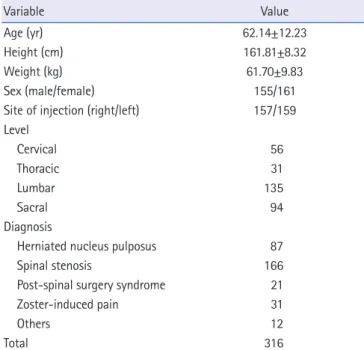
In total, 316 TFEB treatments were performed, with 56 injections (17.7%) at cervical levels, 31 (10.0%) at thoracic levels, 135 (42.7%) at lumbar levels, and 94 (29.7%) at sacral levels. There were no complications associated with TFEB. The 316 TFEB treatments were performed in 218 enrolled participants, with a mean age of 62.1 years. The characteristics of study participants are presented in Table 1. TFEB treatments were performed from
Table 1. Characteristics of 316 injections performed in 218 participants
Variable Value
Age (yr) 62.14±12.23
Height (cm) 161.81±8.32
Weight (kg) 61.70±9.83
Sex (male/female) 155/161
Site of injection (right/left) 157/159 Level
Cervical 56
Thoracic 31
Lumbar 135
Sacral 94
Diagnosis
Herniated nucleus pulposus 87
Spinal stenosis 166
Post-spinal surgery syndrome 21
Zoster-induced pain 31
Others 12
Total 316
Values are presented ad mean±standard deviation or number (proportion).
C3 to S2 spinal levels.
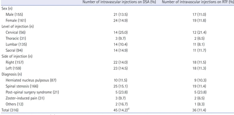
The incidence of intravascular injection at each level detected with DSA and RTF is presented in Table 2. Thirty-six intravascular injections (12 [21.4%] at cervical, 2 [6.5%] at thoracic, 11 [8.1%]
at lumbar, and 11 [11.7%] at sacral levels) were detected, for an overall intravascular injection rate of 11.4% using RTF. Forty- five intravascular injections (14 [25%] at cervical, 3 [9.7%] at thoracic, 14 [10.4%] at lumbar, and 14 [14.9%] at sacral levels) were detected, for an overall intravascular injection rate of 14.2%
using DSA. The intravascular injection detection rate using DSA was statistically different from that using RTF (p=0.004). All intravascular injections detected using RTF were also observed using DSA. RTF missed 9 cases of intravascular injection (2 at cervical, 1 at thoracic, 3 at lumbar, and 3 at sacral levels) that were detected using DSA (RTF sensitivity, 80.0%).
Table 3 shows the adjusted OR and 95% CI for each variable during intravascular injection using DSA. Only the spinal level showed a significant association with intravascular injection.
The incidence of intravascular injection was significantly higher at the cervical level than at the thoracic (p=0.009) and lumbar (p=0.011) levels. Patient age, sex, height, weight, procedure side, and diagnosis had no effect on the incidence.
Discussion
Our results indicate that intravascular injection was sequentially
detected using RTF and DSA during TFEB at the whole spine
level. The overall incidence of intravascular injection was 14.2%
using DSA and 11.4% using RTF. Nine intravascular injections were missed (2 at cervical, 1 at thoracic, 3 at lumbar, and 3 at sacral levels) with RTF compared to DSA. A 25% improvement was observed using DSA compared to RTF. Therefore, DSA had a better detection rate for intravascular injection during TFEB.
DSA is a radiological technique that can be used to clearly visualize and distinguish blood vessels from surrounding tissues;
this is done by subtracting the pre-contrast image from the post- contrast injection image [12,15]. Visnjevac et al. [16] reported the efficacy of DSA in detection of intravascular penetration compared with RTF during TFEB in a recent meta-analysis. They included 1,290 TFEB cases (3.2% at cervical, 76.3% at lumbar, and 20.5% at sacral levels) and demonstrated that DSA showed a 32% improvement in detection of intravascular penetration during TFEB, compared to that using RTF. However, only 3.2%
of cases in their study were performed at cervical levels and did not include any thoracic cases. Therefore, their study could not represent TFEB at the whole spine level. In contrast, the present study attempted to include more cervical and thoracic TFEB cases (17.7% at cervical and 10.0% at thoracic levels).
Even though previous studies demonstrated DSA to be superior to RTF for vascular detection during TFEB, McLean et al. [15]
indicated that the main vascular uptake observed using DSA was Table 2. Incidence of intravascular injections during transforaminal epidural block
Number of intravascular injections on DSA (%) Number of intravascular injections on RTF (%) Sex (n)
Male (155) 21 (13.5) 17 (11.0)
Female (161) 24 (14.9) 19 (11.8)
Level of injection (n)
Cervical (56) 14 (25.0) 12 (21.4)
Thoracic (31) 3 (9.7) 2 (6.5)
Lumbar (135) 14 (10.4) 11 (8.1)
Sacral (94) 14 (14.9) 11 (11.7)
Side of injection (n)
Right (157) 22 (14.0) 18 (11.5)
Left (159) 23 (14.5) 18 (11.3)
Diagnosis (n)
Herniated nucleus pulposus (87) 10 (11.5) 9 (10.3)
Spinal stenosis (166) 25 (15.1) 19 (11.4)
Post-spinal surgery syndrome (21) 5 (23.8) 5 (23.8)
Zoster-induced pain (31) 3 (9.7) 2 (6.5)
Others (12) 2 (16.7) 1 (8.3)
Total (316) 45 (14.2)
a)36 (11.4)
Values are presented as number (%).
DSA, digital subtraction angiography; RTF, real-time fluoroscopy.
a)
p<0.05 compared with RTF.
Table 3. OR and 95% CI of variables on intravascular penetration using DSA
Variable OR 95% CI
Sex
Male 1.00 -
Female 1.26 0.90-1.82
Age 0.99 0.96-1.01
Height 1.00 0.97-1.04
Weight 1.02 0.99-1.05
Side of injection
Right 1.00 -
Left 0.94 0.50-1.76
Level
Cervical 2.88
a)1.27-6.54
Thoracic 0.93 0.25-3.44
Lumbar 1.00 -
Sacral 1.51 0.69-3.34
Diagnosis
Herniated nucleus pulposus 1.21 0.31-4.73
Spinal stenosis 1.66 0.47-5.86
Post-spinal surgery syndrome 3.11 0.65-14.85
Zoster-induced pain 1.87 0.27-12.85
Others 1.00
OR, odds ratio; CI, confidence interval; DSA, digital subtraction angiography.
a)