INTRODUCTION
Polycyclic aromatic hydrocarbons (PAHs) are ubiquitous- ly found in air and aquatic environments as a result of the incomplete combustion of organic materials from industrial processes and human activities (Hylland 2006). PAHs pre- fer to associate with particulate matter in seawater or marine sediments due to their liphophilic and hydrophobic charac- teristics, which is known to be accumulated in marine organ- isms throughout food web (Adamo et al. 1997; Baumard et al. 1998). Among them, benzo[a]pyrene (BaP), dibenzo [a,h]anthracene (DBA), benzo[a]anthracene (BaA), benzo [b]fluoranthene (BbF), benzo(k)fluoranthene (BkF), and
indeno[1,2,3-c, d]pyrene (InP) were known as carcinogenic PAHs (cPAHs) due to their potent carcinogenic and muta- genic activities (IARC 1984).
It is widely accepted that aquatic contamination is caused by the mixture of various organic compounds. These toxic compounds cause the onset of biological disorders associated with development and reproduction of aquatic organisms by interacting with target macromolecules such as proteins or nucleic acids (Guillette et al. 1995; Fielden and Zacharew- ski 2001; Waring et al. 2001; Aravindakshan et al. 2004;
Moggs 2005; Williams et al. 2007). Although cytochrome P450 (CYP450) family has been applied to evaluate the toxicological effects of PAHs on fish in field and laboratory studies, risk assessments for the compounds remain elusive (Peters et al. 1997; Gravato and Santos 2002; Nacci et al.
2002; Carlson et al. 2004; Greytak et al. 2005; Hoffmann et al. 2006; Jonsson et al. 2006; Patel et al. 2006). Thus, iden-
─
─ 391 ──
Changes in Differentially Expressed Genes in the Liver of Oryzias latipes by Binary Exposure to
Carcinogenic Polycyclic Aromatic Hydrocarbons
Jeong Hwan Oh, Hyo-Bang Moon1and Eun Sang Choe*
Department of Biological Sciences, Pusan National University, Busan 609-735, Korea
1Marine Environment Research Team, National Fisheries Research and Development Institute, Busan 619-705, Korea
Abstract -- The biological effects of carcinogenic polycyclic aromatic hydrocarbons (cPAHs) includ- ing benzo[a]pyrene (BaP), dibenzo[a,h]anthracene (DBA), benzo[a]anthracene (BaA), benzo[b]
fluoranthene (BbF), benzo[k]fluoranthene (BkF), and indeno[1,2,3-c, d]pyrene (InP) on transcrip- tomic changes were determined in the liver of Oryzias latipes. Differentially expressed genes (DEGs) by binary exposure to cPAHs (BaP++BaA, BaP++BbF, BaP++BkF, BaP++DbA, BaP++InP) were screened by annealing control primers-based polymerase chain reaction followed by sequence analysis and BLAST searching. The results showed that four DEGs were commonly expressed by cPAHs and they were identified as ribosomal protein S4, coagulation factor II, elongation factor 1 beta, and a predicted protein similar to human immunodeficiency virus type I enhancer binding protein 3. This finding suggests that binary exposure to cPAHs interferes protein synthesis requir- ed for fundamental liver functions in fish.
Key words : organic pollutant, transcription, medaka, aquatic pollution
* Corresponding author: Eun Sang Choe, Tel. 051-510-2272, Fax. 051-581-2962, E-mail. [email protected]
tification of altered genes in fish using genomic analysis would be a way to assess the biological effects of the com- pounds on marine environment.
In the present study, the differentially expressed genes (DEGs) were identified to investigate the effects of cPAHs on the change of transcripts in the liver of Oryzias latipes (O. latipes). The DEGs were screened by annealing control primer (ACP)-based polymerase chain reaction (PCR) with 20 arbitrary primers because the ACP system has advantage to improve the specificity of PCR amplification and the eli- mination of false-positive results (Hwang et al. 2003; Kim et al. 2004). O. latipes (Japanese medaka) was used as a model species in this study because it has several advantages for laboratory study. For instance, it is small in size, easy to rear in a laboratory, and has a short life cycle. The fish also can adapt to a wide range of saline condition, although it is a freshwater species (Sakamoto et al. 2001; Inoue and Takei 2003).
MATERIALS AND METHODS
1. Animal maintenance and chemical exposure
O. latipes was maintained at 25�C under a constant pho- toperiod of 16 : 8 hrs (light : dark). Water quality was moni- tored by measuring pH, dissolved oxygen, and temperature (Table 1). During acclimation in seawater, the fish was fed newly hatched brine shrimp and a commercial flake food
twice a day, but the experiments were performed without being fed. All chemicals were purchased from Sigma-Ald- rich (MO, USA). DMSO was used as a solvent control. Male medaka adapted to seawater was exposed to each of cPAHs (BaP, DBA, BaA, BbF, BkF, InP) or binary combination of cPAHs for 24 hrs. The ratio of binary combination was 1 : 1 based on BaP concentration (25 μg L-1). Following water- borne exposure, the fish was sacrificed and the head was im- mediately frozen in liquid nitrogen and then stored at -80�C until RNA isolation.
2. Total RNA isolation and first-strand cDNA synthesis
Total RNAs were isolated from the liver of O. latipes using easy-spinTM[DNA free] Total RNA Extraction Kit (iNtRON Biotechnology, Gyeonggi, Korea). Briefly, pooled liver samples (n==7) was homogenized in 1 mL of lysis buf- fer and then vigorously vortexed in room temperature for 10 sec. After adding 200 μL of chloroform, the solution was centrifuged at 13,000 rpm for 10 min at 4�C. Then the super- natant (400 μL) was transferred to a fresh 1.5 mL tube and 400 μL of binding buffer was added and mixed it well by 2~3 times gentle inverting. The supernatant was loaded to the column and centrifuged at 13,000 rpm for 30 sec. Follow- ing discarding the flow-through, the column was washed by adding washing buffer and centrifuged at 13,000 rpm for 1~2 min to dry the column membrane. The 50 μL of Elu- tion buffer was added directly onto the membrane after the Table 1. Test conditions of binary exposure to cPAHs in the liver of O. latipes
Parameters Conditions
Control Each cPAH BaP++other cPAHs
Exposure type Water-borne 〃 〃
Test organism Oryzias latipes 〃 〃
Weight (g) 0.14±0.04 0.31±0.09 0.39±0.07
Length (cm) 2.84±0.22 3.22±0.48 3.30±0.17
Exposure duration 24 hrs 〃 〃
pH 7.84±0.02 7.82±0.01 7.81±0.02
Dissolved oxygen (mg L-1) 5.39±0.09 5.55±0.13 5.50±0.07
Salinity (psu) 34.51±0.08 34.70±0.08 34.75±0.17
Temperature (�C) 24.68±0.01 24.81±0.13 24.91±0.08
Photoperiod 16 Light : 8 Dark 〃
Feeding regime None 〃
Test water Filterated seawater
(0.22 μm cartridge filter) 〃
Value shown are mean±S.D., n==7.
cPAHs: BaP, benzo[a]pyrene; DBA, dibenzo[a,h]anthracene; BaA, benzo[a]anthracene; BbF, benzo[b]fluoranthene; BkF, benzo(k)fluoranthene; InP, indeno [1,2,3-c, d]pyrene
column was placed in a new 1.5 mL tube. Finally, the column was incubated for 1 min at room temperature and centrifuged 13,000 rpm for 1 min to elute total RNAs. The isolated total RNAs were used for the synthesis of the first-strand cDNAs by reverse transcriptase. Reverse transcription was perform- ed for 1.5 hrs at 42�C in a final reaction volume of 20 μL containing 3 μL of the purified total RNAs, 4 μL of 5 X reaction buffer (Promega, WI, USA), 5 μL of dNTPs (each 2 mM), 2 μL of 10 μM dT-ACP1 [5′-CTGTGAATGCTGC- GACTACGATIIIIIT(18)-3′], 0.5 μL of RNasin® RNase Inhibitor (40 U μL-1; Promega), and 1 μL of moloney murine leukemia virus reverse transcriptase (200 U μL-1; Promega).
The first-strand cDNAs were diluted by the addition of 80 μL of ultra-purified water for the ACP-based PCR, and stored at -20�C until used.
3. ACP-based PCR
DEGs were screened by ACP-based PCR method (Kim et al. 2004) using the GeneFishingTMDEG kits (Seegene, Seoul, South Korea). Briefly, the second-strand cDNA synthesis was conducted at 50�C during one cycle of the first-stage PCR in a final reaction volume of 20 μL containing 3~5
μL (about 50 ng) of diluted first-strand cDNA, 1 μL of dT- ACP2 (10 μM), 1 μL of 10 μM arbitrary ACP, and 10 μL of 2× Master Mix (Seegene). The PCR protocol for the sec- ond-strand synthesis was one cycle at 94�C for 1 min, fol- lowed by 50�C for 3 min, and 72�C for 1 min. After the sec- ond-strand DNA synthesis was completed, the second-stage PCR amplification protocol was 40 cycles of 94�C for 40 sec, followed by 65�C for 40 sec, 72�C for 40 sec, followed by a 5 min final extension at 72�C. The amplified PCR pro- ducts were separated in 2% agarose gel stained with ethid- iumbromide. The differentially expressed band was selected as DEGs based on the size of PCR products (300~1,200 base pairs), intensity (at least more than two folds compared with control), and shape (sharp but not smear).
4. Direct sequencing
The differentially expressed bands were re-amplified and extracted from the gel by using the GENCLEAN®II Kit (Q- BIO gene, CA, USA), and directly sequenced with ABI PRISM®3100-AvantGenetic Analyzer (Applied Biosystems, CA, USA) using universal primer (5′-GTCTACCAGGCA- TTCGCTTCAT-3′).
DEG 1
DEG 2
DEG 3
DEG 4
ControlBaP BaA BaP BaA ControlBaP BbF BaP BbF ControlBaP InP BaP InP ControlBaP DbA BaP DbA
ControlBaP BaA BaP BaA ControlBaP BbF BaP BbF ControlBaP BkF BaP BkF ControlBaP InP BaP InP ControlBaP DbA BaP DbA
ControlBaP BaA BaP BaA ControlBaP BbF BaP BbF ControlBaP BkF BaP BkF ControlBaP InP BaP InP
ControlBaP BkF BaP BkF
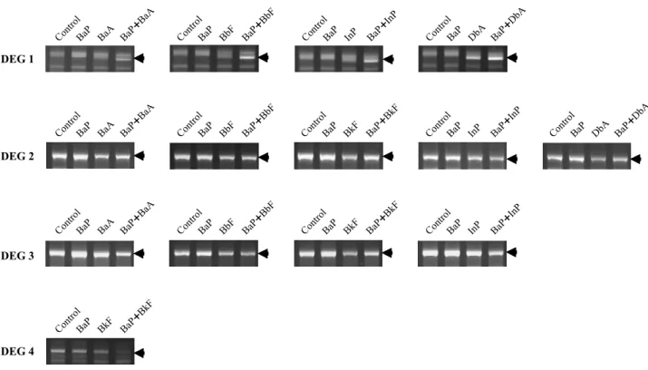
Fig. 1. Identified DEGs in the liver of Japanese medaka were selected by comparing density of bands produced by the control groups (see arrows). DEG 1 was upregulated by all binary exposure to cPAHs excepted BaP+BkF, whereas DEG 2, 3, and 4 were downregulated.
Interestingly, DEG 4 was downregulated by binary exposure to BaP+BkF only.
5. Statistical analysis
The difference of an expression level between each group was determined by their density on agarose gel compared with control (mean±SD). The statistical significance was analyzed with the student’s t-test using GraphPad Prism 4 (GraphPad Software Incorporation, CA, USA) and set at p
⁄0.05.
RESULTS AND DISCUSSION
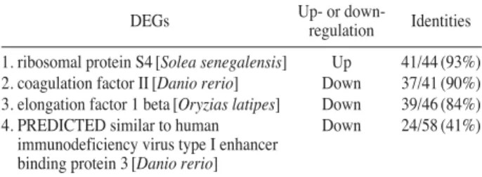
This study was conducted to understand the changes in DEGs by acute binary exposure to cPAHs in the liver of Japanese medaka. The results demonstrated that four DEGs were regulated by following binary exposure to cPAHs, such as BaP++BaA, BaP++BbF, BaP++BkF, BaP++DbA, and BaP++InP, and they were identified as ribosomal protein S4, coagulation factor II, elongation factor 1 beta, and a predict- ed protein similar to human immunodeficiency virus type I enhancer binding protein 3 (Fig. 1) (Tables 2, 3).
DEG 1 was found to be a protein that is highly homolo- gous with ribosomal protein S4 in Solea senegalensis and was upregulated by all binary exposures to cPAHs excepted BaP++BkF. It is well known that ribosomal protein plays a critical role in protein biosynthesis by interacting with trans- lational apparatus including ribosomal RNA (Lodish et al.
2003). Although genetic information stores protein infor-
mation, translation is a critical step for the production of functional proteins that contribute to the variety of biologi- cal activities (Kapp and Lorsch 2004). For this reason, trans- criptomic changes of ribosomal proteins may alter the effi- cient assembly of each ribosome and influence protein bio- synthesis (Liebman et al. 1995). Interestingly, DEG 3 that was downregulated by cPAHs was found to be a protein having structural similarity to the elongation factor 1 beta (EF-1β) in O. latipes involved in the elongation step of protein synthesis (Riis et al. 1990). In eukaryotic cell, EF-1 is composed of 4 subunits (α, β, γ and δ) and the interaction of each subunit helps continuous elongation steps. In parti- cular, the EF-1β, a guanine nucleotide exchange factor, is essential for cell growth and plays an important role in translational rate and fidelity (Hiraga et al. 1993; Carr-sch- mid et al. 1999).
DEG 2 was found to be a downregulated protein that is highly similar to coagulation factor II (prothrombin) in Danio rerio involved in hemostasis. Previous studies have shown that the deficiency of prothrombin in the develop- ment of mouse causes embryonic lethality (Sun et al. 1998).
In addition, cPAHs also interfere with blood coagulation by suppressing prothrombin gene expression in the liver, and causes inflammation by interrupting the aggregation of pla- telet (Coughlin et al. 1992).
Taken together, these findings suggest that cPAHs in the liver of Japanese medaka hinder protein synthesis by pre- venting accurate elongation step as well as blood coagula- tion, and thus causes liver dysfunctions in fish. Although lim- ited primers were used in this study, identified genes may provide an informative insight into assess marine environ- mental risk of cPAHs.
ACKNOWLEDGEMENTS
This research was funded by National Fisheries Research and Development Institute, Busan, Korea.
REFERENCES
Adamo RD, S Pelosi, P Trotta and G Sansone. 1997. Bioac- cumulation and biomagnification of polycyclic aromatic hydrocarbons in aquatic organisms. Mar. Chem. 56:45.
Table 2. Identified DEGs by binary exposure to cPAHs in the liver of O. latipes
DEGs Up- or down-
regulation Identities 1. ribosomal protein S4 [Solea senegalensis] Up 41/44 (93%) 2. coagulation factor II [Danio rerio] Down 37/41 (90%) 3. elongation factor 1 beta [Oryzias latipes] Down 39/46 (84%) 4. PREDICTED similar to human Down 24/58 (41%)
immunodeficiency virus type I enhancer binding protein 3 [Danio rerio]
Table 3. Commonly identified DEGs by binary exposure to cPAHs in the liver of O. latipes
Groups DEG 1 DEG 2 DEG 3 DEG 4
BaP++BaA ● ● ●
BaP++BbF ● ● ●
BaP++BkF ● ● ●
BaP++InP ● ● ●
BaP++DbA ● ●
Aravindakshan J, V Paquet, M Gregory, J Dufresne, M Fourni- er, DJ Marcogliese and DG Cyr. 2004. Consequences of xenoestrogen exposure on male reproductive function in spottail shiners (Notropis hudsonius). Toxicol. Sci. 78:156.
Baumard P, H Budzinski, P Garrigues, JC Sorbe, T Burgeot and J Bellocq. 1998. Concentrations of PAHs (Polycyclic aromatic hydrocarbons) in various marine organisms in relation to those in sediments and to trophic level. Mar. Pol- lut. Bull. 36:951.
Carr-schmid A, L Valente, VI Loik, T Williams, LM Starita and TG Kinzy. 1999. Mutations in elongation factor 1, a guanine nucleotide exchange factor, enhance translational fidelity. Mol. Cell. Biol. 19:5257.
Carlson EA, Y Li and JT Zelikoff . 2004. Benzo[a]pyrene-in- duced immunotoxicity in Japanese medaka (Oryzias latip- es): relationship between lymphoid CYP1A activity and humoral immune suppression. Toxicol. Appl. Pharmacol.
201:40.
Coughlin SR, TK Vu, DT Hung and VI Wheaton. 1992. Cha- racterization of a functional thrombin receptor. Issues and opportunities. J. Clin. Invest. 89:351.
Fielden MR and TR Zacharewski. 2001. Challenges and limi- tations of gene expression profiling in mechanistic and pre- dictive toxicology. Toxicol. Sci. 60:6.
Gravato C and MA Santos. 2002. Juvenile sea bass liver P450, EROD induction and erythrocytic genotoxic responses to PAH and PAH-like compounds. Ecotoxicol. Environ. Saf.
51:115.
Greytak SR, D Champlin and GV Callard. 2005. Isolation and characterization of two cytochrome P450 aromatase forms in killifish (Fundulus heteroclitus): Differential expression in fish from polluted and unpolluted environments. Aquat.
Toxicol. 71:371.
Guillette JLJ, DA Crain, AA Ronney and DB Pickford. 1995.
Organization versus activation: The role of endocrine dis- rupting contaminants (EDCs) during embryonic develop- ment in wildlife. Environ. Health Perspect. 103:157.
Hiraga K, K Suzuki, E Tsuchiya and T Miyakawa. 1993. Clon- ing and characterization of the elongation factor EF-1b homologue of Saccharomyces cerevisiae. EF-1b is essential for growth. FEBS Lett. 316:165.
Hoffmann JL and JT Oris. 2006. Altered gene expression: A mechanism for reproductive toxicity in zebrafish exposed to benzo(a)pyrene. Aquat. Toxicol. 78:332.
Hwang IT, YJ Kim, SH Kim, CI Kwak, YY Gu and JY Chun.
2003. Annealing control primer system for improving specificity of PCR amplification. BioTechniques 35:1.
Hylland K. 2006. Polycyclic aromatic hydrocarbon (PAH) eco- toxicology in marine ecosystems. J. Toxicol. Environ.
Health Part A 69:109.
IARC. 1987. IARC monographs on the evaluation of the car- cinogenic risk of chemicals to humans. Overall evaluation of carcinogenicity: an updating of IAPC monographs. vol.
1-42. Suppl. 7. International Agency for Research on Can- cer, Lyon, France.
Inoue K and Y Takei. 2003. Asian medaka fishes offer new models for studying mechanisms of seawater adaptation.
Comp. Biochem. Physiol. B 136:635.
Jonsson EM, A Abrahamson, B Brunstorm and I Brandt. 2006.
Cytochrome P4501A induction in rainbow trout gills and liver following exposure to waterborne indigo, benzo(a) pyrene and 3,3′,4,4′,5-pentachlorobiphenyl. Aquat. Toxi- col. 79:226.
Kapp LD and JR Lorsch. 2004. The molecular mechanics of eukaryotic translation. Annu. Rev. Biochem. 73:657.
Kim YJ, CI Kwak, YY Gu, IT Hwang and JY Chun. 2004. An- nealing control primer system for identification of differen- tially expressed genes on agarose gels. BioTechniques 36:1.
Liebman SW, YO Chernoff and R Liu. 1995. The accuracy cen- ter of a eukaryotic ribosome. Biochem. Cell Biol. 73:1141.
Lodish H, A Berk, P Matsudaira, CA Kaiser, M Krieger, MP Scott, SL Zipursky and J Darnell. 2003. Molecular cell bio- logy. 5thed. pp. 119-130. W.H. Freeman and Company, New York.
Moggs JG. 2005. Molecular responses to xenoestrogens: Mech- anistic insights from toxicogenomics. Toxicology 213:177.
Nacci DE, M Kohan, M Pelletier and E George. 2002. Effects of benzo(a)pyrene exposure on a fish population resistant to the toxic effects of dioxin-like compounds. Aquat. Toxi- col. 57:203.
Patel MR, BE Scheffler, L Wang and KL Willett. 2006. Effects of benzo(a)pyrene exposure on killifish (Fundulus hetero- clitus) aromatase activities and mRNA. Aquat. Toxicol.
77:267.
Peters LD, HR Morse, R Waters and DR Livingstone. 1997.
Responses of hepatic cytochrome P4501A and formation of DNAadducts in juveniles of turbot (Scophthalmus maxi- mus L.) exposed to water-borne benzo[a]pyrene. Aquat.
Toxicol. 38:67.
Riis B, SIS Rattan, BFC Clark and WC Merrick. 1990. Euka- ryotic protein elongation factors. Trends Biol. Chem. 15:
420.
Sakamoto T, T Kozaka, A Takahashi, H Kawauchi and M Ando.
2001. Medaka (Oryzias latipes) as a model for hypoosmo- regulation of euryhaline fishes. Aquaculture 193:347.
Sun WY, DP Witte, JL Degen, MC Colbert, MC Burkart, K Holmbäck, Q Xiao, TH Bugge and SJ Degen. 1998 Proth- rombin deficiency results in embryonic and neonatal letha- lity in mice. 1: Proc. Natl. Acad. Sci. U.S.A. 95:7597.
Waring JF, R Ciurlionis, RA Jolly, M Heindel and RG Ulrich.
2001. Microarray analysis of hepatotoxins in vitro reveals a correlation between gene expression profiles and macha- nisms of toxicity. Toxicol. Lett. 120:359.
Williams TD, AM Diab, SG George, V Sabine and JK Chip- man. 2007. Gene expression responses of European floun-
der (Platichthys flesus) to 17-beta estradiol. Toxicol. Lett.
168:236.
Manuscript Received: November 5, 2009 Revision Accepted: November 25, 2009 Responsible Editor: Kyung Jin Lee