127
Lab Anim Res 2013: 29(2), 127-130 http://dx.doi.org/10.5625/lar.2013.29.2.127
Uterine leiomyosarcoma in a wild rat (Rattus norvegicus):
usefulness of Ki-67 labeling index for diagnosis
Byung-Suk Jeon
1,2#, Hyung-Gi Kim
1#, Byung-Woo Lee
1, Jeong-Hee Han
1, Byung-Il Yoon
1*
1
College of Veterinary Medicine and Institute of Veterinary Science, Kangwon National University, Chuncheon, Korea
2
Toxicologic Pathology Divison, Korea Institute of Toxicology, Daejeon, Korea
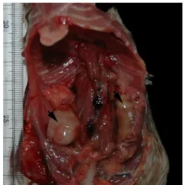
Uterine smooth muscle tumor is very rare in laboratory rats and, there has been no report in the wild rodents. Among a total of 400 wild rats captured in Gyeonggi, Gangwon, and Chungbuk provinces of Korea in 2007, 2010, and 2011, we found a uterine spindle cell tumor, diagnosed as smooth muscle cell origin based on differential features of histology and immunohistochemistry. Its incidence was very low, like in the laboratory rats, as under 0.5% for female. Considering generally applied histological and cellular criteria, this case was difficult in differential diagnosis between benign and malignant. Ki-67 labeling index was therefore further investigated, and it ranged from 26.4 to 37.6% in the 10 different areas, representing an average of 32.9±0.05%. The Ki-67 labeling index of neoplastic cells near the necrotic area was recorded as 83.5%. According to such high Ki-67 labeling index, it was more likely a malignant leiomyosarcoma, assenting to the previous proposal that Ki-67 labeling index is a significant criterion to differentiate between malignant and benign in the smooth muscle tumors.
Key words: Ki-67, leiomyosarcoma, Rattus norvegicus, rat
Received 7 March 2013; Revised version received 23 May 2013; Accepted 4 June 2013
Leiomyoma and leiomyosarcoma are respectively benign and malignant tumor originating from smooth muscle cells [1]. They can occur anywhere since smooth muscle is widely dispersed throughout the body, such as the gastrointestinal and genital tracts, respiratory and vascular systems, hair follicle-associated arrector pilae muscle, and the uveal tract of the eye [1]. Smooth muscle tumor is relatively common in dogs, compared to other domestic animals including the cat and the horse.
In the dog, leiomyoma frequently occurs in the stomach (25%), vagina and cervix (19%), urinary bladder (9%) and uterus (8%), whereas the small intestine (29%), cecum (15%), spleen (13%), urinary bladder (10%) and stomach (7%) are the frequently affected sites of leiomyosarcoma [1]. In dogs, leiomyosarcoma incidence was 0.5% in the Cornel files from 1977 to 1997 [1].
Smooth muscle tumors are less common in laboratory animals, with the incidence being under only 0.5% in an untreated group in a 2-year carcinogenicity studies in the NTP technical report [2]. Bullock and Curtis reported only 4 (0.01%) cases of leiomyoma and 2 cases (<0.01%) of leiomyosarcoma in 31,868 rats, and Crain (1958) and Bullock and Curtis (1930) reported only 2 cases of leiomyoma and no incidence of leiomyosarcoma in 369 Wistar rats, respectively [3,4].
Leiomyosarcoma is differentiated from the benign smooth muscle tumor, leiomyoma, based on several clinical and pathological criteria such as metastasis, invasion, cellular atypia, relatively high mitotic index, areas of necrosis, and so on [1]. However, despite these criteria, it is difficult in many cases to clearly differentiate leiomyosarcoma from the well-differentiated form of
Letter
#