157 DOI: 10.4196/kjpp.2010.14.3.157
ABBREVIATIONS: SCI, spinal cord injury; HSP, heat shock protein;
BBB, Basso-Beattie-Bresnahan motor rating scale.
Received May 6, 2010, Revised May 18, 2010, Accepted May 19, 2010
Corresponding to: Junesun Kim, Department of Physical Therapy, Korea University College of Health Science, 1, Jungneung-3-dong, Sungbuk-gu, Seoul 136-703, Korea. (Tel) 82-2-940-2834, (Fax) 82-2-940-2830, (E-mail) junokim@korea.ac.kr
Loss of hsp70.1 Decreases Functional Motor Recovery after Spinal Cord Injury in Mice
Hyun Jeong Kim1, Ji-In Jung2, Youngkyung Kim2, Jae-Seon Lee4, Young Wook Yoon2, and Junesun Kim3
1Department of Dental Anesthesiology and Dental Research Institute, Seoul National University School of Dentistry, Seoul 110-744,
2Department of Physiology, Korea University College of Medicine, Seoul 136-705, 3Department of Physical Therapy, Korea University College of Health Science, Seoul 136-703, 4Divisions of Radiation Cancer Research and Korea Institute of Radiological and Medical Sciences, Seoul 139-706, Korea
Heat shock proteins (HSPs) are specifically induced by various forms of stress. Hsp70.1, a member of the hsp70 family is known to play an important role in cytoprotection from stressful insults.
However, the functional role of Hsp70 in motor function after spinal cord injury (SCI) is still unclear.
To study the role of hsp70.1 in motor recovery following SCI, we assessed locomotor function in hsp70.1 knockout (KO) mice and their wild-type (W T) mice via the Basso, Beattie and Bresnahan (BBB) locomotor rating scale, before and after spinal hemisection at T13 level. W e also examined lesion size in the spinal cord using Luxol fast blue/cresyl violet staining. One day after injury, KO and WT mice showed no significant difference in the motor function due to complete paralysis following spinal hemisection. However, when it compared to WT mice, KO mice had significantly delayed and decreased functional outcomes from 4 days up to 21 days after SCI. KO mice also showed significantly greater lesion size in the spinal cord than WT mice showed at 21 days after spinal hemisection. These results suggest that Hsp70 has a protective effect against traumatic SCI and the manipulation of the hsp70.1 gene may help improve the recovery of motor function, thereby enhancing neuroprotection after SCI.
Key Words: Spinal cord injury, Neuroprotection, Heat shock protein, Mice
INTRODUCTION
Spinal cord injury (SCI) results in devastating events for patients such as loss of motor function and neuropathic pain. As a consequence of primary mechanical injury to the cord, various pathologic events trigger subsequent secon- dary injury that can aggravate spinal cord damage [1-3].
Thus, controlling for the secondary injury following SCI is an important technique for limiting the extent of tissue damage and consequent functional impairment [4-6]. Previ- ous studies have shown a number of interrelated factors that may contribute to the secondary injury process, includ- ing heat shock proteins [1,7].
The heat shock proteins (HSPs) are a highly conserved proteins that act as molecular chaperones to aid protein transport and assembly of newly synthesized polypeptides [8]. Of the HSPs, Hsp70 is not usually detectable under normal condition and is rapidly induced by various stresses such as hyperthermia, oxidative stress and amino acid ana- logues [9-12]. Previous studies, both in vivo and in vitro studies, have suggested HSP70 induction is associated with
cell protection from various lethal insults [13-15]. Experi- mental studies using transgenic mice have shown that Hsp70 overexpression protected neuronal cells from ische- mic insult [16,17]. Plumier et al. [16] found that Hsp70 overexpression following permanent focal cerebral ischemia significantly protected the pyramidal neurons in the hippo- campus but did not affect the overall infarct area. Rajdev et al. [17] reported that cerebral infarct volume following brain ischemia was significantly lower in Hsp70 overexpre- ssing transgenic mice than in wild-type mice. However, an- other study using transgenic mice showed no significant dif- ference in infarct size or hippocampal cell survival [18].
Taken together, these may suggest that Hsp70 protects neuronal cells against some, but not all types of central nervous system injury. Recently, several studies demon- strated that Hsp70 plays an important role in the secon- dary injury cascade after SCI [19,20]. A number of in vitro [21,22] and in vivo studies [23-26] have suggested Hsp70 has cytoprotective effects in the spinal cord. Because neuro- protection involves several genes including Hsp70 [27], the neuroprotective effects of Hsp70 itself after SCI are difficult to determine. Thus, in the present study, we used hsp70.1 knockout mice (KO) to investigate whether Hsp70 plays a neuroprotective role in SCI.
Experimental animals
All experimental procedures were conducted in accord- ance with guidelines set by the Korea University College of Health Science Animals Research Policies Committee.
We obtained hsp70.1 KO mice used in this study from Dr.
Jeong-Sun Seo, at Seoul National University. A previous study has described these KO mice bearing a null-allele at hsp70.1 and confirmed the absence of hsp70.1 mRNA ex- pression in them [47]. For this experiment, we used 50 adult mice (8 weeks-old) weighing 25∼30 gm at the time of their operation: Thirty hsp70.1 KO mice and twenty lit- termate wild type mice (WT). The animals were kept on a 12-hour light /12-hour dark cycle with lights on at 7:00 A.M.
Surgical procedures
Under enflurane anesthesia (by mixture of 3% enflurane and 95% O2), we shaved the skin overlying the thoracic ver- tebrae of each subject and disinfected with povidone iodine.
After the skin was incised along the midline, a laminectomy on the T12 vertebrae was performed to expose the T13 spi- nal cord. With a no. 11 blade scalpel, the cord was hemi- sected on the left side. The wound was closed in anatomical layers, the skin with stainless wound clips. After surgery, the mice displayed contralateral hindlimb paralysis were excluded from the study.
Behavioral tests
Behavioral test for motor function was performed pre- operatively and postoperatively (PO) for hindlimbs. The test was performed on each mouse 1 day prior to surgery and 1, 4, 7, 10, 13, 17, and 21 days PO. Hindlimb motor function was assessed using the Basso-Beattie-Bresnahan (BBB) motor rating scale [28]. The BBB has 22 levels from 0 to 21 that systematically and logically follow recovery of hindlimb function, from a score of 0, indicative of no ob- served hindlimb movements, to a score of 21, representing a normally ambulating mouse. Testing procedures were as follows. Briefly, animals were allowed to move freely on a paper-covered mattress, which makes a noise if the mouse drags its feet. Test sessions were 4 min in duration, and scores were obtained from 2 blinded observers according to the criteria. Left and right hind limbs were assessed sepa- rately due to potential asymmetrical recovery.
Analysis of histology
After the tests, the animals were randomly selected from both the KO and WT groups and were subjected to the com- parison of histological differences in the spinal segments epicenter to the hemisection. Histological study was con- ducted on 6 mice in each group 3 weeks after spinal hemisection. During this period, the hemisected mice fully recovered motor function and showed well-established signs of mechanical allodynia, as described in our previous report using a rat model [29]. Mice were deeply anesthetized with sodium pentobarbital and perfused with heparinized saline followed by 4% paraformaldehyde in 0.1 M phosphate buf- fer (PB, pH 7.4). The spinal segments including epicenter were removed, post-fixed for 6∼8 hr and stored overnight
8 μm thick longitudinal sections. The sections were treated with xylene and 95% alcohol. The slices on the slides were incubated in Luxol fast blue and then counterstained with cresyl violet. To quantify the degree of tissue damage fol- lowing SCI, size of lesion of each section were measured by using a computer-assisted image analysis system (NIH image software). All assessments were performed in a blinded fashion.
Statistical analysis
All values were expressed as mean±SEM. The Mann- Whitney U test was used to compare scores obtained on a given experimental day between 2 groups. The lesion size in the KO and WT groups was compared with t-test.
Differences between groups were considered statistically significant if p<0.05.
RESULTS
Functional motor recovery after SCI in 70.1 KO and WT mice
Prior to hemisection, locomotor function in all animals was evaluated in both ipsi- and contralateral hindlimbs us- ing the BBB scale via an open field test (Fig. 1). The scores from hsp70.1 KO mice were not different from those of the WT mice. After hemisection, all mice showed paralysis on the ipsilateral hindlimb, corresponding to a BBB score 0.
However, progressive motor recovery was observed with hindlimb joint movement on 4 d PO, and then relatively rapid recovery proceeded until 13 d PO. At that time, the WT mice showed coordinated walking. The mice achieved maximal recovery on 21 d PO. On 1 d PO, mice also showed mild disturbance in contralateral hindlimb, due to walking imbalance resulting from ipsilateral hindlimb paralysis.
However, as shown in Fig. 1, the locomotor function of the contralateral hindlimb improved as the ipsilateral hindlimb recovered locomotor function.
Throughout the entire recovery period, there was no sig- nificant difference in contralateral side motor function be- tween the two groups. However, on the ipsilateral side, the hsp70.1 KO mice showed delayed motor function recovery compared to the WT mice. This difference in motor recovery between hsp70.1 KO and WT mice was statistically sig- nificant from 4 d PO up to 21 d PO, except on 17 d PO after spinal hemisection (p<0.05). Functionally prominent difference in motor function between hsp70.1 KO and WT mice was noticed from 7 d PO. At 7 d PO, the hsp70.1 KO mice scored 10.5±0.65 on the BBB scale, whereas the WT mice scored 14.1±0.31 (p<0.05). Although this score differ- ence does not seem great, the difference in terms of func- tional gain is significant. The hsp70.1 KO mice exhibited frequent weight-supported plantar stepping, with no fore- limb-hindlimb coordination. In contrast, the WT mice showed consistent weight-supported plantar steps and con- sistent forelimb- hindlimb coordination. Although, the hsp70.1 KO and WT mice did not show significant locomo- tor function differences at 17 d PO, the difference retuned on 21 d PO, the last day of the study. At this time, the KO mice displayed frequent to consistent weight-supported plantar steps with consistent forelimb-hindlimb coordina- tion, whereas the WT mice showed consistent weight-sup-
Fig. 1. Motor recovery of hindlimb. Hindlimb motor function on the contralateral (A) and the ipsilateral side (B) was evaluated before SCI and on days 1, 4, 7, 10, 13, 17 and 21 after SCI. Asterisks indicate the values significantly different between the two groups (*p<0.05).
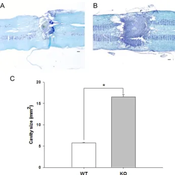
Fig. 2. A representative section of spinal cord segment including epicenter in WT (A) and KO (B) mice. Longitudinal spinal cord sections double-stained with Luxol fast blue and cresyl violet show the injury and atrophy area. The lesion size is significantly different between WT and KO mice (C) (*p<0.05). Scale bars, 400 μm.
ported plantar steps, with consistent forelimb-hindlimb coordination.
Lesion size
To assess the lesion size, the longitudinal cord sections were double-stained with Luxol fast blue and cresyl violet.
The lesions exhibited scar formation and complete loss of myelinated fiber. Scar formation appearances at the lesion site differed between KO and WT mice 3 weeks after SCI.
In the KO mice, scar tissue extended toward the contra- lateral side of the spinal cord and extended rostrocaudally from the epicenter in the spinal cord. In contrast, the WT mice had less scar formation at the lesion site (Fig. 2). In WT and KO mice, the cresyl violet stained areas were 6.70±
0.21 mm2 and 17.5±024 mm2, respectively (Fig. 2). There were significant lesion area differences between hsp70.1 KO and WT mice following spinal hemisection (p<0.05).
DISCUSSION
The present data demonstrated that mice lacking hsp70.1 experienced a poorer functional motor recovery than did
WT mice after spinal cord hemisection. We also found that lesion size was significantly larger in hsp70.1 KO mice than in WT mice. As suggested by several previous studies show- ing the beneficial effects of pharmacological upregulation of Hsp70 on cell survival, the present study indicates Hsp70 possesses neuroprotective potential after SCI. To our know- ledge, this is the first study evaluating the protective role of Hsp70 in SCI by using an hsp70.1 KO mouse model.
In this study, we examined motor function up to 21 d PO after spinal hemisection because previous reports showed motor function recovery plateaued at 3 weeks after SCI [29,30]. The present results show that hsp70.1 KO mice ex- perienced delayed and decreased functional motor recovery as compared to WT mice. As a result, the significant differ- ence in locomotor function between hsp70.1 KO and WT mice was occurred from 4 d PO. However, the prominent functional difference between hsp70.1 KO and WT mice ap- peared from 7 d PO. The difference in hindlimb function quality between the two groups was huge at this time point, although the score difference on BBB expressed in numbers could say. KO mice merely placed their limbs in a weighted fashion and showed occasional weighted plantar steps, whereas WT mice displayed nearly consistent plantar step- ping with consistent forelimb-hindlimb coordination.
Our findings are consistent with previous studies showing Hsp70 upregulation produced beneficial effects after SCI.
Upregulation of Hsp70 via anti-inflammatory drug treat- ment has improved functional outcomes after SCI [31,32].
Previously, Shin et al. [32] reported that cyclosporine A re- duced neurological injury due to spinal cord ischemia in a rabbit model. They concluded this improved outcome after spinal ischemia correlated to overexpression of Hsp70. A significant improvement in neurological function using modified Tarlov’s scores was more evident on day 7 PO.
in motor function at 7 d after SCI in rats pretreated with pioglitazone, a peroxisome proliferator-activated receptor inhibitors. The pioglitazone treated group showed a prom- inent enhanced expression of Hsp 70. Our result is strongly supported by the earlier study [33] on ischemic insult to the spinal cord. Their study showed that Hsp70 expression in motor neurons created ischemic tolerance after spinal ischemia in rats. Taken together, the preservation of motor functions after SCI may be partially mediated by upregula- tion of Hsp70.
It is well known that SCI leads to a progressive series of degenerative processes induced by the original insult to the spinal cord. These secondary degenerative processes, which contribute to progressive tissue loss and cavitation at the injury site, are a major cause of motor dysfunction [34]. Accordingly, recent studies on neuroprotection have focused on the inhibition of secondary injury after SCI [5,35-37]. It has been suggested that the induction of Hsp70 is associated with cell protection from various lethal insults both in vivo and in vitro studies [13-15]. Therefore, in the realm of neuroprotection, Hsp70 might be a useful target molecule for the therapeutic treatment. Previous studies have demonstrated that the induction of Hsp70 may be re- sponsible for secondary processes after SCI [38,39]. Recent experimental evidence showed that upregulation of Hsp 70 with pharmacological inhibition of inflammation achieved a neuroprotective effect [31,32]. In particular, Shin et al.
[32] found that neuroprotective effect of cyclosporine A against ischemia was related to overexpression of neural nitric oxide synthase and Hsp70, indicating the role of HSPs in modulating secondary injury. The present finding is consistent with these studies and with the body of work showing neuroprotective role of Hsp70 in ischemic brain and cardiac injury.
In contrast, other studies have suggested Hsp70 plays a different role of in neuroprotection. Reportedly, intrathecal administration of bupivacaine and hypothermia protected neuronal cells in rat SCI and this effect was most likely due to Hsp70 downregulation [40]. Such contradictory re- ports on the role of Hsp70 in SCI are explicable. First, pre- vious studies have suggested that HSPs have a multi- faceted modulatory role in inflammation, from proinflamm- atory to anti-inflammatory functions [41,42]. Second, the role of Hsp70 seems to depend on the time it is induced [33]
and the level of HSP expression [43]. Whereas most studies on HSP roles in SCI have focused on Hsp70, they limited themselves to certain SCI models, particularly ischemic SCI produced by thoracic aorta occlusion. Most of all, it is difficult to determine whether hsp70, itself, has a neuro- protective effect after SCI, because several other genes are involved in neuroprotective paradigms [27]. Different sub- types of HSP such as Hsp27 are involved in neuropro- tection. Tachibana et al. [44] found that the level of HSP27 gene expression level increased more than 2 times at 24 hours after SCI in a traumatic SCI rat model. Others have similar results [45] suggesting 17-allylamino-17-demethoxy- geldanamycin, a potent Hsp90 inhibitor, produced neuro- protective effects by inhibiting HSP90, indicating that Hsp70 may act in concert with other HSPs.
Currently, gene transfer techniques and transgenic ani- mal strain have made it possible to selectively overexpress HSP, to better understand the precise role of Hsp70 in cel- lular injury. Several studies using transgenic mice have suggested role of Hsp70 in neuroprotection is still con-
tion seems to have a neuroprotective effect on neuronal cells against some, but not all types of central nervous sys- tem injury. Most previous studies using transgenic mice were limited to certain brain injury produced by ischemia.
Thus, less is known about the precise influence of Hsp 70 on neuroprotection and functional outcomes after traumatic SCI. In the present study, hsp70 KO mice had larger lesion size than WT mice had. Inducible Hsp70 is encoded from both hsp70.1 and hsp70.3 genes, which show high similarity in their coding sequences. The fact that the two inducible hsp70 genes differ from each other in the 5’- and 3’-untran- slated regions [46], suggests they might be under differ- ential regulation. However, the deletion of hsp70.1 results in a remarkable decrease in Hsp70 expression [47]. Taken together, these findings also strongly suggest that Hsp70 is a cytoprotective protein within the spinal cord and may be responsible for neuroprotection after SCI.
In summary, the present results demonstrated hsp70.1 deletion results in a remarkable decrease in functional out- come and an increase in lesion size after SCI. The present report supports the ideas that Hsp70 mainly has a neuro- protective effect after SCI. Further molecular mechanisms must be investigated for the clinical application of HSP.
ACKNOWLEDGEMENTS
This workwas supported by a grant of the Korea University College of Health Science (K1031191).
REFERENCES
1. Kwon BK, Tetzlaff W, Grauer JN, Beiner J, Vaccaro AR.
Pathophysiology and pharmacologic treatment of acute spinal cord injury. Spine J. 2004;4:451-464.
2. Taoka Y, Okajima K. Spinal cord injury in the rat. Prog Neurobiol. 1998;56:341-358.
3. Tator CH, Fehlings MG. Review of the secondary injury theory of acute spinal cord trauma with emphasis on vascular mechanisms. J Neurosurg. 1991;75:15-26.
4. Bethea JR. Spinal cord injury-induced inflammation: a dual- edged sword. Neural Plast Regen. 2000;128:33-42.
5. Gris D, Marsh DR, Oatway MA, Chen Y, Hamilton EF, Dekaban GA, Weaver LC. Transient blockade of the CD11d/CD18 integrin reduces secondary damage after spinal cord injury, improving sensory, autonomic, and motor function. J Neurosci. 2004;24:
4043-4051.
6. Hall ED. The neuroprotective pharmacology of methylpredni- solone. J Neurosurg. 1992;76:13-22.
7. Kunz S, Tegeder I, Coste O, Marian C, Pfenninger A, Corvey C, Karas M, Geisslinger G, Niederberger E. Comparative pro- teomic analysis of the rat spinal cord in inflammatory and neuropathic pain models. Neurosci Lett. 2005;381:289-293.
8. Hartl FU. Molecular chaperones in cellular protein folding.
Nature. 1996;381:571-580.
9. Abe T, Konishi T, Hirano T, Kasai H, Shimizu K, Kashimura M, Higashi K. Possible correlation between DNA damage induced by hydrogen peroxide and translocation of heat shock 70 protein into the nucleus. Biochem Biophys Res Comm.
1995;206:548-555.
10. Fukamachi Y, Karasaki Y, Sugiura T, Itoh H, Abe T, Yamamura K, Higashi K. Zinc suppresses apoptosis of U937 cells induced by hydrogen peroxide through an increase of the Bcl-2/Bax ratio. Biochem Biophys Res Comm. 1998;246:364-369.
11. Morimoto RI, Sarge KD, Abravaya K. Transcriptional regu- lation of heat shock genes - a paradigm for inducible genomic responses. J Biol Chem. 1992;267:21987-21990.
12. Wagner M, Hermanns I, Bittinger F, Kirkpatrick CJ. Induction of stress proteins in human endothelial cells by heavy metal ions and heat shock. Am J Phsyio Lung Cell Mol Physiol.
1999;277:L1026-L1033.
13. Kabakov AE, Gabai VL. Heat shock induced accumulation of 70 kDa stress protein (HSP70) can protect ATP depleted tumor cells from necrosis. Exp Cell Res. 1995;217:15-21.
14. Marber MS, Mestril R, Chi SH, Sayen MR, Yellon DM, Dillmann WH. Overexpression of the rat inducible 70kDa heat stress protein transgenic mouse increases the resistance of the heart to ischemic injury. J Clin Invest. 1995;95:1446-1456.
15. Turner CP, Panter SS, Sharp FR. Anti-oxidants prevent focal rat brain injury as assessed by induction of heat shock proteins (HSP70, HO-1/HSP32, HSP47) following subarachnoid injec- tions of lysed blood. Mol Brain Res. 1999;65:87-102.
16. Plumier JCL, Krueger AM, Currie RW, Kontoyiannis D, Kollias G, Pagoulatos GN. Transgenic mice expressing the human inducible Hsp70 have hippocampal neurons resistant to ische- mic injury. Cell Stress Chaperones. 1997;2:162-167.
17. Rajdev S, Hara K, Kokubo Y, Mestril R, Dillmann W, Weinstein PR, Sharp FR. Mice overexpressing rat heat shock protein 70 are protected against cerebral infarction. Ann Neurol. 2000;47:
782-791.
18. Lee JE, Yenari MA, Sun GH, Xu L, Emond MR, Cheng D, Steinberg GK, Giffard RG. Differential neuroprotection from human heat shock protein 70 3roverexpression in in vitro and in vivo models of ischemia and ischemia-like conditions. Exp Neurol. 2001;170:129-139.
19. Awad H, Suntres Z, Heijmans J, Smeak D, Bergdall-Costell V, Christofi FL, Magro C, Oglesbee M. Intracellular and extra- cellular expression of the major inducible 70 kDa heat shock protein in experimental ischemia-reperfusion injury of the spinal cord. Exp Neurol. 2008;212:275-284.
20. Brown IR. Heat shock proteins and protection of the nervous system. Stress Resp Biol Med. 2007;1113:147-158.
21. Kikuchi S, Shinpo K, Takeuchi M, Tsuji S, Yabe I, Niino M, Tashiro K. Effect of geranylgeranylaceton on cellular damage induced by proteasome inhibition in cultured spinal neurons.
J Neurosci Res. 2002;69:373-381.
22. Manabe Y, Wang JM, Murakami T, Warita H, Hayashi T, Shoji M, Abe K. Expressions of nitrotyrosine and TUNEL immunore- activities in cultured rat spinal cord neurons after exposure to glutamate, nitric oxide, or peroxynitrite. J Neurosci Res. 2001;
65:371-377.
23. Cizkova D, Carmel JB, Yamamoto K, Kakinohana O, Sun D, Hart RP, Marsala M. Characterization of spinal HSP72 induction and development of ischemic tolerance after spinal ischemia in rats. Exp Neurol. 2004;185:97-108.
24. Perdrizet GA, Lena CJ, Shapiro DS, Rewinski MJ. Preoperative stress conditioning prevents paralysis after experimental aortic surgery: Increased heat shock protein content is associated with ischemic tolerance of the spinal cord. J Thorac Cardiovasc Surg.
2002;124:162-170.
25. Willoughby DS, Priest JW, Nelson M. Expression of the stress proteins, ubiquitin, heat shock protein 72, and myofibrillar protein content after 12 weeks of leg cycling in persons with spinal cord injury. Arch Phys Med Rehabil. 2002;83:649-654.
26. Sasara T, Cizkova D, Mestril R, Galik J, Sugahara K, Marsala M. Spinal heat shock protein (70) expression: effect of spinal ischemia, hyperthermia (42 degrees C)/hypothermia (27 degrees C), NMDA receptor activation and potassium evoked depolari- zation on the induction. Neuroschem Intern. 2004;44:53-64.
27. Kalmar B, Burnstock G, Vrbova G, Urbanics R, Csermely P, Greensmith L. Upregulation of heat shock proteins rescues motoneurones from axotomy-induced cell death in neonatal rats. Exp Neurol. 2002;176:87-97.
28. Basso DM, Beattie MS, Bresnahan JC. A sensitive and reliable locomotor rating scale for open field testing in rats. J Neurotrauma.
1995;12:1-21.
29. Kim J, Yoon YW, Hong SK, Na HS. Cold and mechanical allodynia in both hindpaws and tail following thoracic spinal cord hemisection in rats: Time courses and their correlates.
Neurosci Lett. 2003;343:200-204.
30. Tanabe M, Ono K, Honda M, Ono H. Gabapentin and pre- gabalin ameliorate mechanical hypersensitivity after spinal cord injury in mice. Eur J Pharmacol. 2009;609:65-68.
31. Park SW, Yi JH, Miranpuri G, Satriotomo I, Bowen K, Resnick DK, Vemuganti R. Thiazolidinedione class of peroxisome pro- liferator-activated receptor gamma agonists prevents neuronal damage, motor dysfunction, myelin loss, neuropathic pain, and inflammation after spinal cord injury in adult rats. J Pharmacol Exp Ther. 2007;320:1002-1012.
32. Shin YC, Choi KY, Kim WG. Cyclosporin A has a protective effect with induced upregulation of Hsp70 and nNOS on severe spinal cord ischemic injury in rabbits. J Invest Surg. 2007;
20:113-120.
33. Matsumoto M, Ohtake K, Wakamatsu H, Oka S, Kiyoshima T, Nakakimura K, Sakabe T. The time course of acquisition of ischemic tolerance and induction of heat shock protein 70 after a brief period of ischemia in the spinal cord in rabbits. Anesth Anal. 2001;92:418-423.
34. Hill CE, Beattie MS, Bresnahan JC. Degeneration and sprouting of identified descending supraspinal axons after contusive spinal cord injury in the rat. Exp Neurol. 2001;171:153-169.
35. Stirling DP, Khodarahmi K, Liu J, McPhail LT, McBride CB, Steeves JD, Ramer MS, Tetzlaff W. Minocycline treatment reduces delayed oligodendrocyte death, attenuates axonal die- back, and improves functional outcome after spinal cord injury.
J Neurosci. 2004;24:2182-2190.
36. Sribnick EA, Matzelle DD, Ray SK, Banik NL. Estrogen treatment of spinal cord injury attenuates calpain activation and apoptosis. J Neurosci Res. 2006;84:1064-1075.
37. Sribnick EA, Wingrave JM, Matzelle DD, Wilford GG, Ray SK, Banik NL. Estrogen attenuated markers of inflammation and decreased lesion volume in acute spinal cord injury in rats. J Neurosci Res. 2005;82:283-293.
38. Song GQ, Cechvala C, Resnick DK, Dempsey RJ, Rao VLR.
GeneChip((R)) analysis after acute spinal cord injury in rat. J Neurochem. 2001;79:804-815.
39. Hecker JG, Sundram H, Zou S, Praestgaard A, Bavaria JE, Ramchandren S, McGarvey M. Heat shock proteins HSP70 and HSP27 in the cerebral spinal fluid of patients undergoing thoracic aneurysm repair correlate with the probability of postoperative paralysis. Cell Stress Chaperones. 2008;13:435-446.
40. Lee JR, Han SM, Leem JG, Hwang SJ. Effects of intrathecal bupivacaine in conjunction with hypothermia on neuronal protection against transient spinal cord ischemia in rats. Acta Anaesthesiol Scand. 2007;51:60-67.
41. Chen Y, Voegeli TS, Liu PP, Noble EG, Currie RW. Heat shock paradox and a new role of heat shock proteins and their recep- tors as anti-inflammation targets. Inflamm Allergy Drug Targets.
2007;6:91-100.
42. Robinson MB, Tidwell JL, Gould T, Taylor AR, Newbern JM, Graves J, Tytell M, Milligan CE. Extracellular heat shock protein 70: A critical component for motoneuron survival. J Neurosci. 2005;25:9735-9745.
43. Sharma HS, Gordh T, Wiklund L, Mohanty S, Sjoquist PO.
Spinal cord injury induced heat shock protein expression is reduced by an antioxidant compound H-290/51. An experi- mental study using light and electron microscopy in the rat.
J Neural Transm. 2006;113:521-536.
44. Tachibana T, Noguchi K, Ruda MA. Analysis of gene expression following spinal cord injury in rat using complementary DNA microarray. Neurosci Lett. 2002;327:133-137.
45. Waza M, Adachi H, Katsuno M, Minamiyama M, Sang C, Tanaka F, Inukai A, Doyu M, Sobue G. 17-AAG, an Hsp90 inhibitor, ameliorates polyglutamine-mediated motor neuron degeneration. Nat Med. 2005;11:1088-1095.
46. Walter L, Rauh F, Gunther E. Comparative analysis of the 3 major histocompatiblity complex linked heat shock protein 70 (HSP70) genes of the rat. Immunogenetics. 1994;40:325-330.
47. Lee SH, Kwon HM, Kim YJ, Lee KM, Kim M, Yoon BW. Effects of hsp70.1 gene knockout on the mitochondrial apoptotic pathway after focal cerebral ischemia. Stroke. 2004;35:2195-2199.