Gasless Transaxillary Robot-Assisted Neck Dissection:
A Preclinical Feasibility Study in Four Cadavers
Yoo Seob Shin,1 Hyun Jun Hong,2 Yoon Woo Koh,2* Woong Youn Chung,3* Hye Yeon Lee,4 Jae Min Hong,2 Chi Sang Hwang,2 Jae Won Chang,2 and Eun Chang Choi2
1Department of Otolaryngology, School of Medicine, Ajou University, Suwon;
Departments of 2Otorhinolaryngology, 3General Surgery, and 4Anatomy, Yonsei University College of Medicine, Seoul, Korea.
Received: June 24, 2011 Revised: August 26, 2011 Accepted: August 31, 2011
Co-corresponding authors: Dr. Yoon Woo Koh, Department of Otolaryngology,
Yonsei University College of Medicine, 50 Yonsei-ro, Seodaemun-gu, Seoul 120-752, Korea.
Tel: 82-2-2228-3607, Fax: 82-2-393-0580 E-mail: ywkohent@yuhs.ac and Dr. Woong Youn Chung Department of General Surgery, Yonsei University College of Medicine, 50 Yonsei-ro, Seodaemun-gu, Seoul 120-752, Korea.
Tel: 82-2-2228-2126, Fax: 82-2-313-8289 E-mail: woungyounc@yuhs.ac
* These authors contributed equally to the paper as corresponding authors.
∙ The authors have no financial conflicts of interest.
© Copyright:
Yonsei University College of Medicine 2012 This is an Open Access article distributed under the terms of the Creative Commons Attribution Non- Commercial License (http://creativecommons.org/
licenses/by-nc/3.0) which permits unrestricted non- commercial use, distribution, and reproduction in any medium, provided the original work is properly cited.
Purpose: We hypothesized that comprehensive neck dissection could be achieved via a gasless transaxillary approach using a robotic system. We intended to evalu- ate the accessibility of level I, IIB and VA nodes with transaxillary robot-assisted neck dissection of four cadavers. Materials and Methods: Transaxillary robotic neck dissection was performed in four cadavers through a 7-cm longitudinal inci- sion at the anterior axilla and a 0.8-cm-sized incision in the chest wall. Results:
We successfully performed neck dissection from level II to V in all four cadavers.
However, dissection of levels IIB and VA, which lie on the cephalic portion of the spinal accessory nerve, was difficult. Vital structures, including the internal jugular vein, carotid artery, vagus nerve, phrenic nerve, superior thyroid artery and hypo- glossal nerve, were successfully identified and preserved. Conclusion: Our results demonstrate the feasibility of robot-assisted neck dissection using a transaxillary approach. We suggest that gasless, transaxillary robotic neck dissection is a prom- ising technique for treating nodal metastasis in thyroid cancers or in selected squa- mous cell carcinomas of the head and neck. However, some modification of the approach might be needed when performing comprehensive neck dissections of all levels of the neck.
Key Words: Trans-axillary, robotic, neck dissection, cadaver, squamous cell car- cinoma, endoscope
INTRODUCTION
Technological advances such as the introduction of the endoscope and surgical ro- botic system have brought great change to surgery. Such progress has led to mini- mally invasive surgery and allowed for a better postoperative cosmetic status for patients without compromising oncologic principles.1,2 The 3-dimensional magni- fied surgical view provided by the da Vinci robotic system (Intuitive Surgical, Mountain View, CA, USA) allows surgeons to overcome the limitations of conven- tional endoscopic surgery, such as 2-dimensional views, lack of a third arm or a limited range of motion of instruments. Furthermore, multi-articulated instruments and downscaling systems provides an opportunity to perform more minute and pre-
approach using the da Vinci robotic system. We intended to assess whether Level I, IIB and VA nodes can be dissected using a robot assisted endoscopic approach for HNSCC with nodal metastases in four cadavers.
MATERIALS AND METHODS
Cadavers
Four human cadavers were obtained from the Department of Anatomy of Yonsei University College of Medicine after approval by the Severance Robot and Minimally Invasive Surgical Training Center. Three of the cadavers were male and one was female. The cause of death were hepatocellu- lar carcinoma in two cadavers, myocardial infarction in one, and uterine cancer in the remaining one. Dissection was done on the left side in three of the cadavers and the right side in the other. The choice of dissection side was made de- pending on the condition of the cadavers, and neck dissec- tion was only performed on the selected side.
Flap elevation
With a cadaver placed in the supine position, the neck was slightly extended, and the dissection-side arm was abducted 45° and fixed (Fig. 1). A 7-cm longitudinal skin incision along the anterior axillary line was made and the skin flap was elevated under direct vision above the pectoralis major muscle to access the subplatysmal plane. After exposing the lateral border of the sternocleidomastoid muscle, we cut its clavicular head and retracted the muscle anteriorly and su- periorly. Then, the dissection continued posteriorly along the anterior border of the trapezius muscle, and the spinal accessory nerve (SAN) was preserved after being identified at about 1 cm above Erb’s point. Initially, we intended to el- evate the skin flap up to the inferior border of the mandible, while preserving the marginal mandibular branch of the fa- cial nerve. However, it was difficult to reach the mandible across the submandibular gland, and the external self re- tractor we used was too short to elevate and suspend the skin flap over level I. As a result, the flap was superiorly el- evated up to the inferior border of the submandibular gland (SMG) and until the posterior belly of the digastric muscle was noted.
Docking of robotic system
To create a working space, we inserted an external retractor (Sejong Medical Corporation, Paju, Korea) through the skin cise dissections and to more successfully preserve vital
structures.
In the field of head and neck surgery, there have been several studies about trans-oral robotic surgery (TORS), in which upper aero-digestive tract tumors are removed using the robotic surgical system.3-5 Our team has also reported at length the feasibility of TORS in various head and neck squamous cell carcinoma (HNSCC) cases.6,7 In addition, at our institution, robotic endoscopic thyroidectomy via trans- axillary approach has been performed in over 3,000 patients by Chung’s research team.8,9 The transaxillary approach provides enough surgical view to perform not only thyroid- ectomies but also dissections of the lateral compartment of the neck, as Chung has shown in his report on the feasibili- ty of transaxillary robotic neck dissections (ND) in differ- entiated thyroid cancer patients with nodal metastasis.10
However, as of yet, studies regarding robotic neck dis- section in HNSCC have not been performed. The need for this separate study is important because the comprehensive- ness of ND in thyroid cancer and HNSCC might be some- what different. The prognosis of patients with HNSCC is much poorer than those of thyroid cancer patients, and sur- vival rates sharply decrease when patients have nodal me- tastasis in HNSCC. The control of neck disease in HNSCC is widely accepted as more important than in differentiated thyroid cancer. In Chung’s study, instead of modified radi- cal ND, they usually only performed selective neck dissec- tion of levels IIA, III, IV, and VB.10 Hence, for cases of HN- SCC, we needed to verify the feasibility of robot-assisted transaxillary comprehensive ND of all neck levels, includ- ing level I, IIB and VA.
Therefore, we aimed to investigate whether a compre- hensive ND for HNSCC could also be performed with this
Fig. 1. Position and neck incision. With a cadaver placed in the supine posi- tion, the neck was slightly extended, and the dissection-side arm was ab- ducted 45° and fixed; a 7 cm longitudinal skin incision along the anterior axillary line was made, and a second skin incision (0.8 cm long) was made on the medial side of the anterior chest wall.
incision in the axilla, which was raised using a lifting device.
Four robotic arms were used during the dissection. Three arms including a dual channel endoscope, Harmonic curved shears and a Maryland dissector (the latter two designed by Intuitive Surgical) were inserted through the axillary port.
The endoscope was placed in the center of the three robotic arms, accompanied with the Harmonic scalpel and Mary- land dissector on both sides. Another 0.8-cm-sized skin in- cision for the fourth robotic arm was made on the anterior chest wall, 2 cm superior and 6-8 cm medial of the dissec- tion-side nipple (Fig. 1). Prograsp forceps (Intuitive Surgi- cal) were then inserted through this anterior chest port.
RESULTS
We successfully performed neck dissection from levels II to V in all four cadavers. After the flap elevation and docking of the robotic arms, dissection was initiated from level IV and V, and then progressed upwards. With the Prograsp for- ceps, we continuously retracted or rolled the surgical speci- men. The dissection was performed with a Harmonic scal- pel under the control of the dominant hand and a Maryland dissector under the non-dominant hand.
At level V, we carefully dissected fibrofatty tissue along the anterior border of the trapezius muscle, preserving the previously identified SAN during skin flap elevation (Fig.
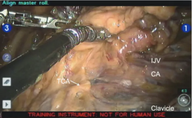
2). The external jugular vein and omohyoid muscle were divided. The transverse cervical artery was identified and preserved (Fig. 3). Fibroadipose tissue at level IV was re- tracted upward, exposing the brachial plexus and the phren- ic nerve (Fig. 3). At level IV, the thoracic duct or lymphatic duct was carefully identified and divided with the Harmon- ic scalpel. As the dissection continued superiorly, the cuta- neous branches of the cervical plexus were divided and the carotid sheath was incised. The vagus nerve, common ca- rotid artery and internal jugular vein were exposed. The su- perior thyroid artery was identified anterior to the carotid sheath and was preserved (Fig. 4).
After level III dissection, the axis of the robotic arms was re-adjusted superiorly for level II dissection (Fig. 5). The inferior border of the SMG was dissected and the posterior belly of the digastric muscle was identified (Fig. 4). The dissection continued posteriorly, and the hypoglossal nerve was identified and preserved. The upper end of the internal jugular vein and the SAN were exposed under the posterior belly of the digastric muscle. The specimen was removed
Fig. 4. Surgical view. STA, superior thyroid artery; digastric muscle, posteri- or belly of the digastric muscle; CA, carotid artery; IJV, internal jugular vein;
SH, sternohyoid muscle.
Fig. 2. Surgical view. SAN, spinal accessory nerve; BP, brachial plexus; CA, carotid artery.
Fig. 5. The axis of robotic system (A) for level III, IV, V dissection; (B) for lev- el II dissection.
Fig. 3. Surgical view. TCA, transverse cervical artery; IJV, internal jugular vein, CA, carotid artery.
A B
was difficult to dissect completely because of a limited view.
Third, in the presence of a prominent clavicle, the lower por- tion of level IV was difficult to dissect completely because the Harmonic shears have a limitation in bending motion.
On the other hand, the concept of super-selective neck dissection has been gradually accepted as a reasonable neck treatment for HNSCC patients. In the treatment of clinical- ly node-negative HNSCC, limited neck levels have been removed for elective neck management and the possible exclusion of several sub-levels has also been reported.14 For example, there are several reports that level I or IIB could be excluded in elective neck dissection or therapeutic neck dissection in selected larynx, hypopharynx or tonsillar can- cer patients.15-17 Using the robotic system via the transaxil- lary approach, surgeons could successfully dissect selected neck levels. Therefore, we presume that robotic neck dis- section could be applied for neck treatment in selected HN- SCC patients after validation of surgical or oncologic stud- ies. In cases of thyroid cancer, there are not many cases where level I or IIB dissection is needed, as level I metasta- sis is hardly observed,18 and the incidence of level IIB me- tastasis is reported to be low in some studies.19
In oral cavity cancer, in which level I dissection is man- datory, we postulate that several additional ports combined with the transaxillary approach would allow for the com- plete dissection of level I. For example, the floor of the mouth (FOM) could be a good additional route for level I dissection. As several authors have reported on trans-oral robot assisted FOM thyroidectomy,20 level I can be accessed through the FOM. Primary tumors in the oral cavity could be removed simultaneously by lingual release or a pull- through approach. However, even with the FOM approach, level IIB dissection still remains a pitfall of the transaxillary approach.
Another plausible additional route is a retroauricular ap- proach, which some surgeons have used for endoscopic submandibular gland excision.21 By combining the retroau- ricular approach, we might be able to dissect not only level I but also level IIB. Hence, we are currently investigating the feasibility of a transaxillary and retroauricular approach for comprehensive neck dissection in human cadavers, and we are planning a follow-up report.
In conclusion, our results demonstrate the limited feasi- bility of robot-assisted endoscopic neck dissection using a gasless, transaxillary approach. We suggest that robotic neck dissection using the transaxillary approach might be applied for treating nodal metastasis only in selected squa- through the axillary incision.
The side of dissection did not have any significant effect on the performance of ND. Dissection of levels IIB and VA, which lie on the cephalic portion of the SAN, was dif- ficult on both sides, as full exposure and complete dissec- tion was hard to obtain due to the angle of the robotic arm and endoscope.
DISCUSSION
Conventional neck dissection requires a long incision and leaves a prominent scar on the neck. There has been a lot of effort to reduce postoperative scars in head and neck sur- gery, such as minimally invasive video assisted thyroidec- tomy11 and endoscopic thyroidectomy.9,12 In addition, very recently, the introduction of the surgical robotic system, da Vinci, has spurred the development of new surgical tech- niques to minimize visible postoperative scars.13 Among these various new surgical techniques, the transaxillary ap- proach has the advantage of cosmesis. As the incidence of thyroid cancer is much higher in females and the proportion of young patients is increasing, endoscopic or robot-assist- ed transaxillary thyroidectomy is now accepted as a good alternative to conventional open thyroidectomy. Moreover, if we could remove the lateral neck nodes through the axil- lary port successfully, thyroid cancer patients with positive neck nodes could also be treated through this approach leaving no visible scars on their neck. Robotic neck dissec- tion could be an alternative operative method, in low risk, well-differentiated thyroid cancer patients with lateral neck metastasis.
However, even though reducing the surgical extent of neck dissection in HNSCC has become an important aspect in surgery, HNSCC still necessitates a wider and more thor- ough removal of neck nodes than differentiated thyroid cancer. In this respect, the transaxillary approach has some limitations, which we need to overcome for appropriate neck treatment. First, dissection at level I was extremely difficult.
During the cadaver dissection, we elevated the skin flap and tried to dissect level I; however, level I was too far from the axillary skin port to dissect efficiently with robotic arms un- der endoscopic view. Furthermore, vital neural structures such as the lingual nerve, the hypoglossal nerve beneath the mylohyoid muscle or the marginal mandibular branch of the facial nerve were difficult to identify and preserve. Second, level IIB, which lies outside of the spinal accessory nerve,
flation for unilateral micropapillary thyroid carcinoma: prelimi- nary report. Surg Endosc 2010;24:188-97.
10. Kang SW, Lee SH, Ryu HR, Lee KY, Jeong JJ, Nam KH, et al.
Initial experience with robot-assisted modified radical neck dis- section for the management of thyroid carcinoma with lateral neck node metastasis. Surgery 2010;148:1214-21.
11. Miccoli P, Berti P, Raffaelli M, Conte M, Materazzi G, Galleri D.
Minimally invasive video-assisted thyroidectomy. Am J Surg 2001;181:567-70.
12. Ikeda Y, Takami H, Niimi M, Kan S, Sasaki Y, Takayama J. Endo- scopic thyroidectomy by the axillary approach. Surg Endosc 2001;
15:1362-4.
13. Ballantyne GH, Moll F. The da Vinci telerobotic surgical system:
the virtual operative field and telepresence surgery. Surg Clin North Am 2003;83:1293-304.
14. Ferlito A, Rinaldo A, Silver CE, Gourin CG, Shah JP, Clayman GL, et al. Elective and therapeutic selective neck dissection. Oral Oncol 2006;42:14-25.
15. Shah JP. Patterns of cervical lymph node metastasis from squa- mous carcinomas of the upper aerodigestive tract. Am J Surg 1990;160:405-9.
16. Kim YH, Koo BS, Lim YC, Lee JS, Kim SH, Choi EC. Lymphat- ic metastases to level IIb in hypopharyngeal squamous cell carci- noma. Arch Otolaryngol Head Neck Surg 2006;132:1060-4.
17. Lim YC, Lee JS, Koo BS, Choi EC. Level IIb lymph node metas- tasis in laryngeal squamous cell carcinoma. Laryngoscope 2006;
116:268-72.
18. Scheumann GF, Gimm O, Wegener G, Hundeshagen H, Dralle H.
Prognostic significance and surgical management of locoregional lymph node metastases in papillary thyroid cancer. World J Surg 1994;18:559-67.
19. Koo BS, Yoon YH, Kim JM, Choi EC, Lim YC. Predictive factors of level IIb lymph node metastasis in patients with papillary thy- roid carcinoma. Ann Surg Oncol 2009;16:1344-7.
20. Witzel K, von Rahden BH, Kaminski C, Stein HJ. Transoral access for endoscopic thyroid resection. Surg Endosc 2008;22:1871-5.
21. Song CM, Jung YH, Sung MW, Kim KH. Endoscopic resection of the submandibular gland via a hairline incision: a new surgical approach. Laryngoscope 2010;120:970-4.
mous cell carcinomas of the head and neck, which do not require level I or IIB dissection. Some modification of the approach might be needed when performing the compre- hensive ND of all levels of the neck.
REFERENCES
1. Franklin ME Jr, Rosenthal D, Abrego-Medina D, Dorman JP, Glass JL, Norem R, et al. Prospective comparison of open vs. lap- aroscopic colon surgery for carcinoma. Five-year results. Dis Co- lon Rectum 1996;39:S35-46.
2. Clinical Outcomes of Surgical Therapy Study Group. A compari- son of laparoscopically assisted and open colectomy for colon cancer. N Engl J Med 2004;350:2050-9.
3. Weinstein GS, O’Malley BW Jr, Cohen MA, Quon H. Transoral robotic surgery for advanced oropharyngeal carcinoma. Arch Oto- laryngol Head Neck Surg 2010;136:1079-85.
4. Moore EJ, Olsen KD, Kasperbauer JL. Transoral robotic surgery for oropharyngeal squamous cell carcinoma: a prospective study of feasibility and functional outcomes. Laryngoscope 2009;119:
2156-64.
5. Park YM, Lee WJ, Lee JG, Lee WS, Choi EC, Chung SM, et al.
Transoral robotic surgery (TORS) in laryngeal and hypopharyn- geal cancer. J Laparoendosc Adv Surg Tech A 2009;19:361-8.
6. Park YM, Kim WS, Byeon HK, De Virgilio A, Jung JS, Kim SH.
Feasiblity of transoral robotic hypopharyngectomy for early-stage hypopharyngeal carcinoma. Oral Oncol 2010;46:597-602.
7. Park YM, Lee JG, Lee WS, Choi EC, Chung SM, Kim SH. Feasi- bility of transoral lateral oropharyngectomy using a robotic surgi- cal system for tonsillar cancer. Oral Oncol 2009;45:e62-6.
8. Kang SW, Lee SC, Lee SH, Lee KY, Jeong JJ, Lee YS, et al. Ro- botic thyroid surgery using a gasless, transaxillary approach and the da Vinci S system: the operative outcomes of 338 consecutive patients. Surgery 2009;146:1048-55.
9. Koh YW, Park JH, Kim JW, Lee SW, Choi EC. Endoscopic hemithyroidectomy with prophylactic ipsilateral central neck dis- section via an unilateral axillo-breast approach without gas insuf-