CONSERVATIVE MANAGEMENT OF PLACENTA
INCRETA TREATED WITH SELECTIVE UTERINE ARTERY EMBOLIZATION: REVIEW OF TWO CASES
Hee Suk Chae, MD, Eun Ji Lim, MD, Dong Hyun Lee, MD, Won Ku Choi, MD, Young Ju Jeong, MD, PhD, Sung Nam Cho, MD, PhD
Department of Obstetrics and Gynecology, Chonbuk National University Medical School, Jeonju, Korea
Placenta increta is a condition characterized by abnormal adherence of the placenta owing to entire or partial absence of the decidua basalis. It is generally recognized during difficult placental removal at the time of delivery. It is a life-threatening complication of pregnancy due to massive hemorrhage, uterine perforation, and infection and thus often a hysterectomy is required. We review two cases of placenta increta conservative managed successfully by selective uterine artery embolization.
Keywords:
Placenta increta; Conservative; Selective uterine artery embolization
Received: 2011.12.27. Revised: 2012.3.7. Accepted: 2012.4.2.
Corresponding author: Hee Suk Chae, MD
Department of Obstetrics and Gynecology, Chonbuk National University Medical School, 20 Geonji-ro, Deokjin-gu, Jeonju 561-712, Korea
Tel: +82-63-250-1360 Fax: +82-63-254-4833 E-mail: [email protected]
Th is is an Open Access article distributed under the terms of the Creative Commons Attribution Non-Commercial License (http://creativecommons.org/licenses/
by-nc/3.0/) which permits unrestricted non-commercial use, distribution, and reproduction in any medium, provided the original work is properly cited.
Copyright © 2012. Korean Society of Obstetrics and Gynecology http://dx.doi.org/10.5468/KJOG.2012.55.6.403
pISSN 2233-5188 · eISSN 2233-5196
Placenta increta is a serious complication of pregnancy character- ized by entire or partial defect of the decidua basalis, and by the incomplete development of the fi brinoid (Nitabuch’s) layer; it re- sults in abnormally invasive implantation of placenta. Although it is considered a rare occurrence with a prevalence of approximately 1 in 2,500-7,000, it is associated with high morbidity and some- times with a lethal outcome, mainly as a result of massive hemor- rhage, uterine rupture and infections [1].
Placenta increta associated with increased maternal morbidity and mortality, generally requires Cesarean hysterectomy. Since this re- sult in loss of fertility preservation, especially in cases who want to preserve future fertility, conservative approaches are desirable.
Case Reports
1. Case 1
A 27-year-old woman, gravid 2, para 1, was referred to the De- partment of Obstetrics and Gynecology at the University of Chon- buk with a diagnosis of placenta percreta. She had a history of a fi rst trimester surgical termination of pregnancy and no signifi cant previous medical history.
She underwent elective low segment Cesarean section 2 hours ago at a private hospital and delivered a healthy infant weigh-
ing 2,370 g at 40 weeks of gestation. Intraoperatively following the delivery of her fetus, placenta was identifi ed to have grown through the uterine wall with an intact uterine serosa (Fig. 1).
Therefore a manual removal of placenta was not performed. She
was referred for further management. On admission, her blood
pressure was 120/80 mm Hg, body temperature 36.7
oC, and pulse
rate 108 beats/min. Physical examination showed minimal vaginal
bleeding and uterine good contraction. Her haemoglobin was 10.3
g/dL, hematocrit 32.4%. Considering the hemodynamically stable
condition, we decided to manage conservatively. Both uterine ar-
teries embolization (UAE) was performed immediately. Supportive
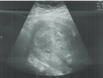
measures, like blood transfusion and broad spectrum antibiotics, initiated. Uterotonic drugs, including oxytocin and methylergono- vine were given continuously for 2 days. Transabdominal sono- gram on the second day after operation showed retained placenta accompanied with a thin uterine wall and the anterior and pos- terior uterine wall was deeply invaded by the placenta at fundus (Fig. 2). Follow-up ultrasonography showed gradual shrinkage of the placenta in the uterus. Her condition remained stable and her postoperative recovery was uneventful. Fourteen days after opera- tion, uterine exploration and curettage procedure was performed to take out a retained placenta and a small fragment of placenta was extracted. The patient was discharged and followed-up regu-
larly under the close observation including clinical examination and pelvic ultrasonography. The patient remained asymptomatic.
Fifty days after operation, because ultrasonography revealed the placental remnant showed no signifi cant reduction in size, uterine exploration and curettage procedure was repeated. A bulky pla- cental tissue was expelled. All tissue was examined microscopically for confi rmation of placental tissue. Seven months after operation, she was feeling well and menstruating regularly. A follow-up ultra- sound examination showed a linear endometrial cavity.
2. Case 2
A 28-year-old woman, gravid 1, para 1, who was undergone an emergency low segment Cesarean section due to induction failure 2 hours ago at a local maternity hospital was referred to the De- partment of Obstetrics and Gynecology at the Chonbuk National University with massive vaginal bleeding from a intraoperatively diagnosed placenta percreta. She had no past history of abor- tion and no signifi cant previous medical history. She delivered a healthy infant weighing 3,240 g at term. Intraoperatively follow- ing the delivery of her fetus, the placenta was retained and fi rmly adherent to the uterus. In addition, the uterus was identifi ed to have grown through left-sided fundal wall with an intact uterine serosa (Fig. 3). Therefore, a manual removal of placenta was not performed.
On admission to our clinic two hours after operation, she was re- ceiving 1,000 mL of fl uid infusion and 1 unit of packed red blood cells after operation. Two hours after operation, estimated blood loss was about 1,000 mL. She was pale with a blood pressure of 90/60 mm Hg and a pulse rate of 108 per minute. Laboratory da-
Fig. 1. Uterine abnormal bulging during Cesarean section (arrows).Fig. 2. Ultrasound examination obtained 2 days postoperation shows a thin uterine wall (arrowheads) at fundus of uterus and the loss of the echolucent space behind the placenta (PL) (arrows).
Fig. 3. View of uterus after baby out during Cesarean section: an en- larged uterus with extension out towards the left cornu (arrows).
tas were as follows: hemoglobin 12.5 g/dL, white blood cell count 28.5 ×10
3/μL and platelet count 226 ×10
3/μL. Vaginal examination revealed active vaginal bleeding. After counseling, conservative management to retain her uterus was chosen. Immediately, she was received both UAE. High dose antibiotics (cefminox, astromi- cin and metronidazole) were administered to prevent secondary infection. Uterotonic drugs, including oxytocin and methylergo- novine were given continuously for 3 days. After UAE procedure, she remained afebrile with no evidence of infection and had no vaginal bleeding after a few days of minimal vaginal bleeding.
Transabdominal sonogram on the eighth day after operation showed retained placenta and deeply invasion by the placenta at the left fundus of uterus (Fig. 4). Fifteen days after operation,
uterine exploration and curettage procedure was performed to evacuate a retained placenta. All expelled tissue was examined microscopically for confi rmation of placental tissue. Her condition remained stable, and she made a favorable progress. She was dis- charged and followed-up on twenty days after operation. Follow- up ultrasonography showed complete uterine involution and no intrauterine placental fragments (Fig. 5).
Discussion
Placenta accreta is defi ned as an abnormally fi rm attachment of the placenta to the uterine wall with a defect of the decidua ba- salis. The three variants of this abnormal placentation are placenta accreta vera, increta and percreta, depending on the degree of the spectrum of abnormal placentation. Placenta accrete vera is de- fi ned as attachment to the myometrium without invasion into or through uterine muscle. Placenta increta involves invasion of the placenta into the myometrium. Placenta percreta represents the greatest degree of severity, including deep invasion through the entire myometrium and uterine serosa or other pelvic structures.
Placenta increta is the rare form of placental abnormalities, with a 15% incidence among all placenta accreta cases [2]. Placenta accrete occurs in approximately 1 in 2,500 deliveries, and its prevalence seems to be increasing. The incidence has increased ten times over the past fifty years, reflecting the rapidly rising numbers of Cesarean section performed [3]. Other risk factors include placenta previa, advanced maternal age, multiparity, previ- ous uterine curettage, or previous myomectomy scars [4]. Because this form of placental abnormalities can cause life-threatening severe bleeding or insuffi cient hemostasis requiring hysterectomy or death, antepartum recognition of abnormal adherent placenta- tion is important for planning an appropriate management to a safe delivery and minimizing the risks of postpartum hemorrhage.
Ultrasonography, power Doppler, and magnetic resonance imag- ing for prenatal diagnosis of placenta accrete are used [5,6].
Ultrasonography is now an established fi rst-line investigation for the diagnosis of abnormal placentation and its extent. Magnetic resonance imaging is superior to ultrasonography in cases with posterior accreta or in delineating the extent of placental invasion [7,8]. Therefore, the use of ultrasound with color Doppler imaging together with MRI in combination may be potentially effective for a diagnosis of placenta accrete. These examinations can improve the accuracy in prediction of placenta accreta and lead to more favorable outcomes. However, there is currently no consensus
Fig. 4. Ultrasound examination showed the left uterine wall was deeplyinvaded by the placenta.
Fig. 5. Transvaginal sonogram 4 weeks after operation showed a thin linear endometrial cavity and no remnant placental tissue.
about these diagnostic maneuvers of placenta accreta, and the predictive value of these examinations is low [7]. Therefore, adher- ent placenta is often diagnosed after a failed attempt to separate the placenta from the uterus. In the case of life-threatening severe bleeding, such as placenta accreta, any delay in management may result in catastrophic complications. Hysterectomy immedi- ately after delivery is usually preferred in an emergent situation.
However, over recent decades, the conservative managements for morbidly adherent placenta have been reported, such as supple- mental methotrexate or UAE. The successful treatment of placenta accrete with methotrexate by Arulkumaran et al. [9] in 1986 was fi rst reported. Henrich et al. [10] reported a complicated case of a placenta percreta in a woman infected with the human immuno- defi ciency virus, which was successfully treated with the placenta left in situ and methotrexate therapy. Mussalli et al. [11] reported conservative management with methotrexate treatment resulted in uterine preservation of two of three patients and did not pre- vent signifi cant delayed hemorrhage.
The use of UAE as an alternative management for uncontrollable post-partum hemorrhage was fi rst reported in 1979 [12]. It per- mits the reduction of hysterectomy rate and the preservation of fertility. Descargues et al. [13] reported that the success rate of UAE for abnormal placentation is 71%, which is lower than the success rate for postpartum bleeding. The author suggested that the conservative management with the placenta left in situ and UAE procedure in cases of abnormal placentation permits more favorable outcomes.
However, because of limitation of evidence, there is no consensus of the optimal management of placenta accreta. Presently, the reported results showed varying degrees of success. Over the past 20 years, the conservative management of abnormally invasive placentation has been reviewed. In 5 of 22 women received adju- vant methotrexate, therapy failed. In 3 of twelve women managed with arterial embolization, therapy failed [14]. Therefore, it should be only considered for highly selected cases when the patient has a strong desire to retain her uterus or when blood loss is minimal [14,15].
We describe two cases of placenta increta in which the placenta was left in situ, UAE performed and a delayed uterine exploration and curettage successfully performed.
In our experience, the use of the strategy of leaving placenta in situ with adjuvant UAE as conservative management of adherent placenta is effective. However, it is important to emphasize that those women should be followed-up closely to detect complica- tions and further studies are needed to investigate efficacy and
effects of conservative techniques.
References
1. Berchuck A, Sokol RJ. Previous cesarean section, placenta in- creta, and uterine rupture in second-trimester abortion. Am J Obstet Gynecol 1983;145:766-7.
2. Breen JL, Neubecker R, Gregori CA, Franklin JE Jr. Placenta accreta, increta, and percreta. A survey of 40 cases. Obstet Gynecol 1977;49:43-7.
3. Committee on Obstetric Practice. ACOG committee opinion.
Placenta accreta. Number 266, January 2002. American Col- lege of Obstetricians and Gynecologists. Int J Gynaecol Obstet 2002;77:77-8.
4. Miller DA, Chollet JA, Goodwin TM. Clinical risk factors for placenta previa-placenta accreta. Am J Obstet Gynecol 1997;177:210-4.
5. Gielchinsky Y, Rojansky N, Fasouliotis SJ, Ezra Y. Placenta ac- creta: summary of 10 years: a survey of 310 cases. Placenta 2002;23:210-4.
6. Suh YH, Song EH, Kim DH, Lee YH, Park HY, Koh KS, et al. A clinical study of placental adhesions: placenta accreta, pla- centa increta, and placenta percreta. Korean J Obstet Gynecol 2003;46:81-8.
7. Levine D, Hulka CA, Ludmir J, Li W, Edelman RR. Placenta ac- creta: evaluation with color Doppler US, power Doppler US, and MR imaging. Radiology 1997;205:773-6.
8. Legro RS, Price FV, Hill LM, Caritis SN. Nonsurgical manage- ment of placenta percreta: a case report. Obstet Gynecol 1994;83:847-9.
9. Arulkumaran S, Ng CS, Ingemarsson I, Ratnam SS. Medical treatment of placenta accreta with methotrexate. Acta Obstet Gynecol Scand 1986;65:285-6.
10. Henrich W, Fuchs I, Ehrenstein T, Kjos S, Schmider A, Duden- hausen JW. Antenatal diagnosis of placenta percreta with planned in situ retention and methotrexate therapy in a wom- an infected with HIV. Ultrasound Obstet Gynecol 2002;20:90-3.
11. Mussalli GM, Shah J, Berck DJ, Elimian A, Tejani N, Manning FA. Placenta accreta and methotrexate therapy: three case reports. J Perinatol 2000;20:331-4.
12. Brown BJ, Heaston DK, Poulson AM, Gabert HA, Mineau DE,
Miller FJ Jr. Uncontrollable postpartum bleeding: a new ap-
proach to hemostasis through angiographic arterial emboliza-
tion. Obstet Gynecol 1979;54:361-5.
13. Descargues G, Douvrin F, Degré S, Lemoine JP, Marpeau L, Clavier E. Abnormal placentation and selective emboliza- tion of the uterine arteries. Eur J Obstet Gynecol Reprod Biol 2001;99:47-52.
14. Timmermans S, van Hof AC, Duvekot JJ. Conservative manage-
ment of abnormally invasive placentation. Obstet Gynecol Surv 2007;62:529-39.
15. Kayem G, Davy C, Goffi net F, Thomas C, Clément D, Cabrol D.
Conservative versus extirpative management in cases of pla- centa accreta. Obstet Gynecol 2004;104:531-6.
선택적 자궁동맥색전술을 이용한 감입태반의 보존적 치료 2예
전북대학교 의과대학 산부인과교실
채희숙, 임은지, 이동현, 최원구, 정영주, 조성남
감입태반은 비록 흔하지는 않지만 발생할 경우 과거에는 자궁절제술과 같은 수술적 방법이 많이 이용되었는데, 이 경우 불가피하게 임신 능력의 소실이 초래되어 최근에는 이런 수술적 방법에서 자궁을 보존하려는 다양한 방법들이 시도되고 있다. 이에 본 저자들은 감입태반 환 자에서 보존적 방법인 선택적 자궁동맥색전술을 시행하여 성공적으로 치료한 2예를 경험하였기에 문헌고찰과 함께 보고하는 바이다.
중심단어: 감입태반, 보존적 방법, 선택적 자궁동맥색전술