Introduction
The annual incidence of cervical artery dissection is 2.6-3.0 per 100,000 population, and that of vertebral artery dissection, in particular, is 1-1.5 per 100,000 population. The mean age of patients with a cervical artery dissection is 46.3 years, and their male-to-female ratio is equivalent [1,2]. Cervical artery dissection may arise spontaneously or traumatically, even due to trivial events such as a sudden rotation of the neck. In addition, many studies have reported various pathophysiological risk factors for cervical artery
dissection, such as connective tissue disorders [2].
Neurofibromatosis 1 (NF1) is a common autosomal dominant disorder (incidence of 1 per 3,000 population) that leads to systemic conditions including neurofibromas, gliomas of the optic nerve, hamartomatous lesions, mental retardation, skeletal defects, and cutaneous hyperpigmented macules (café au lait spots) [3]. Many studies have been reported that NF1 is associated with intrathoracic meningoceles and scoliosis [4,5].
Here, we repor t an autopsy case of massive intrathoracic hemorrhage due to spontaneous vertebral artery dissection in a patient with NF1, who had http://dx.doi.org/10.7580/kjlm.2016.40.1.14
Spontaneous Extracranial Vertebral Artery Dissection in a Neurofibromatosis 1 Patient with Bilateral Intrathoracic Spinal Meningoceles around the Scoliosis: Report of an Autopsy Case
Ji Hye Park, Jaehong Park
Department of Forensic Investigation, National Forensic Service Seoul Institute, Seoul, Korea
Neurofibromatosis 1 (NF1) is a common autosomal dominant disorder that causes several systemic diseases. Many studies have reported that NF1 is associated with intrathoracic meningoceles and scoliosis. The incidence of vertebral artery dissection is estimated to be 1-1.5 per 100,000 population.
We experienced an autopsy case of massive intrathoracic hemorrhage due to spontaneous vertebral artery dissection in a patient with NF1, who had intrathoracic spinal meningoceles and scoliosis. A 47-year-old man was found dead at his home in the morning. He had a history of NF1 including numerous cutaneous neurofibromas and hyperpigmented macules, scoliosis, and deformity of the leg. The autopsy revealed the dissection and rupture of the left vertebral artery, and a pseudocyst that had formed due to arterial leakage on the wall of the meningocele on the left side. The pseudocyst had eventually ruptured and leaked blood, resulting in a massive hemothorax on the left side. Thus, it was revealed that the patient had suffered from NF1-associated intrathoracic meningoceles and scoliosis, and we concluded that the cause of his death was a massive hemothorax on the left side, caused by the dissection and rupture of the left vertebral artery.
Key Words: Vertebral artery; Dissection; Neurofibromatosis 1; Meningocele;
Scoliosis Received: October 23, 2015
Accepted: November 16, 2015 Correspondence to Jaehong Park
Department of Forensic Investigation, National Forensic Service Seoul Institute, 139 Jiyang- ro, Yangcheon-gu, Seoul 08036, Korea
Tel: +82-2-2600-4819 Fax: +82-2-2600-4828 E-mail: [email protected]
pISSN 2383-5702 ⓒCopyright 2016 by the Korean Society for Legal Medicine
intrathoracic spinal meningoceles and scoliosis. Case Report
A 47-year-old Korean man was found dead, lying in the supine position on his bed at home in the morning.
A B
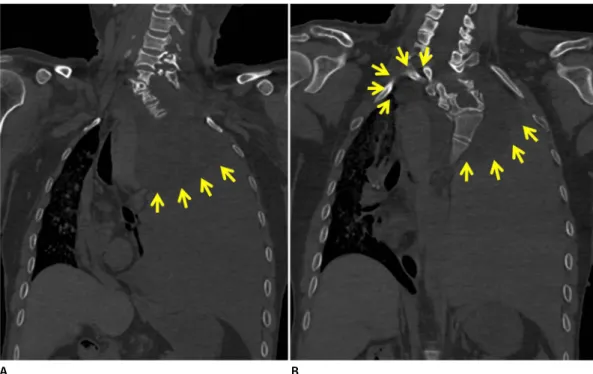
Fig. 2. Postmortem computed tomography findings. (A) A ‘white out’ of the left thorax with a mediastinal shift to the right was revealed. (A, B) Bilateral round masses were seen at the superior intrathoracic area (masses are indicated by yellow arrows).
A B
Fig. 1. External and internal autopsy findings consistent with neurofibromatosis. (A) Numerous cutaneous neurofibromas and hyperpigmented macules were noticed in the patient’s body. (B) Skeletal deformity of the right leg and scars from a previous operation were observed. (C) Scoliosis was observed at the cervical and thoracic spine.
C
The patient’s day previous to death had reportedly been uneventful, and he had gone to bed as usual.
The patient had a history of NF1 including numerous cutaneous neurofibromas and hyperpigmented macules (Fig. 1A), deformity of the leg (Fig. 1B), and scoliosis (Fig.
1C).
Postmortem computed tomography (CT) performed before the autopsy revealed a ‘white-out’ of the left thorax, with a mediastinal shift to the right (Fig. 2), and bilateral round masses in the superior intrathoracic area (Fig. 2B).
Autopsy revealed a massive hemorrhage with blood clots (more than 2,300 mL) in the left thoracic cavity, and the leakage of blood from the left intrathoracic round mass (Fig. 3A). Upon further dissection, the bilateral intrathoracic masses at the T2-T3 intervertebral
foramen were revealed to be spinal meningoceles filled with clear cerebrospinal fluid. A blood and hematoma- filled pseudocyst was found at the wall of the intrathoracic meningocele on the left side (Fig. 3B-D).
Upon close examination of the neck, bilateral thoracic cavities, and the area of the meningoceles, we found a dissection and rupture of the prevertebral segment of the left vertebral artery as the focus of the bleeding (Fig. 4). We concluded that a dissection and rupture of the left vertebral artery had occurred, leading to arterial leakage, which, in turn, had led to the formation of the pseudocyst on the wall of the meningocele on the left side. The pseudocyst had eventually ruptured and leaked blood into the thorax, resulting in a massive hemothorax on the left side.
The autopsy further revealed a 0.5×0.4 cm mass
A B
C D
Fig. 3. Intrathoracic meningoceles found upon internal examination. (A) Blood had leaked out of the round mass in the left intrathoracic area. (B, C) The left intrathoracic round mass was revealed to be a spinal meningocele filled with clear cerebrospinal fluid. A pseudocyst filled with blood and hematoma was found at the wall of the intrathoracic meningocele on left side. (D) The bilateral intrathoracic round masses at the bilateral T2-T3 intervertebral foramen were revealed to be spinal meningoceles (bilateral meningoceles are indicated by yellow arrows).
in the left lobe of the thyroid gland, which was confirmed to be an adenomatous hyperplasia. The left anterior descending coronary artery showed mild coronary atherosclerosis. The dissected surface of the collapsed left lung showed a pale appearance due to the hemothorax. The liver showed a mild fatty change, and a 1.8×1.5 cm mass in the small bowel, which was confirmed to be a gastrointestinal stromal tumor. Histopathological examination was performed to confirm all of the pathological findings. The toxicological findings were negative for toxins. The blood alcohol concentration was less than 0.010%.
In summary, the postmortem diagnoses by CT, autopsy, and histopathological examination revealed that the patient had suffered from NF1 with intrathoracic meningoceles and scoliosis. We concluded that the cause of his death was the dissection and rupture of the left vertebral artery resulting in a massive hemothorax on the left side, based on a comprehensive assessment of the findings from the autopsy and the histopathological, toxicological, and blood alcohol tests.
Discussion
Many studies (including case reports, autopsy reports, original articles, etc.) have reported the occurrence of ruptured aneurysms, intrathoracic meningoceles, or scoliosis in NF1 patients [6]. However, reports about the dissection of arteries in NF1 patients with
meningoceles or scoliosis are rare [7,8]. Miura et al. [7]
reported spontaneous hemothorax due to the dissection and rupture of the subclavian artery in a patient with NF1. They reviewed 12 Japanese studies related to spontaneous hemothorax in NF1 patients, and found no cases of vertebral artery dissection [7]. Fohrding et al. [8]
reported spontaneous hemothorax in a patient with NF1 who had an intrathoracic meningocele; however, they could not find a distinct bleeding focus.
The diagnostic criteria for NF1 per the guidelines of the National Institutes of Health are as follows: (1) six or more café au lait macules (>0.5 cm in children and >1.5 cm in adults); (2) two or more cutaneous/
subcutaneous neurofibromas, or one plexiform neurofibroma; (3) axillary or groin freckling; (4) optic pathway glioma; (5) two or more Lisch nodules (iris hamartomas seen on slit lamp examination); (6) bony dysplasia (sphenoid wing dysplasia, bowing of the long bones, and pseudarthrosis); and (7) a first degree relative with NF1. A diagnosis of NF1 may be confirmed if two or more of these criteria are satisfied. In our case, NF1 was confirmed based on the fulfillment of criteria 1 and 2.
The etiology of vertebral artery dissection is controversial; however, it is known that spontaneous conditions as well as traumatic ones can cause dissection [2]. In our case, there was no evidence of trauma in the patient’s living circumstances or his autopsy findings, and therefore, we determined that the
A B
Fig. 4. (A, B) Upon close examination, dissection and rupture of the prevertebral segment of the left vertebral artery were noticed (the ruptured areas are indicated by yellow arrows).
vertebral artery dissection had occurred spontaneously.
Some reports show histological abnormalities in the arterial walls of NF1 patients [9,10]. These studies have reported that intimal proliferation and weakness of the internal elastic lamina cause cerebrovascular aneurysms in NF1 patients. We hypothesized that those abnormalities could also cause dissection of the arteries in NF1 patients.
In conclusion, we experienced a rare autopsy case of vertebral artery dissection in an NF1 patient with intrathoracic spinal meningoceles and scoliosis. It is our opinion that all NF1 patients must be carefully examined for the possible presence of vertebral artery dissection during routine autopsy.
Conflicts of Interest
No potential conflict of interest relevant to this article was reported.
References
1. Lee VH, Brown RD Jr, Mandrekar JN, et al. Incidence and outcome of cervical artery dissection: a population-based study. Neurology
2006;67:1809-12.
2. Haneline MT, Rosner AL. The etiology of cervical artery dissection.
J Chiropr Med 2007;6:110-20.
3. Kumar V, Abbas AK, Aster JC. Robbins and Cotran pathologic basis of disease. 9th ed. Philadelphia, PA: Elsevier/Saunders; 2015.
4. Ueda K, Honda O, Satoh Y, et al. Computed tomography (CT) findings in 88 neurofibromatosis 1 (NF1) patients: Prevalence rates and correlations of thoracic findings. Eur J Radiol 2015;84:1191-5.
5. Swetz KM, Spinner RJ. Large intrathoracic meningocele associated with neurofibromatosis type 1. Mayo Clin Proc 2009;84:769.
6. Na JY, Park JP, Kim DW, et al. Aneurysmal rupture of the internal carotid artery in a presumed neurofibromatosis type I patient.
Korean J Leg Med 2013;37:34-7.
7. Miura H, Taira O, Uchida O, et al. Spontaneous haemothorax associated with von Recklinghausen's disease: review of occurrence in Japan. Thorax 1997;52:577-8.
8. Fohrding LZ, Sellmann T, Angenendt S, et al. A case of lethal spontaneous massive hemothorax in a patient with neurofibromatosis 1. J Cardiothorac Surg 2014;9:172.
9. Uranishi R, Ochiai C, Okuno S, et al. Cerebral aneurysms associated with von Recklinghausen neurofibromatosis: report of two cases. No Shinkei Geka 1995;23:237-42.
10. Sobata E, Ohkuma H, Suzuki S. Cerebrovascular disorders associated with von Recklinghausen's neurofibromatosis: a case report. Neurosurgery 1988;22:544-9.