6 Copyright © 2015 Journal of Rhinology
INTRODUCTION
Headache associated with rhinogenic symptoms pres- ents a diagnostic dilemma that commonly confronts phy- sicians. Diagnostic clarity is essential because primary headache disorders, such as migraine, and secondary headaches, such as rhinosinusitis, require very different treatments. Headaches secondary to inflammatory sinus disease can be treated surgically. Both the International Headache Society (IHS) and the American Academy of
Otolaryngology-Head and Neck Surgery (AAO-HNS) have described conditions that can cause headaches of rhinogenic origin.1) However, these criteria represent the consensus of expert opinion rather than scientific evidence- based evaluation, and focus on acute rhinosinusitis and do not consider chronic rhinosinusitis, as a cause of headache or facial pain.2)
Few prospective studies have investigated the improve- ment of headaches after nasal or sinus surgery. Mariotti and Setliff3) evaluated patient history and computed to- mography (CT) scans in a prospective study designed to predict the outcome of headaches after surgery. They re- ported that history and CT parameters could not distin- guish between the patients who improved and those who did not.
The present prospective study was performed to exam-
Prospective Study on the Characteristics and Postoperative Improvement of Rhinogenic Headache
Jee Hye Wee, MD1, Ji-Eun Lee, MD2, Sung-Lyong Hong, MD3, Jae Min Shin, MD4 and Dong-Young Kim, MD, PhD1,5
1Department of Otorhinolaryngology-Head and Neck Surgery, Seoul National University College of Medicine, Seoul; and
2Department of Otorhinolaryngology-Head and Neck Surgery, Chosun University College of Medicine, Gwang-ju; and
3Department of Otorhinolaryngology-Head and Neck Surgery, Pusan National University College of Medicine, Pusan; and
4Department of Otorhinolaryngology-Head and Neck Surgery, Soon Chun Hyang University College of Medicine, Seoul; and
5Research Center for Sensory Organs, Seoul National University College of Medicine, Seoul, Korea
ABSTRACT
Background and Objectives:Headache secondary to sinonasal disease can improve after surgery, but few prospective studies have investigated this outcome. We aimed to evaluate the characteristics of headaches, such as clinical features, underlying disease, and postoperative improvement in patients who underwent nasal surgery, and to identify the characteristics that reliably predict rhinogenic headache. Materials and Method:Of 356 patients who underwent nasal surgery between March and December 2009, 41 patients with headaches were enrolled in this prospective study. Clinical features of headache, such as onset, time of day, duration, frequency, nature, side and location, existence of aura, aggravating and relieving factors and accompanying nasal symptoms, underlying diseases, endoscopic findings, and computed tomography scans of the paranasal sinuses were evaluated. Headache intensity was graded based on a 10-point visual analog scale (VAS) pre- and post-operatively.
Results:The most common characteristics of rhinogenic headache included a stabbing or squeezing nature, frontal area location, accompanying nasal obstruction or rhinorrhea, and underlying sinusitis or septal deviation. The subjective intensity of the headache, measured using the VAS score, improved in 80% (33/41) of the patients after surgery. Conclusion:Nasal surgery should be considered when rhinogenic headache is suspected and there are definite nasal pathologies.
KEY WORDS:HeadacheㆍNasal diseaseㆍProspective studyㆍNasal surgeryㆍPostoperative pain.
J Rhinol 2015;22(1):6-10 www.ksrhino.or.kr
Received: March 26, 2015 / Revised: April 23, 2015 Accepted: May 22, 2015
Address for correspondence: Dong-Young Kim, MD, PhD
Department of Otorhinolaryngology-Head and Neck Surgery, Seoul Na- tional University College of Medicine, 101 Daehak-ro, Jongno-gu, Seoul 110-744, Korea
Tel: +82-2-2072-2440, Fax: +82-2-745-2387 E-mail: [email protected]
ine the characteristics of headache, such as clinical fea- tures, underlying disease, and postoperative improvement in patients with headache who underwent nasal surgery, and to identify characteristics that reliably predict rhino- genic headache.
MATERIALS AND METHODS
We screened 356 patients scheduled for nasal surgery at the Seoul National University Hospital between March and December 2009. Of the 356 patients, 41 who had ac- companying headaches were enrolled in our prospective study. The patients were 21 men and 20 women with a mean age of 40.2 years (range, 14-73 years). Inclusion criteria were an age of at least 14 years, a history of head- ache that had never been satisfactorily relieved by any treatment, and no obvious cause of the headache. Exclu- sion criteria included a history of nasal or sinus surgery, sinonasal neoplasm, pregnancy, and a history of migraine or other definitive cause of the headache.
Prior to surgery, all patients were asked to complete a questionnaire on the clinical features of their headache in- cluding onset, time of day, duration, frequency, nature, side of the head and location, existence of an aura, aggra- vating and relieving factors, and accompanying nasal symptoms. Furthermore, underlying diseases, nasal endo- scopic findings, and CT scans of the paranasal sinuses were evaluated. An endoscopic examination was performed to detect the presence of septal deviation, septal spur, infe- rior turbinate hypertrophy, nasal discharge, contact point, nasal polyp, and postnasal drip. The CT scan was per- formed to reveal concha bullosa, paradoxical middle tur- binate, and rhinosinusitis. The Lund-Mackay score4) was calculated for patients who had rhinosinusitis, and the correlation between the side of the lesion and side of the headache was determined.
Preoperative headache intensity was graded based on a 10-point visual analog scale (VAS), in which 0 indicated the absence of headache and 10 represented the most in- tense pain.
After obtaining written informed consent, surgery was performed under general anesthesia. One surgeon operat- ed on all patients. At the end of the surgical procedure, the nasal cavity was packed with Merocel and gauze. Antibi- otics and analgesics were administered for 7 days follow- ing surgery.
Postoperative follow-up evaluations were scheduled at 2 weeks, 1, 2, 3, and 6 months. The evaluations included an endoscopic examination of the nasal cavity and VAS
score grading of headache intensity. During the follow-up period, the patients were examined for the development of other diseases and whether a neurological evaluation was necessary.
For further analysis, we classified the patients into four categories. The ‘resolved’ group consisted of patients who rated headache pain as 0 on the VAS, the ‘improved’
group was defined as patients whose postoperative VAS score was lower than their preoperative score. The ‘no change’ group comprised patients who showed no change in the VAS score, and the ‘worsened’ group included pa- tients whose postoperative VAS score was higher than their preoperative score. The VAS scores of the four groups were compared using a non-parametric Wilcoxon signed- rank test. The SPSS 12.0 software (SPSS Inc., Chicago, IL) was used to conduct the statistical tests. p values <0.05 (2-sided) were deemed to indicate statistical significance.
The Institutional Review Board of the Clinical Research Institute at Seoul National University Hospital approved this study protocol (H-1012-081-344).
RESULTS
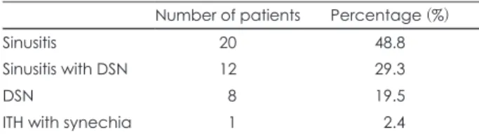
Of the 356 patients who underwent nasal surgery, 41 (11.5%) complained of headache. Underlying diseases in the 41 patients are summarized in Table 1. The follow-up duration averaged 10.7 months (range, 7-16 months). Of the 41 patients, five visited the neurology clinic after the surgery: none was diagnosed with migraine headaches by the neurologist, but two were diagnosed with tension-type headaches, and one patient was diagnosed with depres- sion. No definite diagnosis could be made in the remaining two patients.
The average initial onset of headaches was 16 months, ranging from 2 weeks to 8 years. The mean headache du- ration was 6.85 hour per headache, ranging from 40 sec to 24 hour, and the frequency of headaches ranged from one episode per month to every day. The headache occurred on both sides of the head in 14 patients, on the right side in 11 patients, centrally in 8 patients, and on the left side in 8 patients. Two patients experienced photophobia as an aura of headache and no patient reported phonophobia.
Table 1. Underlying disease
Number of patients Percentage (%)
Sinusitis 20 48.8
Sinusitis with DSN 12 29.3
DSN 8 19.5
ITH with synechia 1 2.4
DSN: Deviated nasal septum, ITH: Inferior turbinate hypertrophy
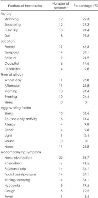
Table 2 summarizes the clinical features of the partici- pants’ headaches. The nature of the headache most com- monly reported was stabbing or squeezing, and the most common location was the frontal area of the head. The time of the attack varied. Stress was the most common ag- gravating factor, and nasal obstruction and rhinorrhea were commonly accompanied by headache.
The endoscopic examination of the nasal cavity revealed septal deviation in 76% (31/41) of patients, septal spur in 61% (25/41), hypertrophy of the inferior turbinate in 51%
(21/41), nasal discharge in 49% (20/41), contact point in 42% (17/41), nasal polyp in 27% (11/41), and postnasal drip in 10% (4/41). Of the 35 patients who had a CT scan, bilat- eral concha bullosa was revealed in 4, right concha bullo- sa was found in 4, and left concha bullosa in 5 patients.
Paradoxical middle turbinate was observed bilaterally in 1 patient, on the right side in 2 patients and on the left side in 1 patient. Sinusitis was observed bilaterally in 14 patients, on the right side in 10 patients, and on the left side in 8 patients. The mean preoperative CT scan Lund- Mackay score was 8.15 (range, 2-21). The surgery was well tolerated and no patient experienced postoperative complications.
Table 3 shows the correlation between the side of the lesion and that of the headache. The lesion and headache were on the same side of the head in 65.6% (21/32) of the patients with sinusitis and in half of the patients with par- adoxical middle turbinate. However, no correlation was found between lesion side and headache side in patients with concha bullosa, septal deviation, septal spur, or con- tact point. When groups were divided same and different (opposite and nonspecific) side of the lesion and head- ache, complete relief from headache showed in 61.9%
(13/21) of same side group and 36.4% (4/11) of the differ- ent side group.
The overall success rate of the surgery in relieving head- aches, measured by the VAS score, was approximately 80%. Complete relief from headaches was achieved in 24 of 41 patients (58.5%), and 9 patients (22.0%) reported a decrease in headache intensity after surgery. In contrast, 6
patients reported that headache intensity was unchanged after surgery (14.6%), and 2 patients (4.9%) found that their headaches were worse after surgery. Among 2 patients
Table 2. Clinical features of headache Feature of headache Number of
patients* Percentage (%) Nature
Stabbing 12 29.3
Squeezing 12 29.3
Pulsating 10 24.4
Dull 8 19.5
Location
Frontal 19 46.3
Temporal 14 34.1
Parietal 9 21.9
Occipital 6 14.6
Periorbital 4 9.8
Time of attack
Whole day 11 26.8
Afternoon 11 26.8
Morning 10 24.4
Evening 10 24.4
Sleep 0 0
Aggravating factor
Stress 15 36.6
Routine daily activity 6 14.6
Allergy 4 9.8
Other 4 9.8
Light 1 2.4
Sound 0 0
None 11 26.8
Accompanying symptom
Nasal obstruction 22 53.7
Rhinorrhea 17 41.5
Postnasal drip 14 34.1
Facial pain/pressure 14 34.1
Itching/sneezing 14 34.1
Hyposmia 8 19.5
Cough 5 12.2
Fever 1 2.4
*: Numbers are not mutually exclusive Table 3. Correlation between the side of the lesion and side of the headache
Lesion N* Same (%) Opposite (%) Not specific (%)
Sinusitis 32 21 (65.6) 1 (3.1) 10 (31.3)
Paradoxical middle turbinate 4 2 (50.0) 2 (50.0) 0 (0)
Concha bullosa 13 4 (30.8) 3 (23.1) 6 (46.1)
Septal deviation 31 6 (19.4) 6 (19.4) 19 (61.2)
Septal spur 25 2 (8.0) 7 (28.0) 16 (64.0)
Contact point 17 6 (35.3) 1 (5.9) 10 (58.8)
*: Numbers are not mutually exclusive. N: number of patients
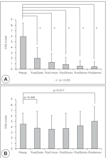
diagnosed with tension type headache, one patient resolved and the other improved headaches. One patient with de- pression showed improvement. The pre- and post-surgery VAS scores are shown in Fig. 1. for the resolved and im- proved groups (A) and for the no change and worsened groups (B). The postoperative VAS score was significantly decreased in the resolved and improved groups compared with the preoperative score. However, the pre- and post- surgery VAS scores were not significantly different in the no change and worsened groups.
DISCUSSION
A rhinogenic headache can mimic primary headaches such as migraine, tension-type, and cluster headaches5); however, headaches secondary to nose and sinus disor- ders can be treated by correcting the disorder.6) Schonsted- Madsen et al.7) followed 157 patients who had headache and were treated with septoplastic surgery, reconstruction
of the nasal pyramids, or submucosal conchotomy. Chron- ic headache was relieved in 60% of the patients who un- derwent surgery. Low and Willatt8) reported that 63% of 116 patients who underwent submucous resection for a deviated nasal septum experienced complete or partial re- lief from headaches. In 1998, Parsons and Batra9) reported a 91% improvement in headache intensity in 19 adults and 15 children after endoscopic sinus surgery. Rama- dan10) showed their success rate to be 60%. Our overall surgical success rate of 80% in patients with headache is consistent with the rates reported in these previous studies.
Several studies have examined contact point, a well- known cause of rhinogenic headache. Stammberger and Wolff11) suggested that intranasal mucosal contact re- leased substance P, causing pain and headache. Substance P, which is associated with the inflammatory process, has a potent vasodilator effect. Vasodilatation and perivascu- lar inflammation are the final common pathways in pain.
Surgical treatment for contact point-induced headaches has had good success. Clerico et al.12) found that after en- doscopic nasal surgery, 79% of the patients reported a de- crease in pain severity or headache frequency. Mohebbi et al.,6) Tosun et al.,13) and Welge-Luessen et al.,14) Lee et al., reported 83%, 90%, 85%, and 80% success rates, respec- tively, following surgery to correct contact point-induced headaches. Furthermore, Parsons and Batra9) reported an 85% success rate following surgery to correct contact point- induced headaches. However, a number of patients with obvious contact points revealed on the CT scan do not complain of headaches. Contact points revealed by an en- doscopic examination or by a CT scan are not believed to be pathognomonic for headache.9) Contact points can in- duce concurrent chronic sinusitis in addition to headache.
The cause of headache in the presence of contact point can be attributed to malventilation of the sinuses resulting in hypoxia, reduced pH, reduced ciliary beat, thick vis- cous mucus, and increased vulnerability to infections.16) In our study, 17 of 41 patients (42%) had contact point and complained of headaches. The headaches improved in 14 of the 17 patients (82%) after surgery. We found no corre- lation between side of contact point and side of the head- ache in 59% (10/17) of the patients. Thus, the mechanism by which headache improved after surgery may be related to other factors. The patients had lesions other than con- tact point, such as sinusitis. The headache and sinusitis were on the same side of the head in eight of the 17 pa- tients (47.1%) with contact point. The pathogenesis of rhinogenic headache may not be limited to mucosal con- tact, but rather, may be related to poor sinus ventilation.3)
9 8 7 6 5 4 3 2 1 0
Preop Poet2wks Post1mon Post2mon Post3mon Post6mon
*: p<0.05
VAS score
* * * * *
A
B
9 8 7 6 5 4 3 2 1 0
Preop Poet2wks Post1mon Post2mon Post3mon Post6mon p=0.445
p=0.317
VAS score
Fig. 1. Pre-and post-operative visual analog scale score for headache in the resolved and improved groups (A) and in the no-change and worsened groups (B). *: p<0.05. VAS: visual analog scale, Preop: Preoperative, Post: Postoperative, wks:
weeks, mon: months.
Stammberger and Wolf11) suggested that hypoxia second- ary to pressure differentials within the sinuses was a possi- ble mechanism for the release of substance P.
Anatomical abnormalities in the nasal cavity such as septal deviation, concha bullosa, and paradoxical middle turbinate have been reported to cause headaches.10) How- ever, few studies have investigated a correlation between side of the lesion and side of the headache. Tarabichi17) ex- amined the correlation between the severity and site of pain and the extent or location of mucosal disease. The re- sults showed that the pain score did not correlate with the preoperative Lund-Mackay score, and the site of pain did not correlate with the location of the disease. In contrast, the present study demonstrated a 66% concordance rate between the side of sinusitis and the side of the headache.
In a prospective, randomized, controlled study, Ragab et al.18) reported significant subjective and objective im- provements in headache with no significant difference be- tween patients who received medical treatment and those who underwent surgery. The authors suggested using maximum medical therapy as the primary treatment for chronic rhinosinusitis, and reserving surgical treatment for cases refractory to medical therapy. Our study included only patients with headache that had not been satisfactori- ly relieved by any treatment, including medical therapy. Thus, according to the criteria of Ragab et al.,18) our subjects would be classified as candidates for surgical therapy to relieve their headache. Previous studies have applied topical anes- thetics to the lesion in the nasal cavity and observed wheth- er this relieved the headache as a test to identify patients who would benefit from surgery. However, this test is not reliable because patients who failed the test have been re- ported to improve after surgery, whereas others who passed the test continued to have symptoms after sur- gery.9)12) At present, no consensus exists as to whether sur- gery relieves the rhinogenic headache.
This study has some limitations. First, small numbers of patients were included. Therefore, the overall success rate of the surgery in relieving headaches was evaluated. Fur- ther studies including large samples are needed to obtain success rate according to the each underlying disease or to the correlation of the side of headache and lesion. Sec- ond, follow up duration was relatively short. In the no
change and worsened groups, further evaluation for other cause of headache should be needed through longer fol- low up.
Acknowledgments
This work was supported by Research Resettlement Fund for the new faculty of SNU.
REFERENCES
1) Cady RK, Dodick DW, Levine HL, Schreiber CP, Eross EJ, Setzen M, et al. Sinus headache: A neurology, otolaryngology, allergy, and pri- mary care consensus on diagnosis and treatment. Mayo Clin Proc 2005;
80:908-16.
2) Chung SK. Headache and facial pain related to the paranasal sinus- es. J Rhinol 2012;19:83-6.
3) Mariotti LJ, Setliff RC, Ghaderi M, Voth S. Patient history and CT findings in predicting surgical outcomes for patients with rhinogenic headache. Ear Nose Throat J 2009;88:926-9.
4) Lund VJ, Mackay IS. Staging in rhinosinusitus. Rhinology 1993;31:
183-4.
5) Blumenthal HJ. Headaches and sinus disease. Headache 2001;41:883-8.
6) Mohebbi A, Memari F, Mohebbi S. Endonasal endoscopic manage- ment of contact point headache and diagnostic criteria. Headache 2010;
50:242-8.
7) Schonsted-Madsen U, Stoksted P, Christensen PH, Koch-Henriksen N.
Chronic headache related to nasal obstruction. J Laryngol Otol 1986;
100:165-70.
8) Low WK, Willatt DJ. Headaches associated with nasal obstruction due to deviated nasal septum. Headache 1995;35:404-6.
9) Parsons DS, Batra PS. Functional endoscopic sinus surgical out- comes for contact point headaches. Laryngoscope 1998;108:696-702.
10) Ramadan HH. Nonsurgical versus endoscopic sinonasal surgery for rhinogenic headache. Am J Rhinol 1999;13:455-7.
11) Stammberger H, Wolf G. Headaches and sinus disease: the endoscop- ic approach. Ann Otol Rhinol Laryngol Suppl 1988;134:3-23.
12) Clerico DM, Evan K, Montgomery L, Lanza DC, Grabo D. Endoscop- ic sinonasal surgery in the management of primary headaches. Rhi- nology 1997;35:98-102.
13) Tosun F, Gerek M, Ozkaptan Y. Nasal surgery for contact point head- aches. Headache 2000;40:237-40.
14) Welge-Luessen A, Hauser R, Schmid N, Kappos L, Probst R. Endo- nasal surgery for contact point headaches: A 10-year longitudinal study. Laryngoscope 2003;113:2151-6.
15) Lee JH, Ahn TJ, Ahn SY, Bae WY. Surgical treatment of contact point headache. J Rhinol 2010;17:29-32.
16) Mahajan CS, Kochhar AC, Gupta AK. Sinugenic headache and na- sal endoscopy. Armed Forces Med J India 2003;59:121-4.
17) Tarabichi M. Characteristics of sinus-related pain. Otolaryngol Head Neck Surg 2000;122:842-7.
18) Ragab SM, Lund VJ, Scadding G. Evaluation of the medical and surgical treatment of chronic rhinosinusitis: a prospective, random- ized, controlled trial. Laryngoscope 2004;114:923-30.