ABSTRACT
Purpose: The present study investigated the impact of 2 different suture techniques, the conventional crossed mattress suture (X suture) and the novel hidden X suture, for alveolar ridge preservation (ARP) with an open healing approach.
Methods: This study was a prospective randomized controlled clinical trial. Fourteen patients requiring extraction of the maxillary or mandibular posterior teeth were enrolled and allocated into 2 groups. After extraction, demineralized bovine bone matrix mixed with 10%
collagen (DBBM-C) was grafted and the socket was covered by porcine collagen membrane in a double-layer fashion. No attempt to obtain primary closure was made. The hidden X suture and conventional X suture techniques were performed in the test and control groups, respectively. Cone-beam computed tomographic (CBCT) images were taken immediately after the graft procedure and before implant surgery 4 months later. Additionally, the change in the mucogingival junction (MGJ) position was measured and was compared after extraction, after suturing, and 4 months after the operation.
Results: All sites healed without any complications. Clinical evaluations showed that the MGJ line shifted to the lingual side immediately after the application of the X suture by 1.56±0.90 mm in the control group, while the application of the hidden X suture rather pushed the MGJ line slightly to the buccal side by 0.25±0.66 mm. It was demonstrated that the amount of keratinized tissue (KT) preserved on the buccal side was significantly greater in the hidden X suture group 4 months after the procedure (P<0.05). Radiographic analysis showed that the hidden X suture had a significant effect in preserving horizontal width and minimizing vertical reduction in comparison to X suture (P<0.05).
Conclusions: Our study provided clinical and radiographic verification of the efficacy of the hidden X suture in preserving the width of KT and the dimensions of the alveolar ridge after ARP.
Keywords: Alveolar process; Bone regeneration; Bone resorption; Suture techniques; Tooth extraction
INTRODUCTION
The substantial reduction of alveolar bone dimension following tooth extraction has been reported in a number of studies [1,2], and this phenomenon can have negative consequences for further restorative treatment. To prevent this reduction or at least compensate for the loss
Research Article
Received: Oct 7, 2016 Accepted: Nov 25, 2016
*Correspondence to Jung-Chul Park
Department of Periodontology, Dankook University College of Dentistry, 119 Dandae-ro, Dongnam-gu, Cheonan 31116, Korea.
E-mail: periopark@dankook.ac.kr Tel: +82-41-550-0263
Fax: +82-303-3442-7364
Copyright © 2016 Korean Academy of Periodontology
This is an Open Access article distributed under the terms of the Creative Commons Attribution Non-Commercial License (http://
creativecommons.org/licenses/by-nc/3.0/).
ORCID Jung-Chul Park
http://orcid.org/0000-0002-2041-8047 Ki-Tae Koo
http://orcid.org/0000-0002-9809-2630 Hyun-Chang Lim
http://orcid.org/0000-0001-7695-1708 Conflict of Interest
No potential conflict of interest relevant to this article was reported.
Jung-Chul Park,1,* Ki-Tae Koo,2 Hyun-Chang Lim3
1Department of Periodontology, Dankook University College of Dentistry, Cheonan, Korea
2Department of Periodontology, Seoul National University School of Dentistry, Seoul, Korea
3Department of Periodontology, Kyung Hee University School of Dentistry, Seoul, Korea
The hidden X suture: a technical
note on a novel suture technique for
alveolar ridge preservation
of bony dimension, the alveolar ridge preservation (ARP) technique was developed [3-6]. The initial stages of research into ARP focused on the extent to which hard tissue collapse could be prevented [7,8], and a clear benefit for counteracting hard tissue resorption was demonstrated compared to natural healing. However, investigators have recently started to holistically evaluate the effects of ARP not only on the hard tissue, but also on the soft tissue profile [9,10].
Although a variety of studies of ARP have been published, methodological heterogeneity has been consistently pointed out in previous systematic reviews [2,11]. The heterogeneity mainly occurred due to different types of biomaterials and surgical techniques. Numerous biomaterials and various combinations thereof have been used, but no material or combination of materials has been established as superior. Surgically, no consensus exists regarding whether the flap should be elevated, whether primary flap closure is mandatory, and so on. Moreover, insufficient evidence exists regarding the duration of healing before the implantation following ARP.
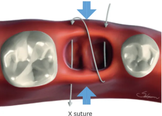
An issue that is usually neglected, but is very important in the opinion of the authors, is the suture technique following ARP. In most previous studies, the conventional crossed mattress suture (X suture) [12] was generally applied following ridge preservation, especially when primary closure was not intended (Figure 1). Other studies used the criss-cross suture, which is essentially a horizontal external mattress suture (Figure 2) [13]. These suture techniques take advantage of pulling vectors to the center of socket to narrow the socket entrance and keep the biomaterial in the socket. However, the authors of the present study have observed soft tissue profiles following ARP sutured using the criss-cross technique or conventional X suture with large losses of facial keratinized tissue (KT) and have noted that the mucogingival junction (MGJ) can be shifted to the lingual side due to the pulling of buccal tissue, especially when the buccal bone is damaged.
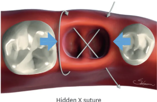
Meanwhile, applying the hidden X suture on the grafted extraction socket may successfully secure the grafted biomaterials and minimally retract the buccal tissue. The hidden X suture, which was first presented in a plastic surgery study, is a modification of the conventional X suture (Figure 3) [14]. In plastic surgery, it has also been reported that the hidden X suture has certain advantages over the conventional X suture. The latter is more harmful to skin
X suture
Figure 1. X suture or conventional X suture. The needle passes through over the extraction socket twice as if performing a continuous suture. A large crossed X is created over the socket after suturing. The blue arrows indicate the pulling vectors created by the X suture.
X suture, crossed mattress suture.
healing, leaving a very visible X mark after healing, whereas the hidden X suture has only 2 minor suture scars far apart. To the best of our knowledge, the hidden X suture has not been discussed in the dental literature, and its benefit in comparison to other suture techniques has not been properly assessed especially for ARP.
Therefore, the aim of this study was to provide proof-of-concept data about the effects of the hidden X suture technique in preserving the width of KT and the dimensions of the alveolar ridge following ARP procedures with a double-layered open healing approach.
MATERIALS AND METHODS
Study population and design
This study was a single-blinded, prospective, randomized controlled clinical trial, and was carried out from January 2016 to July 2016 at the Department of Periodontology, Dankook University Dental Hospital, Cheonan, Korea. The research protocol was approved by the Ethical Committee of Dankook University Dental Hospital, Korea (H-1412/012/002).
Criss-cross suture
Figure 2. Criss-cross suture or crossed horizontal external suture. The needle engages the buccal and lingual flaps in the same direction (mesial to distal or distal to mesial), then a knot is created. A large crossed X is created over the socket, as in the X suture.
X suture, crossed mattress suture.
Hidden X suture
Figure 3. Hidden X suture. The needle enters the buccal flap and passes to the opposite side in a diagonal direction, then it passes again from the buccal to the lingual side, also in a diagonal direction. A crossed X is created under the flap, unlike the X suture or criss-cross suture. The blue arrows indicate the vectors created by the hidden X suture.
X suture, crossed mattress suture.
The inclusion and exclusion criteria were as follows:
Inclusion criteria
• Patients' age between 18 years old and 65 years old
• Presence of a single periodontally compromised molar in the mandible or the maxilla requiring extraction and expected to be suitable for replacement by a dental implant
• Residual extraction sockets with less than 50% bone loss in all dimensions
• Ability to fully understand the nature of the proposed operation and ability to sign an Ethics Committee- approved informed consent form
Exclusion criteria
• Uncontrolled or untreated periodontal disease
• History of systemic diseases that would contraindicate surgical treatment
• Allergy to collagen and bone substitute
• Requirement of antibiotic prophylaxis
• Heavy smoking (>10 cigarettes per day)
• Pregnancy or lactation
• Inability to consent to participation in the study and/or to accept the proposed treatment plan
Experimental groups
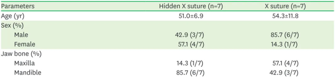
A total of 14 patients (7 control and 7 test) were enrolled in this study (Table 1). Random numbers for group assignment were generated by a statistician. A sequentially numbered, opaque, sealed envelope containing the group allocation was created for each participant.
After the completion of bone substitute filling and membrane coverage, an assistant opened the envelope to identify the group assignment.
Group 1 (open healing and hidden X suture; test)
The sockets were filled with demineralized bovine bone mineral with 10% collagen (DBBM-C; Bio-Oss® Collagen, Geistlich Pharma, Wolhusen, Switzerland) and covered with double-layers of a collagen membrane (DL-CM; Bio-Gide®, Geistlich Pharma). No attempt to obtain primary closure was made. The hidden X suture was performed.
Group 2 (open healing and X suture; control)
The sockets were filled with DBBM-C (Bio-Oss® Collagen, Geistlich Pharma) and covered with DL-CM (Bio-Gide®, Geistlich Pharma). No attempt to obtain primary closure was made.
The conventional X suture was performed.
Table 1. Demographic information of the enrolled patients
Parameters Hidden X suture (n=7) X suture (n=7)
Age (yr) 51.0±6.9 54.3±11.8
Sex (%)
Male 42.9 (3/7) 85.7 (6/7)
Female 57.1 (4/7) 14.3 (1/7)
Jaw bone (%)
Maxilla 14.3 (1/7) 57.1 (4/7)
Mandible 85.7 (6/7) 42.9 (3/7)
Values for age are presented as mean±standard deviation.
X suture, crossed mattress suture.
Suture techniques Hidden X suture procedure
The needle enters one of the flaps, passes under the flap, and reaches the opposing side of flap in an oblique direction (Figure 3). The needle then enters again on the initial side of the flap and passes obliquely under the flaps, leaving the opposing side flap. It ends up leaving 2 parallel silk threads over the soft tissue on the mesial and distal sides, with the crossed silk threads under the flaps.
X suture procedure
The overall process is similar to that of the hidden X suture; however, the needle enters the buccal flap and engages the opposing flap in a perpendicular direction (Figure 1). The needle then enters again on the buccal flap and passes the opposite flap again. Essentially, it is involved 2 turns of interrupted sutures, and the large X is created after making a knot.
Outcomes
The primary outcome was the change of KT width, as measured by the MGJ shift.
The secondary outcomes were as follows:
• Change in ridge width 1 mm (HW1), 3 mm (HW3), and 5 mm (HW5) below the ridge crest
• Change in ridge height at the buccal and lingual crest (VHB and VHL, respectively)
• Vertical reduction measured at the mid-crestal area (VMC)
Surgical procedure
After local anesthesia with 2% lidocaine containing 1:80,000 epinephrine, the teeth were extracted and meticulous debridement by surgical curettage was performed. For both groups, the sockets were filled with DBBM-C with gentle pressure. A collagen membrane was then placed over the bone substitute in a double-layered fashion [15,16]. The flaps were immobilized with minimal tension using a hidden X suture for the test group and an X suture for the control group (Ethilon® 4-0, Ethicon, Cincinnati, OH, USA). The membrane was not engaged with the suture material, and no attempts for primary flap closure, such as a releasing incision, were made. Immediately after surgery, a cone-beam computed tomography (CBCT) scan was taken with a resolution of 1 mm (scan time, 17 seconds;
exposure time, 17 seconds; 80 kV, 7 mA) using an Alphard 3030 apparatus (Asahi Roentgen Ind. Co., Ltd., Kyoto, Japan). Patients were instructed to rinse twice a day with mouthwash (GUM gargle, Osaka, Japan), and received analgesics (Somalgen, Keunhwa, Seoul, Korea) and antibiotics (Sultamox, Keunhwa) for 5 days. All patients were recalled 7–10 days later for a check-up and suture removal. The patients then received follow-up care 2, 4, 8, and 16 weeks post-ARP before implant placement. Four months after the initial procedure, the same surgeon saw the patients for the measurement and placement of the implant (Figure 4).
The location of MGJ was measured at the facial level immediately after the extraction, after suturing, and 4 months post-ARP by a single investigator (Jung-Chul Park). Using a rolling technique, MGJ was determined and marked on a stent with a notch [10]. A negative value was given if the MGJ has shifted to the lingual side.
Re-entry procedure
Four months later, the operation for implant placement was scheduled, and a second CBCT scan was taken before the implant placement. After local anesthesia, mucoperiosteal flaps were elevated, and the implants (Luna®, Shinhung, Seoul, Korea) were placed. To maximize the primary stability after placement, the final drills were one size smaller than the actual implant diameter. The tissues were sutured with 4-0 nylon (Ethilon®, Ethicon).
CBCT analysis
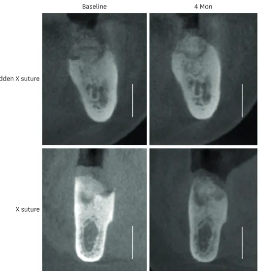
Two CBCT scans were taken at baseline and at 4 months post-ARP (Figure 5). The data were processed in the Digital Imaging and Communications in Medicine format. The 2 scans were superimposed using stable reference points (the cranial base for the maxilla and the inferior border for the mandible, respectively), and an additional manual correction was performed in the best-matched cuts. Subsequently, CBCT measurements of the cross-sectional images were made at baseline and 4 months using the same reference points and lines.
Statistical analysis
The data are presented as mean±standard deviation and median. The Shapiro-Wilk test was used to test normality. For the KT change, HW1, HW3, and HW5, the data were not normally distributed (P<0.05), while a normal distribution was found for VHB, VHL, and VMC (P>0.05). The Mann-Whitney U test was used to assess statistical significance in the KT change, HW1, HW3, and HW5, and the independent t-test was used for VHB, VHL, and VMC. All analyses were performed using SPSS version 21.0 (IBM Corp., Armonk, NY, USA).
Statistical significance was set at P<0.05.
X suture Hidden X suture
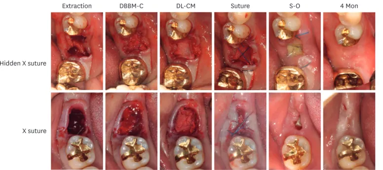
Extraction DBBM-C DL-CM Suture S-O 4 Mon
Figure 4. The clinical process from baseline to 4 months after ARP.
ARP, alveolar ridge preservation; X suture, crossed mattress suture; DBBM-C, demineralized bovine bone matrix mixed with 10% collagen; DL-CM, double- layered collagen membrane; S-O, stitch-out.
RESULTS
All patients healed without any adverse events, and no cases of graft loss or infection were recorded. Rapid epithelial migration was observed in both groups. The patients underwent implant surgery 4 months after surgery, at which point most sites were covered with thick and firm KT. Minimal changes were observed in the gingival level or papilla height on the adjacent teeth. The incision for the implant placement was not compromised at all in any case, and no invagination of the soft tissue was observed. All implants (Luna®, Shinhung) were placed in a non-submerged fashion, with satisfactory initial stability.
The MGJ line shifted to the lingual side immediately after the application of the X suture by 1.56±0.90 mm, while the application of the hidden X suture slightly pushed the MGJ line to the buccal side by 0.25±0.66 mm (Table 2). The difference between the groups was statistically significant (P=0.003). At 4 months, the width of the facial KT decreased in comparison to before the placement of sutures in both groups, but this reduction was different between the 2 groups to a statistically significant extent (P=0.007).
X suture Hidden X suture
Baseline 4 Mon
Figure 5. CBCT analysis. The horizontal and vertical dimensional changes were measured by comparing the CBCT images taken immediately after the graft (baseline) and before implant surgery (4 months). Scale bar=1 cm.
CBCT, cone-beam computed tomographic; X suture, crossed mattress suture.
Measurements of dimensional changes of the alveolar ridge after ARP using CBCT images revealed that the hidden X suture resulted in significantly less resorption in both the horizontal and vertical aspects than the X suture. Additionally, statistical significance was observed for the HW1 (P=0.016) and VMC (P=0.034) parameters. While minimal resorption was noted 1 mm below the crest in the hidden X suture group (−0.53±0.66 mm; median, −0.35 mm), the X suture group had significantly greater resorption (−5.55±6.63 mm; median, −1.75 mm) (Tables 3 and 4).
DISCUSSION
In the present study, the authors applied an open healing approach following ARP using the double membrane technique, which has been evaluated and demonstrated to have comparable or better results than conventional primary closure [9]. Previous studies have consistently reported that complete preservation was not achieved even after obtaining primary closure [17], as well as recession and loss of the KT of the adjacent teeth [18]. In contrast to the common knowledge that bone grafts should be covered by primary intention, Barone et al. [9]
demonstrated that an intentional open healing approach to ridge preservation did not affect the results of ridge preservation in comparison to closure with primary intention. Moreover, it has been shown that open healing can substantially increase the width of KT [10].
The results of this study corroborate the finding that the open healing approach for ridge preservation can successfully preserve the alveolar bone dimension for implant placement.
An interesting finding was that the suture technique significantly affected the soft tissue Table 2. Change in KT width from the extraction to 4 months
Parameters Hidden X suture (mm; n=7) X suture (mm; n=7) P value
Baseline to post-suture 0.25±0.66 (0.0) −1.56±0.90 (−1.5) 0.003a)
Baseline to 4 mon −1.05±1.07 (−1.0) −2.83±1.26 (−2.5) 0.007a)
Values are presented as mean ± standard deviation (median); The Mann-Whitney U test was used to compare differences in KT changes between the 2 groups.
KT, keratinized tissue; X suture, crossed mattress suture.
a)Statistically significant difference.
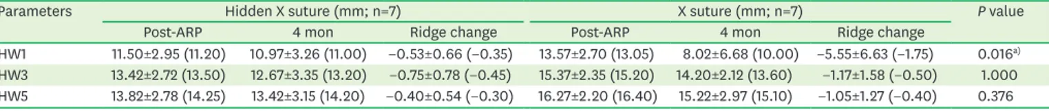
Table 3. Horizontal changes of the alveolar ridge
Parameters Hidden X suture (mm; n=7) X suture (mm; n=7) P value
Post-ARP 4 mon Ridge change Post-ARP 4 mon Ridge change
HW1 11.50±2.95 (11.20) 10.97±3.26 (11.00) −0.53±0.66 (−0.35) 13.57±2.70 (13.05) 8.02±6.68 (10.00) −5.55±6.63 (−1.75) 0.016a) HW3 13.42±2.72 (13.50) 12.67±3.35 (13.20) −0.75±0.78 (−0.45) 15.37±2.35 (15.20) 14.20±2.12 (13.60) −1.17±1.58 (−0.50) 1.000 HW5 13.82±2.78 (14.25) 13.42±3.15 (14.20) −0.40±0.54 (−0.30) 16.27±2.20 (16.40) 15.22±2.97 (15.10) −1.05±1.27 (−0.40) 0.376 Values are presented as mean ± standard deviation (median); The Mann-Whitney U test was used to compare differences in the horizontal changes of the alveolar ridge between the 2 groups.
X suture, crossed mattress suture; Post-ARP, immediately after alveolar ridge preservation; HW1, change in ridge width 1 mm below the ridge crest; HW3, change in ridge width 3 mm below the ridge crest; HW5, change in ridge width 5 mm below the ridge crest.
a)Statistically significant difference.
Table 4. Vertical changes in the alveolar ridge
Parameters Hidden X suture (mm; n=7) X suture (mm; n=7) P value
VHB −0.30±0.64 (−0.45) −0.50±0.51 (−0.40) 0.699
VHL −0.13±0.85 (−0.10) −0.82±0.81 (−0.70) 0.240
VMC −0.42±1.22 (−0.20) −1.47±1.43 (−1.60) 0.034a)
Values are presented as mean ± standard deviation (median); The vertical difference was measured by superimposing the best-matched images taken immediately after ridge preservation and 4 months later. The independent t-test was used to assess statistical differences in the vertical changes of the 2 groups.
X suture, crossed mattress suture; VHB, change in ridge height at the buccal crest; VHL, change in ridge height at the lingual crest; VMC, vertical reduction measured at the mid-crestal area.
a)Statistically significant difference.
healing pattern. The conventional X suture is the most common suture following ridge preservation, and insufficient attention has been paid to the fact that this suture technique can create a pulling vector along the buccolingual axis, decreasing the width of the KT.
Meanwhile, the hidden X suture can minimize the tension along the buccolingual axis, and it has been shown that it can comparably secure bone grafts and membranes. The application of the hidden X suture immediately pushed the KT to the facial side after suturing, and eventually reduced the loss of KT after a 4-month healing period.
In the comparison of the dimensions of the extraction socket, the horizontal width at 1 mm from the crest was significantly smaller in the X suture group. It appears that the X suture created a pulling vector along the buccolingual axis, as well as downward pressure, since it is a variation of the horizontal external suture. Meanwhile, the hidden X suture did not apply a significant pressing force on the buccal tissue, which may have prevented horizontal resorption. The X suture also applied downward pressure onto the grafted material, with statistically significant results.
The importance of KT in implant dentistry cannot be emphasized enough. First, the presence of an adequate keratinized zone enables proper incision placement and easy flap reflection.
Additionally, the substantial thickness of KT in natural teeth has long been a controversial issue, although recent systematic reviews have shown that the presence of keratinized mucosa around implants is much more clinically significant than the presence of keratinized mucosa around the natural teeth [19-22]. The presence of KT around implants has been shown to prevent the accumulation of plaque, reduce inflammation, and result in less marginal bone resorption. Clinically, maintaining or increasing the zone of KT usually requires a free gingival graft, an apically positioned flap, or the use of special stents [23]; these techniques are difficult to perform and involve the possibility of significant morbidity for patients. Moreover, none of these approaches can be performed concomitantly with tooth extraction. The soft tissue created by secondary healing over the extraction socket shows satisfactory epithelialization and the connective tissue has a well-structured network of collagen fibers (manuscript in preparation).
Within the limitations of this study, we demonstrated that the hidden X suturing technique significantly decreased the reduction of the width of KT in comparison to the conventional X suture, and showed that the dimensional change of the alveolar ridge after tooth extraction was minimized by using the hidden X suture after ARP.
ACKNOWLEDGEMENTS
This present research was conducted by the research fund of Dankook University in 2014 (R- 0001-27206).
REFERENCES
1. Schropp L, Wenzel A, Kostopoulos L, Karring T. Bone healing and soft tissue contour changes following single-tooth extraction: a clinical and radiographic 12-month prospective study. Int J Periodontics Restorative Dent 2003;23:313-23.
PUBMED
2. Wang RE, Lang NP. Ridge preservation after tooth extraction. Clin Oral Implants Res 2012;23 Suppl 6:147-56.
PUBMED | CROSSREF
3. Araújo MG, Liljenberg B, Lindhe J. Dynamics of Bio-Oss Collagen incorporation in fresh extraction wounds: an experimental study in the dog. Clin Oral Implants Res 2010;21:55-64.
PUBMED | CROSSREF
4. Araújo MG, Lindhe J. Ridge preservation with the use of Bio-Oss collagen: a 6-month study in the dog.
Clin Oral Implants Res 2009;20:433-40.
PUBMED | CROSSREF
5. Fickl S, Zuhr O, Wachtel H, Bolz W, Huerzeler MB. Hard tissue alterations after socket preservation: an experimental study in the beagle dog. Clin Oral Implants Res 2008;19:1111-8.
PUBMED | CROSSREF
6. Jung RE, Philipp A, Annen BM, Signorelli L, Thoma DS, Hämmerle CH, et al. Radiographic evaluation of different techniques for ridge preservation after tooth extraction: a randomized controlled clinical trial. J Clin Periodontol 2013;40:90-8.
PUBMED | CROSSREF
7. Araújo MG, Lindhe J. Ridge alterations following tooth extraction with and without flap elevation: an experimental study in the dog. Clin Oral Implants Res 2009;20:545-9.
PUBMED
8. Rothamel D, Schwarz F, Herten M, Chiriac G, Pakravan N, Sager M, et al. Dimensional ridge alterations following tooth extraction. An experimental study in the dog. Mund Kiefer Gesichtschir 2007;11:89-97.
PUBMED | CROSSREF
9. Barone A, Ricci M, Tonelli P, Santini S, Covani U. Tissue changes of extraction sockets in humans: a comparison of spontaneous healing vs. ridge preservation with secondary soft tissue healing. Clin Oral Implants Res 2013;24:1231-7.
PUBMED
10. Engler-Hamm D, Cheung WS, Yen A, Stark PC, Griffin T. Ridge preservation using a composite bone graft and a bioabsorbable membrane with and without primary wound closure: a comparative clinical trial. J Periodontol 2011;82:377-87.
PUBMED | CROSSREF
11. Horváth A, Mardas N, Mezzomo LA, Needleman IG, Donos N. Alveolar ridge preservation. A systematic review. Clin Oral Investig 2013;17:341-63.
PUBMED | CROSSREF
12. Cardaropoli D, Tamagnone L, Roffredo A, Gaveglio L, Cardaropoli G. Socket preservation using bovine bone mineral and collagen membrane: a randomized controlled clinical trial with histologic analysis. Int J Periodontics Restorative Dent 2012;32:421-30.
PUBMED
13. Glocker M, Attin T, Schmidlin PR. Ridge preservation with modified “socket-shield” technique: a methodological case series. Dent J 2014;2:11-21.
CROSSREF
14. Gomes OM, Amaral AS, Gonçalves AJ, Brito AS, Monteiro EL. New suture techniques for best esthetic skin healing. Acta Cir Bras 2012;27:505-8.
PUBMED | CROSSREF
15. Kim SH, Kim DY, Kim KH, Ku Y, Rhyu IC, Lee YM. The efficacy of a double-layer collagen membrane technique for overlaying block grafts in a rabbit calvarium model. Clin Oral Implants Res 2009;20:1124-32.
PUBMED | CROSSREF
16. Kozlovsky A, Aboodi G, Moses O, Tal H, Artzi Z, Weinreb M, et al. Bio-degradation of a resorbable collagen membrane (Bio-Gide) applied in a double-layer technique in rats. Clin Oral Implants Res 2009;20:1116-23.
PUBMED | CROSSREF
17. Darby I, Chen ST, Buser D. Ridge preservation techniques for implant therapy. Int J Oral Maxillofac Implants 2009;24 Suppl:260-71.
PUBMED
18. Ten Heggeler JM, Slot DE, Van der Weijden GA. Effect of socket preservation therapies following tooth extraction in non-molar regions in humans: a systematic review. Clin Oral Implants Res 2011;22:779-88.
PUBMED | CROSSREF
19. Brito C, Tenenbaum HC, Wong BK, Schmitt C, Nogueira-Filho G. Is keratinized mucosa indispensable to maintain peri-implant health? A systematic review of the literature. J Biomed Mater Res B Appl Biomater 2014;102:643-50.
PUBMED | CROSSREF
20. Gobbato L, Avila-Ortiz G, Sohrabi K, Wang CW, Karimbux N. The effect of keratinized mucosa width on peri-implant health: a systematic review. Int J Oral Maxillofac Implants 2013;28:1536-45.
PUBMED | CROSSREF
21. Lin GH, Chan HL, Wang HL. The significance of keratinized mucosa on implant health: a systematic review. J Periodontol 2013;84:1755-67.
PUBMED | CROSSREF
22. Souza AB, Tormena M, Matarazzo F, Araújo MG. The influence of peri-implant keratinized mucosa on brushing discomfort and peri-implant tissue health. Clin Oral Implants Res 2016;27:650-5.
PUBMED | CROSSREF
23. Park JC, Yang KB, Choi Y, Kim YT, Jung UW, Kim CS, et al. A simple approach to preserve keratinized mucosa around implants using a pre-fabricated implant-retained stent: a report of two cases. J Periodontal Implant Sci 2010;40:194-200.
PUBMED | CROSSREF