大힘’i放射線않學 f한悲 Vol.XV, No.2, 1979
- Abstract -
直立後前 및 {fp 歐前後播影 뼈部 X 線像의 比較
홉麗大學校 짧科大學 放射緣科學敎室
鄭圭炳
• 李淑
•朴仁植 • 徐源M.
A Comparision of Upright Chest PA and Supine Chest AP View
Kyoo Byung Chung
,
M.D.,
Sook Lee,
M.D.,
ln Sik Park,
M.D.,
Won Hyuk Suh,
M.D. Department of Radiology,
College of Medicine,
Korea University,
Seoul,
KoreaThe routine chest roentgenogram is standardized as upright postero.anterior teleroentgenogram with half inspiration
,
but in impossible cases,
such as infants,
children and severe illed patients,
the supine chest AP view have to be taken. There are many different points between upright chest PA and supine chest AP view.Authors analysed the 51 cases of normal upright chest PA and supine chest AP views which were
taken in the same people
,
same exposure factors and the same tube.film distance of 72 inches.The results were as follows:
1. The width of the med iastinum was wider in the supine chest AP view than the upright chest PA view. In the chest PA view
,
the mediastinal width was 5.56:!:1.37cm,
and in supine chest AP view,
6.60:!:1.36cm,
respectively.2. The diameter of right descending pulmonary artery was slightly smaller in the supine chest AP view than the upright chest PA view. The distribution of the p 비 monary vasculatures was nearly even in the supine chest AP view.
3. The transverse diameter of the thorax was slightly smalier in supine chest AP view, but the transverse diameter of the heart was significantly increased in the supine chest AP view. The transverse diameter of the heart was 12.12:!:1.5cm in upright chest PA, and 13.12:!:1.63cm in
supine chest AP, respectively.
4. The cardiothoracic ratio was markedly increased in supine chest AP than the upright chest PA view. The cardiothoracic ratio of the upright ιhest PA was 43.72:!:3.97%, and in the supine chest AP, it was 48.19:!:4.73%.
5. The supine chest AP view of this study is different in tube.film distance from the routine portable films of the chest AP view, and the magnification factors are probably different. Further studies are need.
1. 績 등A llftU
益한 放射線學的 檢훌方法이다.
最近 X 線發生裝置의 發達과 그에 附隨되 는 器具 및 필릎둥의 開發로 小뭘의 X 線被i앓고} 短時間에 良質의
x
뼈部 X 線사진은 가장 흔히 施行되는 X線檢좁이며 또 線像을 만들 수 있게 된 것은 周'!;[]외 훨줬이마. 그러나 가장 많은 情趙를 한장으로 提供해 주는 때單하면서도 有 6행部X線像의 경우 被옳部에 가장 높은 X 線吸收性을 가
- 376 -
진 骨格과 가장 낮은 吸收性올 가진 뿔氣, 그러고 중간 면서 I휩立 cheSI PA 와 띠l 베 cheSI AP 1!용을 얻어 比따親 정도의 軟部*.fl織둥이 共存하기 혜문에 判펌에 척당한 사 察한 바 몇가지의 知見을 얻었으므로 文없考察과 아울러 진條件을 맞추기란 용이하지 않다. 뿐만 아니라 恩휴의
없位에 따라 異常有無 確認에 決定的 影響을 미 칠 수도 있어 理想的인 8햄部 X 線餘을 얻기란 여간 어려운 일이 아 니마.
通常 뼈部X 線像은 直立後前像(chest PA) 을 얻은 것 으로 되어. 있으냐 낮은 年敵의 小兒냐 重愚者둥 바로 서 기가 不可能한 惠者에서는 누운 자세에서 前後提影 (Supine
chest AP) 을 하는 수 밖에 없으며 이 때 얻은 사진은
chest PA 와 여 러 모로 마르게 나타나으로 異常有無¥U 定 이 어렵고 경우에 짜라서는 不可能한 해도 있다.
일쩨기 F.I.Jackson은 直立 chesl PA 사진파 直끄 chesl AP 사진의 握影法파 그 差異點을 記述한 척 이 있 고 1971 年 H. Burko 둥은 8市血뿔造影振影으로 협 n
Chesl AP 像과 {대없 chesl AP 像에 서 R퍼 r.$의 8市靜H*直
행告하는 바이다.
n
형象및方法1. 對 象
任룡로 i행擇한 協 fJ,~ 可能한 正常 Á 51 名을 對象으로 하 였으며 그 年敵分布는 13歲에서 60 歲사이였마。 男子가 44名, 女子가 7 名이었으며, 21 歲에서 40歲사이가 36
例로 金顆의 70 %를 차지하였마(Table
1).
2. 方 法
直立 Chest PA는 通常方法과 같이 얻었으며 이때 管 電@경은 60 ~75 KVP, rnA s는 10~25 rnAs , 그러고 管
필름 距離는 180 cm 이 었 다. {J대톰^ Chest AP 사진은 낮은 용台를 X線室에 두고 그 위에 愚者를 똑바로 둡게 한 쩔이 意味있는 變化를 보였다고 報告한 바 있다 1 , 5). 後 同-距離, 同-擬影條件으로 嚴影하였다. 이혜 25f列
直끄 chest PA像과 {대없 cheSI AP 隊사이에는 前述한 에서는 {대없 Chest AP 의 경우 Chest PA보마 2~5 KUP 뼈動服뿐만 아니라 總隔洞, 心職, 師動服 둥에도 差異가 높은 뿔핍뺨을 주어 振影하였다.
난마는 것은 잘 알려진 事웰이다 2, 3). 同 -Á 의 直끄Chest PA 와 {대에 Chest AP를 16 個頂으 著者동은 任풍로 選擇한, 協調可能한 正常짧國人을 對 로 나누어 比較없IJ定하였 으며 그 中 意義가 크다고 생 각 象으로 同-人에서 同-擬影條件과 同- 距離를 維持하 되는 7個項에 關하여 分析檢討하였다( Fig.ll.
r
ιhe났 AP
Chest PA: Upright chest teleroentgenogram of trachea bifurcation.
with tube.film distance of 180cm B. D iameter of the right descending pulm. Chest AP: Supine chest teleroentgenogram onary artery.
with tube-film distance of 180cm C. Transverse diame ter of the heart.
A. Width of the mediastinum at the level D. Transverse diameter of the chest.
::t
1.36 cm로 띠]없Chest AP 의 경우 약 1 cm 넓게 보였IH. ~ 究 成 績 마 (Table
n )
右下行8市動~의 直쩔은 Leinbach IJ:,法파 J.C.Chang 동 {대없 Chest AP 사진이 直立Chest PA 像과 큰 差異點은 이 使用한 方法을 따랴 iflU)'효했으며 이 때 右下行뼈動~
우선 冒뼈骨이 師野를 상당끔ß分 가리게 되고 銷骨陰影이 의 直쩔은 펴[lZ Chest PA에서 1.28
::t
O.14 cm 이었고 때 師것위로 올라가며 또 橫隔陳이 學上된다는 것은 잘 알 없 Chest AP 에서는 1.18::t
0.17 cm로 直끄 Chest PA 얘려진 바와 같았다 (Fig.l). 야 0.1 cm 정도 큰 數順를 냐타내었마 (Table
m ).
測定可 氣管二分部位에 서 測定한 總隔洞의 넓 이 는 直立Chest 能한 40 例만 擇하였 다PA 에서 5.56
::t
1.37 cm 이었고 때없 Chest AP 에서 6.60 뼈鄭橫쩔은 直立Chest PA에서 28.64::t
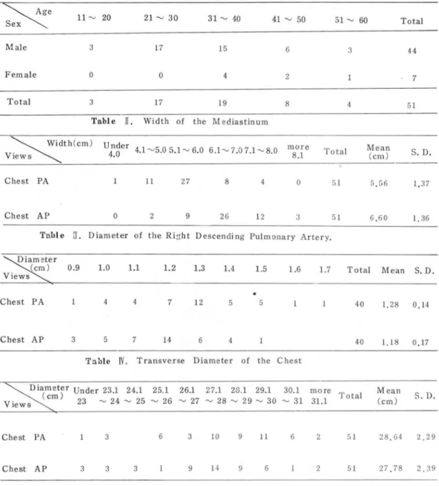
2. 29cm 이었고 Tahle I. Age and Sex Distribution of Materials.Age 11 ~ 20 21 ~ 30 31 ~ 10 41 ~ 50 51 ~ 60 Total Sex
Male 3 17 15 6 3 44
Female 0 0 4 2 7
Total 3 17 19 8 4 51
Table ll. Width of the M ediastinum
Width(cm)
U nder 4.1 ~5.0 5.1 ~ 6.0 6.1-...., 7.07.1-...., 8.0 more Total Mean
S. D.
Views \ \ 4.0 4.l~5.05.1~6.0 6.1-....,7.07 . l-....,8.0 8.1
(cm)
Chest PA 11 27 8 4 0 51 5.56 1.37
Chest AP 0 2 9 26 12 3 51 6.60 1 36
Tnble fJ. Diameter of the Ri~ht Descending Pulmonary Artery.
Diameter
\\{cm) 0.9 1.0 1.1 Views
1.2 1.3 1.4 1.5 1.6 1.7 Total Mean S. D.
Chest PA 4 4 7 12 5 a 40 1.28 0.14
Chest AP 3 5 7 14 6 4 40 1.18 0.17
Table
JV .
Transverse Diameter of the ChestD1a(met )er Under 23.l 24 . 1 25 l 261 27.1 28.l 29.1 30.1 more T l Mean S. D.
cm) Tota
v
lewsX;
23~
24~
25~
26~
27~
28~
29~
30~
31 3l l (cm)Chest PA 3 6 3 10 9 11 6 2 51 28 64 2 29
Chest AP 3 3 3 9 14 9 6 2 51 27 78 2.39
- 378 -
{대없 Chest AP 에서는 27.78
:t
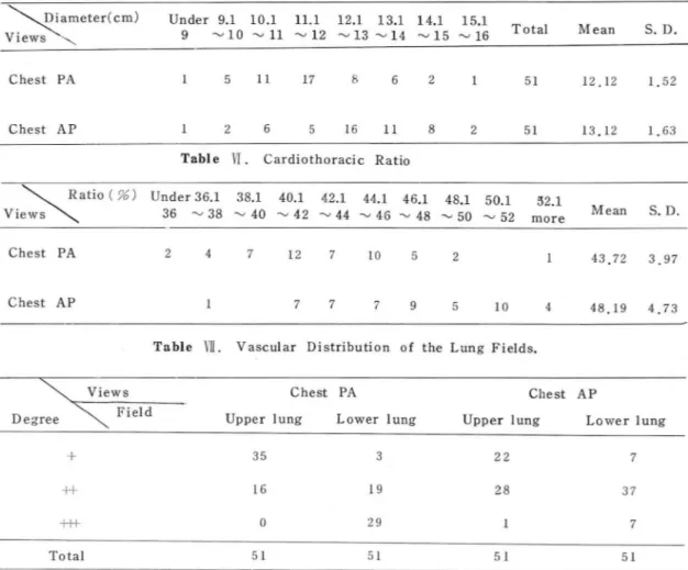
2.39 cm 로 直îJ. Chest 骨格 빛 그 周圍 軟部組織 둥이 마. 그 中 8市는 가장 큰 PA 혜 가 약 1 cm 큰 數順를 냐타내 였 £며 ; 心觸橫쩔은 容협을 차지 하고, 成人ZjS-均容積이 Fraser 둥에 依하면 直끄 Chest PA 에서 12.12:t
1. 52 cm, f대없 Chest AP 에 7240 cc 이 며 이 中 組織이 나 血흉은 10 %인 740 cc 에 서 13.12:t
1.63 cm 로 때없 Chest AP 혜가 약 1cm 증가 不過하고 90 %는 空氣가 차지한다고 한마 4)되었다{Table
N
& TableV
l. X 線像의 陰影은 ~氣含量에 反比例하여 떨어지게 되 心뼈比 (Cardiothoracic ratiol 는 直끄 Chest PA 에 서 고 血쨌이 냐 組織 또는 水分의 增 1]Q에 따라 그 陰影은 43.72 :t 3.97% 였으며 {대없 Chest AP 에서는 48.19:t
增加하게 된마 4)4.73 %로 때봐 Chest AP 에서 약 4.5 % 增加를 보여주 8행部 X 線像은 直立後前投射로 遠距離, 즉 72 인치距雖 었마. 이는 直立 Chest PA 의 心觸比 43.72 %에 對해서 半吸入狀顧에서 찍는 것£로 標準化 되어 있마. 1964年 는 約 10 %의 增加횡을 보인 생이마 (Table
VI
l. F. 1. Jackson둥은 Air -Gap technique 을 써서 直立Chest 또 師血管分布는 直 nCþest PA 에서 보는 上下市野의 AP 像 찍는 법을 考按해 내였으나 現在는 널리 使用되 差異가 {며없 Chest AP 에서는 거의 認知할 수 없었마 지 않는 方法이다 5) 바로 서기가 不可能하거나 協調가(Table Vlll. 어려운 ¥L兒, 小兒 또는 重惠者의 경우엔 부흑이 때없
N.
考 察8행部 X 線像에 包含되는 部分은 뼈, 心職, 總隔洞, n행흘g
Chest AP 를 찍 어 異常有無를 判讀하지 않으연 안된 다.
이때 {띠없 Chest AP 사진은 直立 Chest PA 사진과 많은 差 異가 있어 判調에 어려웅이 많다. 우선 {대없 Chest AP
가 直立Chest PA 와 가장 큰 差異點은 冒뼈骨이 師野의
Table V. Transverse D iameter of the Heart.
Vi、e\wsD
\ia\meter(cm) Under 9.1 10.1 11.1 12.1 13.1 14.1 15.1Total Mean S. D.
9 ~10~ 1l ~12 ~13~14 ~15~16
Chest PA 5 11 17 8 6 2 51 12 12 1 52
Chest AP 2 6 16 11 8 2 51 13.12 1 63
Table
vr.
Cardiothoracic Ratio\ \ Ratio ( %) Under 36.1 38.1 40.1 42.1 44.1 46.1 48.1 50.1 Views \ \ 36 '" 38 '" 40 '" 42 ~ 44 '" 46 '" 48 '" 50 ~ 52
52.1
Mean S. D.
more
Chest PA 2 4 7 12 7 10 5 2 43.72 3 97
Chest AP 7 7 7 9 5 10 4 48.19 4.73
Table
Vll .
Vascular Distribution of the Lung Fields.Views Chest PA Chest AP
Degree \ Field Upper lung Lower lung Upper lung Lower lung
+
35 3 22 7++
16 19 28 37+t+ 0 29 7
Total 51 51 51 51
상당부분을 가리게 되고 또 銷骨陰影이 8힘失위로 올라가 게 되 며 숲般的인 뼈 l합의 陰影은 直 nChest PA 보다 t뽑 加하게 된다. 때없 Chest AP 像이 直立 Chest PA 像보다 陰影度가 增加하는 가장 큰 理田는 {띠며位블 取하면 1Í1l 쨌量이 t업加되 기 때 문인 것 으로 알려 져 있 다 3, 4) Fraser 에 依하연 {대l?M立의 경 우 師숲體에 存在하는 血陳量은 直
V
結 홉움同-距離와 擬影條件으로 同一A에서 擬影한 51名의 直立 Chest PA 와 fJ1J 없Chest AP 사진을 比較觀察 하였던
바 다음과 갇은 結論을 얻었다.
n
Chest PA얘 보마 約
30%의 增加를 보여 준마고 한다
1.{.대없 Chest
AP사진이 直立
ChestPA 와의 큰 差異
4)
따라서 空氣에 對한 血쨌의 比가 t뽑加하여 陰影增tm 는 휩8甲骨이 師野內로 들어 와 있고 銷骨뾰置는 뼈失
위로
를 招來하게 펀다.
*i!t隔 t혀은 兩師사이의 軟部組織을 말하며 心戰, 大血管 食道 임파節 및 其他 軟部組織둥이 包含된마 {띠폼A前後
올라가며 또 橫隔뺑운 -般的으로 짧上되 었 다. 이 들 各 各의 경 우 없u定基準點 定하기 가 어 려 워 統計處理는 鍵하 였다
쳤훌影像에 서 는 이 짧隔1同이 보동 直立Chest PA 보다 鷹大 2. 總隔1同福은 띠1 êÅ Chest AP 에 서 直立Chest PA 보마
되어 냐타냐는데 이의 가장 큰 原因中의 하냐는
總隔洞2ft딩順가 約
1 cmÞ협 1m.를 보였고, 이는約
18.7%의 t협
이 比較的 前方에 {立置하고 있어 {대없 Chest AP 의 경우 加率에 해 당한다.필릎파 總隔洞과의 距離가 直立Chest PA 때보마 크으로 3. 右下師動lI*의 直쯤은 {대동)., Chest AP에서 直끄Chest 없大率이 크기 혜문이며 또 하냐는 心職을 包含한 維隔 PA 보마 약간 減少되었마.
I同 軟部組織이 띠n 동M立 의 경우 울주머니처럼
左右옆으로
4뼈탱lit홈짧은 {LD 닮)., C
hestAP 에서 直立Chest PA 보다주
저앉기때운이다. 約
3% 의 뼈少{直를 나바내었고 心驗의 橫쩔은{따:i!..Che
st右下行뼈動lI*의 直짧은 {띠동)., ChestAP 때에 直立Chest AP 에서 約 1 cm t,뽑 tm , 즉 8.25 %의 增加를 보였다. 따 PA 보
다 多小 작아지는데 이는 血被自體의 靜水慶이 {대 라서 心뼈比는 {대뼈 C
hest AP 에서 直立 ChestPA보마 約
없位의 경우 直끄 Chest PA 만몸 크지 않기 혜운인 것무 4.47% t뽑加를 나타내였마.로 생각된마1)
5師血管 分布樣狀은 때뚫Á
ChestAP 에서는 直끄 Chest
8엠郞의 橫건짚은 따l 톰Á Chest AP 사진에 서 直끄 Chest PA PA 와 같은 上下部差異가 없고 고른 分布를 보여 주었 마.때 보다 작아지 는데 이 것 은 뼈節을 斷面에 서 보면 뒤 쪽이
6.띠}똥ÁDL
Chest AP를 보고 異常有無를
判定하는데는 앞쪽보마 크기 혜 문에 直立
Chest PA에 서 더 많이 擬大 以上과 같은 여 러 要素를 參動하여 判폈하는 것 이 종富
되기 얘운인 것으로 생각펀마 할 것무로 생각된다.또 心廳橫쩔의 경우엔 縮隔 1同파 "1 숫하게 {대êÅ Chest AP 에 서 커 지 게 되 고 따라서 Jl;、뼈比는 {대봐 Chest AP 의 경우 더 增加하게 된다. 著者들의 경우 心뼈比는 直立 Chest PA 에 서 2f均 43.7% 였 는데 띠j 봐 Chest AP에 서 는 48.2 %로 增加하였으며 이는 約 10 % t,협 110한 생이 펀마-
!lilí野의 血管分布는 直立Chest PA 의 경 우 上部와 下部 의 差異가 있 어 t部보마 下部에 더 뚜렷 이 血管陰影이 나타나는데 反하여 {대몽Á Chest AP 의 경우 이런 上下區別 이 거의 없어진다. 이것은 血쨌}경體의 靜水壓혜운이기도 하고 또 한펀」즈로는 {대똥사立의 경우 師野에 모이는 血~
量이 상당히 t뽑加하는 것도 한 原因이 될 수 있다고 한 다4) 血滾품이 t멸加하므로 X線像最影時 때닮ÁChest AP 에 서 는 直立Chest PA 보다 同-mAs 를 주는 정 우 管電 밍용을 2~5 KVP 增 110 시 켜 주어 야 비 슷한 陰影度의 X 線 像을 얻을 수 있다.
以上 著者들이 分析檢討한 {대똥ÁChestAP 사진은 臨j;f 에 서 흔히 擬影하는 Chest AP 사진과는 다릎데 이 떼 에 는 대개 管-필릎 距離를 90~120 cm 로 하고 있어 據
大率에 상당한 差異가 있을 것이다. 이에 關하여는 앞 으로 더욱 追究하여야 할 것으로 생각된다.
REFERENCES
,. Burko, H. M.D., Carwell, G. M.D. and Newman E., M.E. 5ize
,
location and gravitational changes of normal upper lobe pulmonary veins. A merican J ournal of Roentgenology 777: 687-689,
7977.2. Chang
,
C. H. ()oseph),
M.D. : The normal roentgeno graphic measurements of the r,ψ'ht descending pul- monary artery in 7085 casεs. American Journal of Roentgenology 87: 929-935,
7962.3. Felson
,
B. M.D. Chest Roentgenology 72. W.B.5aunders Company
,
Philadelphia,
7973.4. Fraser, R.G., M.D., Peter Pare, ).A., M.D. Diagnosis of diseases of the chest
,
second edition 765. W. B.Saunders company
,
Philadelphia,
7977.5. )ackson, F.I., M.D. The air-gap technique: And an improvement by anterior-posterior positioning for chest roentgenography. American Journal of Roentgenology 92: 688.697
,
7964.- 380