The clivus is a bony surface, and the term itself means
“slope.” It forms the central part of the skull base and is formed by the fusion of the basisphenoidal and basioccip- ital bones.1,2 This fusion is not usually complete before the age of 18 years, until which point it is separated by the spheno-occiptal synchondrosis.1,3
Prabhu et al.4 stated that Testut was the first to describe fossa navicularis magna on the radiographs of a patient. In the literature, it has been referred to using several names, such as fossa pharyngea, large pharyngeal fossa, fossa na- vicularis, canalis basilaris medianus, keyhole defect, and longitudinal or transverse segmentations.3,5,7,8 This is an anatomical variant that appears as a notch-like defect in the basiocciput.1,6-8 Anatomical variants of the basiocciput are generally uncommon. The incidence of fossa navicularis magna was reported on dry skulls to be as low as 5.7%.5 Studies using computed tomography(CT) and magnetic
resonance imaging have reported that this osseous defect may be filled with lymphoid tissue of the pharyngeal ton- sils.9 In the literature, it has been postulated that it may serve as a tract for intracranial infections.3
Cone-beam CT(CBCT) has become immensely popular in dental offices within the past decade and is commonly used in several fields of dentistry, including oral and max- illofacial radiology, orthodontics, oral surgery, periodon- tics, and endodontics.10 Depending of the field of view, CBCT may cover an area larger than the area correspond- ing to the practitioner’s comfort level and expertise. In the large field of view, structures such as the sella and clivus are captured. Hence, an in-depth knowledge of the anato- my of these areas and appropriate training are required to interpret pathological findings and anatomical variants in large-volume CBCT images.
The purpose of this report was to familiarize practicing dentists and specialists with the importance of identifying the anatomical variations in the basiocciput by presenting four cases of fossa navicularis magna. Considering the paucity of the literature, especially in the field of oral and maxillofacial radiology, we also investigated the imaging
Fossa navicularis magna detection on cone-beam computed tomography
Ali Z. Syed1,*, Mel Mupparapu2
1Department of Oral and Maxillofacial Medicine and Diagnostic Sciences, School of Dental Medicine, Case Western Reserve University, Cleveland, OH, USA
2Division of Radiology, University of Pennsylvania School of Dental Medicine, Philadelphia, PA, USA
AbstrAct
Herein, we report and discuss the detection of fossa navicularis magna, a close radiographic anatomic variant of canalis basilaris medianus of the basiocciput, as an incidental finding in cone-beam computed tomography(CBCT) imaging. The CBCT data of the patients in question were referred for the evaluation of implant sites and to rule out pathology in the maxilla and mandible. CBCT analysis showed osseous, notch-like defects on the inferior aspect of the clivus in all four cases. The appearance of fossa navicularis magna varied among the cases. In some, it was completely within the basiocciput and mimicked a small rounded, corticated, lytic defect, whereas it appeared as a notch in others. Fossa navicularis magna is an anatomical variant that occurs on the inferior aspect of the clivus. The pertinent literature on the anatomical variations occurring in this region was reviewed.(Imaging Sci Dent 2016; 46:
47-51)
Key Words: Cone-Beam Computed Tomography; Skull Base; Clivus Fossa, Posterior; Multidetector Computed Tomography
Copyright ⓒ 2016 by Korean Academy of Oral and Maxillofacial Radiology
This is an Open Access article distributed under the terms of the Creative Commons Attribution Non-Commercial License(http://creativecommons.org/licenses/by-nc/3.0) which permits unrestricted non-commercial use, distribution, and reproduction in any medium, provided the original work is properly cited.
Imaging Science in Dentistry·pISSN 2233-7822 eISSN 2233-7830 Received August 21, 2015; Revised September 27, 2015; Accepted October 10, 2015
*Correspondence to : Prof. Ali Z. Syed
Department of Oral and Maxillofacial Medicine and Diagnostic Sciences, School of Dental Medicine, Case Western Reserve University, 10900 Euclid Avenue, Cleve- land, OH 44106-4905, USA
Tel) 1-216-368-6802, Fax) 1-215-573-7853, E-mail) azs16@case.edu
characteristics of fossa navicularis magna. In addition, this report is intended to help the practitioner identify these entities as anomalies rather than invasive lesions. Fossa navicularis magna is an anatomical variant that requires no intervention. No prior studies have investigated fossa navicularis using CBCT, making this the first series to highlight fossa navicularis magna on CBCT. This article also sheds light on the terminology associated with other anatomical variants that have been noted in the basioc- ciput bone in the past.
case report
Case 1
The CBCT scan of a 65-year-old female with a noncon- tributory medical history was acquired using a CS9300 machine(Carestream, Atlanta, GA, USA) for preimplant treatment planning purposes. The patient underwent CBCT scanning at a private dental office. All the data were saved according to the Digital Imaging and Communication in Medicine(DICOM) standards and were sent for evalua- tion on a compact disc. The volume was evaluated by a board-certified oral and maxillofacial radiologist and was reviewed using InVivo 5.4.3(Anatomage, San Jose, CA, USA).
The data were analyzed using multiplanar reconstruc- tions. The inferior aspect of the basiocciput of the clivus
showed a notch-like osseous defect, measuring approxi- mately 4mm at its greatest dimension and outlined by a regular cortical margin(Fig. 1). Based on the radiographic appearance, a diagnosis of fossa navicularis magna was made.
Case 2
The CBCT scan of a 50-year-old male with no signifi- cant medical history was acquired using a CS9300 ma- chine(Carestream, Atlanta, GA, USA) and referred for implant placement evaluation. The scan was taken at a private office, saved using the DICOM standards, and was referred for interpretation via a Health Insurance Porta- bility and Accountability Act(HIPAA)-compliant cloud application with medical-grade encryption. All the data were evaluated using InVivo 5.4.3(Anatomage, San Jose, CA, USA) by a board-certified oral and maxillofacial ra- diologist.
Volumetric image analysis revealed a well-defined and solitary low-attenuation area showing an osseous defect on the inferior surface of the basiocciput. The periphery was generally well-defined and corticated on the superior aspect. It measured approximately 2mm in its greatest di- mension. The osseous defect was filled with a soft-tissue density mass. Based on the radiographic presentation, an impression of fossa navicularis magna was made(Fig. 2).
Fig. 1. Sagittal and axial cone-beam computed tomographic images of case 1 demonstrate a notch-like osseous defect in the clivus(arrows) radiographically consistent with fossa navicularis magna.
Fig. 2. Axial and sagittal images of case 2 show a well-defined, osseous defect in the anterior-inferior aspect of the clivus(arrows), demonstrat- ing fossa navicularis magna.
Case 3
A 12-year-old female was referred from an orthodontist for evaluation of the development of a right maxillary ca- nine that was partially impacted. There was a missing per- manent premolar in the mandible, and several premolars in various stages of eruption were observed. The CBCT volume data were acquired at the orthodontist’s office(I- CAT Next Generation, Imaging Sciences International, Hatfield, PA, USA) and the DICOM images were sent for interpretation on a compact disc. The data were evaluated using InVivo 5.4.3(Anatomage, San Jose, CA, USA) by a board-certified oral and maxillofacial radiologist.
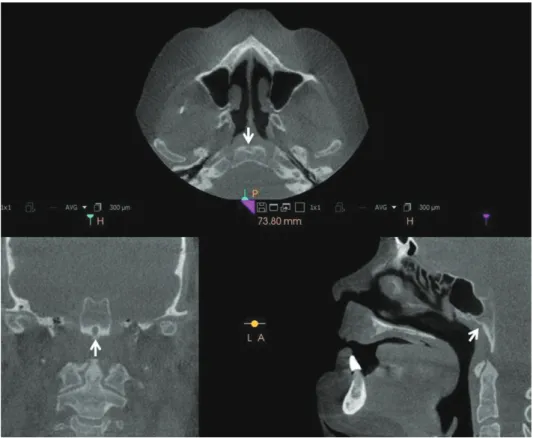
Volumetric image analysis revealed a well-defined soli- tary notch in the inferior part of the basiocciput(Fig. 3).
The notch was approximately 10mm posterior to the spheno-occipital synchondrosis in the sagittal view. Both coronal and axial views clearly showed the notch within the basiocciput. Based on the radiographic features and the fact that the patient was completely asymptomatic, a radiographic diagnosis of fossa navicularis magna was made. The orthodontist was informed of its presence and that it needed no special attention.
Case 4
A 68-year-old female was referred from her oral and maxillofacial surgeon, who was planning to place root- form implants in her lower jaw. The CBCT volume was acquired in the surgeon’s office using a CS9300 machine (Carestream, Atlanta, GA, USA) and was sent as an en- crypted DICOM file over a HIPAA-compliant cloud ap- plication. The files were decrypted and downloaded via a DICOM viewer(CS 3D imaging software; Carestream, Atlanta, GA, USA), and interpreted by a board-certified oral and maxillofacial radiologist.
Volumetric image analysis showed a well-defined, cor- ticated, solitary circular lytic area towards the inferior part of the basiocciput. The diameter of this well-defined lytic area was approximately 4mm. The sagittal image showed notching, but the coronal and axial images showed a clear- er, well-defined radiolucency(Fig. 4). Based on the ra- diographic features and the lack of symptoms related to this finding, a radiographic diagnosis of fossa navicularis magna was given to this entity, and the oral and maxillo- facial surgeon was informed of this diagnosis.
discussion
The skull base is a very complex structure, and the cli- vus forms the mid-portion of the base of the skull. The clivus develops during the second month of intrauterine life. It is the result of endochondral bone formation.8 The clivus remains separated until the age of 16-20 years by the spheno-occiptal synchondrosis.1-3 Ossification of the spheno-occipital synchondrosis begins earlier in females, at approximately the age of 12-13 years, whereas in males, this process begins at the age of 14-15 years.2
It has been hypothesized that fossa navicularis magna is an anatomical variant that may form from either the per- sistence or enlargement of emissary veins or as a result of remnants of the notochordal canal.4,8-10 In the English-lan- guage literature, it has been referred to with synonymous names, such as fossa pharyngea, large pharyngeal fossa, and fossa navicularis, with terminological overlap occurr- ing between this developmental anomaly and canalis basi- laris medianus.3,7,8 Currarino(1988)8 referred to a descrip- tion of the canalis basilaris medianus defect in humans published in the late 19th century. According to the early descriptions, this defect was a well-defined channel, typ-
Fig. 3. Coronal, sagittal, and axial images reveal a well-defined, osseous defect within the basiocciput in case 3, as shown by arrows.
ically more than 2mm in diameter, originating on the in- tracranial surface of the basiocciput in the midline very close to the anterior rim of the foramen magnum. The initial three types of the canalis basilaris were the inferior, superior, and bifurcated types, and other variants, such as the long-channel, superior, and inferior recess types, were added to the literature more recently.8 It is still unclear if the fossa navicularis magna and canalis basilaris medi- anus are anomalies from the same notochord remnants;
however, we theorize that they both are representations of the same notochord remnants with radiographically over- lapping features. It is possible that fossa navicularis magna may actually be a variant of incomplete canalis basilaris medianus(inferior basiocciput type). On a contradictory note, Beltramello et al.9 colleagues argued that fossa navi- cularis magna must be differentiated from canalis basilar- is medianus, which the authors stated represents the per- sistence of the chordal canal. It is known that this notch- like osseous bony defect in the basiocciput can be filled with lymphoid tissue of the nasopharyngeal tonsils.9,11,12 However, this is in contrast with the study of Cankal et al.,13 who found no soft tissue in the bony dehiscence. The fossa navicularis magna reported by Betranello et al.9 was approximately 8mm×6mm in diameter and was outlined by regular cortical margins in the basiocciput. In rare in- stances, fossa navicularis magna may serve as a route for
the spread of an infection from the oropharynx to the base of the skull.3,4 The incidence of fossa navicularis magna in dry skulls was found to be 5.3%,5 and using CT, the reported incidence was 3%.10 The higher incidence in an- atomic studies could be attributed to the slice thickness of CT.9 The incidence of fossa navicularis magna as quoted by Beltramello et al.9 in their study ranged from 0.9% to 2.1% in three studies, all performed on dry skulls.
Currarino’s study8 of the canalis basalis medianus and related defects of the basiocciput highlighted the impor- tance of identifying these anomalies. In that report, a 19- month-old male child with several previous episodes of meningitis was found to have a complete canal, 5mm in diameter, traversing the basiocciput, which protruded into the subarachnoid space. The diagnosis was basioccipital meningocele, which was identified as responsible for the recurrent meningitis. The child had no further bouts of meningitis during a 36-month follow-up period after re- moval of the sac and surgical obliteration of the bony de- fect.
CBCT was introduced into the dental profession at the beginning of the new millennium. CBCT has many ad- vantages over multidetector computed tomography, such as good spatial resolution, a rapid scan time, and, best of all, lower radiation doses.10 CBCT is used in all fields of dentistry, including orthodontics, oral surgery, and im-
Fig. 4. Axial, coronal, and sagittal images in case 4 show a well-de- fined, round osseous defect noted in the inferior aspect of the clivus, radiographically consistent with fossa navicularis magna.
plant treatment planning.9 CBCT scans can be acquired with multiple fields of view, including small, medium, and large. Since large fields of view capture a larger area, extending outside the region of interest, images must be thoroughly evaluated to rule out any pathology or devel- opmental anomalies.
CBCT is a relatively new technology in dentistry. Since CBCT findings include areas within the skull base that are not primarily intended to be imaged, oral and maxillo- facial radiologists have the obligation to identify, review, and report anatomical variations of the skull base and prevent the initiation of unnecessary further imaging by dental practitioners. Fossa navicularis magna is one such anatomical variation affecting the basiocciput, and is sim- ilar to canalis basilaris medianus, which also affects the basiocciput.
references
1. Neelakantan A, Rana AK. Benign and malignant diseases of the clivus. Clin Radiol 2014; 69: 1295-303.
2. Shah A, Goel A. Clival dysgenesis associated with Chiari Type 1 malformation and syringomyelia. J Clin Neurosci 2010; 17: 400-1.
3. Segal N, Atamne E, Shelef I, Zamir S, Landau D. Intracranial
infection caused by spreading through the fossa naviclaris magna - a case report and review of the literature. Int J Pediatr Otorhinolaryngol 2013; 77: 1919-21.
4. Prabhu SP, Zinkus T, Cheng AG, Rahbar R. Clival osteomy- elitis resulting from spread of infection through the fossa na- vicularis magna in a child. Pediatr Radiol 2009; 39: 995-8.
5. Collins HB Jr. Frequency and distribution of fossa pharyngea in human crania. Am J Phys Anthropol 1927; 11: 101-6.
6. Krmpotić-Nemanić J, Vinter I, Ehrenfreund T, Marusić A.
Age-related changes in the anatomical landmarks of the osse- ous epipharynx. Ann Anat 2006; 188: 459-67.
7. Ginat DT, Ellika SK, Corrigan J. Multi-detector-row com- puted tomography imaging of variant skull base foramina. J Comput Assist Tomogr 2013; 37: 481-5.
8. Currarino G. Canalis basilaris medianus and related defects of the basiocciput. AJNR Am J Neuroradiol 1988; 9: 208-11.
9. Beltramello A, Puppini G, El-Dalati G, Girelli M, Cerini R, Sbarbati A, et al. Fossa navicularis magna. AJNR Am J Neu- roradiol 1998; 19: 1796-8.
10. Quereshy FA, Savell TA, Palomo JM. Applications of cone beam computed tomography in the practice of oral and maxil- lofacial surgery. J Oral Maxillofac Surg 2008; 66: 791-6.
11. Ben Salem D, Duvillard C. Fossa navicularis: anatomic varia- tion at the skull base. Clin Anat 2006; 19: 365.
12. Marom T, Russo E, Ben Salem D, Roth Y. Nasopharyngeal cysts. Int J Pediatr Otorhinolaryngol 2009; 73: 1063-70.
13. Cankal F, Ugur HC, Tekdemir I, Elhan A, Karahan T, Sevim A.
Fossa navicularis: anatomic variation at the skull base. Clin Anat 2004; 17: 118-22.