208
Immune Network
Arthritis
Sang-Heon Lee1 and Gary S Firestein2
1Division of Rheumatology, Department of Internal Medicine, College of Medicine, The Catholic University of Korea, Seoul, Korea, 2Division of Rheumatology, Allergy and Immunology, UCSD School of Medicine, La Jolla, CA, U.S.A.
ABSTRACT
Background: Reactive oxygen and nitrogen are produced by rheumatoid arthritis (RA) synovial tissue and can induce mutations in key genes. Normally, this process is prevented by a DNA mismatch repair (MMR) system that maintains sequence fidelity.
Key members of the MMR system include MutSα (comprised of hMSH2 and hMSH6), which can sense and repair single base mismatches and 8-oxoguanine, and MutSβ (comprised of hMSH2 and hMSH3), which repairs longer insertion/deletion loops.
Methods: To provide further evidence of DNA damage, we analyzed synovial tissues for microsatellite instability (MSI). MSI was examined by PCR on genomic DNA of paired synovial tissue and peripheral blood cells (PBC) of RA patients using specific primer sequences for 5 key microsatellites. Results: Surprisingly, abundant MSI was observed in RA synovium compared with osteoarthritis (OA) tissue. Western blot analysis of the same tissues for the expression of MMR proteins demonstrated decreased hMSH6 and increased hMSH3 in RA synovium. To evaluate potential mechanisms of MMR regulation in arthritis, fibroblast-like synoviocytes (FLS) were isolated from synovial tissues and incubated with the nitric oxide donor S-nitroso-N-acetylpeni- cillamine (SNAP). Western blot analysis demonstrated constitutive expression of hMSH2, 3 and 6 in RA and OA FLS. When FLS were cultured with SNAP, the RA synovial pattern of MMR expression was reproduced (high hMSH3, low hMSH6). Conclusion:
Therefore, oxidative stress can relax the DNA MMR system in RA by suppressing hMSH6. Decreased hMSH6 can subsequently interfere with repair of single base mu- tations, which is the type observed in RA. We propose that oxidative stress not only creates DNA adducts that are potentially mutagenic, but also suppresses the mechanisms that limit the DNA damage. (Immune Network 2002;2(4):208-216)
Key Words: Mismatch repair enzyme, rheumatoid arthritis, microsatellite instability
Correspondence to: Sang-Heon Lee, Department of Internal Medicine, Kangnam St. Mary's Hospital, The Catholic University of Korea, 505 Banpo-dong, Seocho-gu, Seoul 137-701, Korea.
(Tel) 02-590-2708, (Fax) 02-599-3589, (E-mail) shlee@catholic.
ac.kr
This work was supported by the postdoctoral fellowship program of Korean Science and Engineering Foundation (KOSEF) and the Catholic University of Korea.
Introduction
Rheumatoid arthritis (RA) is a chronic inflammatory disease characterized by invasion of synovial tissue into cartilage and bone. Fibroblast-like synoviocytes (FLS) contribute to this process and exhibit some features of transformed cells, including anchorage- independent growth in vitro, loss of contact inhibi- tion in vitro and expression of several oncogene in situ (1-3). The most compelling evidence of invasive
behavior of RA synoviocytes was demonstrated in a SCID mouse model using FLS that were coimplanted with cartilage (4). RA FLS invade the cartilage ex- plants, whereas osteoarthritis (OA) and normal FLS do not. The mechanism of autonomous behavior has been investigated and may involve permanent genetic changes in RA synovial cells. For instance, several groups have identified somatic mutations in the p53 tumor suppressor gene in rheumatoid synovium and FLS (5-8). Similarly, mutations have been observed in other genes in RA, including hprt and H-ras (9,10).
The presence of somatic mutations in RA syno- vium suggests that they might result from local pro- duction of genotoxic compounds like reactive oxygen and nitrogen species. Inflammation-induced genetic damage is not unique to RA, and a similar process accounts for mutations in ulcerative colitis that ulti- mately lead to neoplasia (11-13). However, the human
genome is protected by several homeostatic systems that repair mutations, and evidence that reactive oxy- gen alone is sufficient for mutagenesis is sparse. A highly efficient DNA repair mechanisms present in the normal human nucleus protects our genome from sequence alterations, including the DNA mismatch repair (MMR) system (14,15). In eukaryotes, the he- terodimeric complexes MutSα and MutSβ play es- sential roles in this process. Both complexes contain hMSH2, which can pair with either hMSH6 (MutSα) or hMSH3 (MutSβ). The former primarily repairs single base mismatches, while the latter repairs larger insertion/deletion mispairs. Abnormalities of DNA repair could produce a hypermutable state, which can account for the mutations found in some cancers (16,17).
Progress in our understanding of DNA damage and repair can be applied to understanding mutagens in inflammatory diseases. In colorectal cancer, for instance, the presence of microsatellite instability (MSI) is evidence of ongoing mutagenesis (18,19).
MSI is the type of genomic instability that results from a deficient DNA MMR system. Sequencing the DNA from novel bands in microsatellite studies re- vealed that these changes were the result of insertion or deletion mutations at mononucleotide (e.g., An) and dinucleotide repeat (e.g., [CA]n) sequences (20, 21). MSI was subsequently found resulting from de- fective human DNA MMR function (22-25). Well- defined criteria for assessing the MSI phenotype have been developed and can be applied to quantify the degree of genotoxicity and MMR function.
Based on the existence of somatic mutations in RA synovial cells, we sought additional evidence of DNA damage in synovium by assessing MSI. These studies demonstrated evidence of a high degree of MSI in RA compared with non-inflamed synovium. The ex- pression and regulation of DNA mismatch repair enzymes was then evaluated. Because reactive oxygen can suppress DNA repair activity in some tumor cell lines (26), we determined the effect of nitric oxide, the most prevalent reactive intermediate in the joint, on MMR expression in this non-neoplastic tissue.
Most notably, the key MMR enzyme MSH6 was de- creased in RA synovium. Furthermore, the expression of this enzyme was suppressed in cultured FLS after exposure to reactive nitrogen species (RNS). We pro- pose that oxidative stress not only creates DNA ad- ducts that are mutagenic, but also relaxes the mecha- nisms that ordinarily repair DNA damage.
Materials and Methods
Synovial tissue and fibroblast-like synoviocytes. RA and OA synovial tissues were obtained at joint replacement surgery. The diagnoses conformed to the 1987 re-
vised ACR criteria (27). FLS were prepared as previously described (28). Briefly, the tissues were minced and incubated with 1 mg/ml collagenase in serum free DMEM (Life Technologies Inc., Grand Island, NY) for 2 h at 37oC, filtered through a nylon mesh, extensively washed, and cultured in DMEM supplemented with 10% fetal calf serum (FCS) (Gibco, endotoxin content <0.006 ng/ml), penicillin, streptomycin, gentamicin, and L-glutamine in a humi- dified 5% CO2 atmosphere. After overnight culture, non-adherent cells were removed and adherent cells were cultivated in DMEM plus 10% FCS. At con- fluence, cells were trypsinized, split at a 1:3 ratio, and recultured in medium. Synoviocytes were used from passages 3 through 9 where they comprised a homogeneous population of fibroblast-like synovio- cytes (<1% CD11b, <1% phagocytic, and <1% Fc RII receptor positive).
Microsatellite analysis. Microsatellite instability (MSI) sta- tus was determined following DNA extraction from the synovial tissue and paired autologous blood cells collected during joint replacement surgery (6 RA, 6 OA patients) using the genomic DNA separation kit (QIAGEN Inc., Valencia, CA). A panel of five microsatellite markers were utilized comprising 3 dinucleotide repeats (D2S123, D5S346, D17S250) and two mononucleotide repeats (BAT25 and BAT26), which were recommended as reference panel for the detection of MSI at the National Cancer Institute collaborative meeting on colorectal cancer (18).
Changes in the electrophoretic mobility of amplified PCR products were used to assess the microsatellite status. PCR amplification was performed with 100 ng of purified genomic DNA in a final reaction volume of 20μl using an MJ Research Thermocycler (PTC100;
MJ Research, Watertown, MA). The primers for PCR reaction were end-labeled with γ-ATP-P32. Products were denatured, electrophoresed on 8% denaturing polyacrylamide gels, and visualized by autoradio- graphy. MSI was determined by the presence of altered allelic shifts in the PCR products in the syno- vial tissue specimens when compared with the paired autologous control DNA derived from the patient's PBC. MSI status was independently evaluated by two observers. Synovial tissues exhibiting allelic shifts in
≥2 of the 5 markers were defined as microsatellite- high (MSI-H). A low level of MSI (MSI-L) was de- fined by a shift in only 1 of the 5 markers. Synovial tissues that did not exhibit any allelic shifts compared to the control DNA were defined as microsatellite stable (MSS).
Western blot analysis of MMR proteins. Tissue homo- genates (100 mg) or cells (approximately 106 cells) were suspended in cold PBS/0.1% sodium deoxy- cholate, frozen for 2 hr at -20oC and thawed for
PBM ST PBM ST PBM ST PBM ST
D17S250D5S346
RA1 RA2 OA1 OA2
A
PBM ST PBM ST PBM ST PBM ST
D17S250D5S346
RA1 RA2 OA1 OA2
A
0 1 2 3 4 5 6
MSI-H MSI-L MSS
RA OA
# of patients
B
0 1 2 3 4 5 6
MSI-H MSI-L MSS
RA OA
# of patients
B
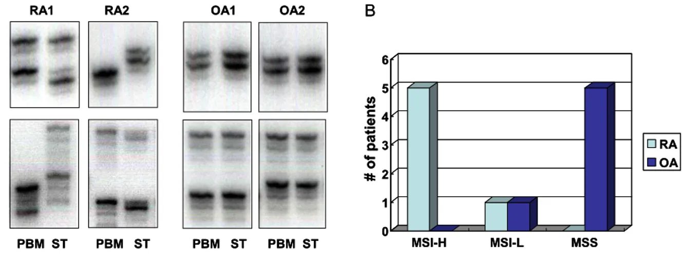
Figure 1. Microsatellite instability (MSI) in RA. A. Microsatellite instability (MSI) analysis using primers for two microsatellite loci, D5S346 and D17S250. Note that patterns for autologous peripheral blood cells (PBC) differ from synovial tissue (ST) from the same patient in rheumatoid arthritis (RA). In contrast, band patterns in osteoarthritis (OA) synovium matched autologous PBC. B. Frequencies of MSI in 6 patients with RA and OA for 5 standard microsatellite markers. MSI-H (MSI in 2 or more loci) is predominantly observed in RA synovium (5 cases), while most of OA exhibit MSS (no MSI) except 1 case showing MSI-L (MSI in 1 locus) (P=0.0025).
30 min on ice. After a second freeze thaw cycle, the membranes were gently broken by mechanical pi- petting (5). Protein concentrations were measured with the BSA protein assay kit (Bio-Rad Laboratories Inc., Hercules, CA). Protein samples from FLS lysates (30μg) or synovial tissue lysates (100μg) were sepa- rated by electrophoresis using a sodium dodecyl sulfate (SDS)-8% polyacrylamide gel, and transferred onto a nitrocellulose membrane at 140 mA in 25 mM Tris Cl (pH 8.3), 200 mM glycine, and 20% metha- nol. Membranes were blocked with 5% nonfat dry milk in phosphate-buffered saline-0.1% Tween 20 (PBS-T) for 1 hr. This was followed by overnight incubation at 4oC with mouse monoclonal anti-hMSH2 (Calbiochem-Novabiochem Corp., La Jolla, CA) at a dilution of 1:1,000, goat polyclonal anti-hMSH6 (Santa Cruz Biotechnology Inc., Santa Cruz, CA) at a dilution of 1:200, rabbit polyclonal anti-hMSH3 at a dilution of 1:1,000, mouse monoclonal anti- hMLH1 (Calbiochem-Novabiochem Corp.) at a dilu- tion of 1:250 and mouse monoclonal anti-hPMS2 (Pharmingen, San Diego, CA) at a dilution of 1:500.
Blots were washed with PBS-T and incubated for 2 hrs at room temperature with 1:2,000 dilution of a horseradish peroxidase-conjugated secondary anti- mouse IgG antibody (Santa Cruz Biotechnology Inc.) for hMSH2, hMLH1, and hPMS2; a 1:5,000 dilu- tion of anti-goat IgG antibody (Santa Cruz Biotech- nology Inc.) for hMSH6; and a 1:2,000 dilution of anti-rabbit IgG antibody (Santa Cruz Biotechnology Inc.) for hMSH3. The proteins were visualized using hydrogen peroxide and luminal as a substrate (Cell Signalling Technology Inc., Beverly, MA), with Kodak
X-Omat AR film (Eastman Kodak Co., Rochester, NY). To quantify the amount of each MMR proteins, each band densities were normalized to actin protein and digitized using NIH image analyzer software (Bethesda, MD).
Induction of oxidative stress in vitro. To induce oxidative stress, FLS were washed with serum-free DMEM and treated with 0.1 mM of H2O2 (Sigma Chemical Co., St. Louis, MO) for 1 hr or 1 mM of S-nitroso- N-acetylpenicillamine (SNAP; Sigma Chemical Co.) for 3 hrs. Cells were then washed with DMEM and allowed to recover in DMEM supplemented with 10% FCS. During recovery, cell lysates were collected at various time points (0, 6, 12, 24, 48 hr after the initial stress) for analysis and stored at -70oC.
Statistics. Data are expressed as mean±SEM. Statis- tical analysis was performed by Fisher's exact test for MSI analysis. Student's t-test was used to compare relative MMR expression in synovial tissues. Repeated measure ANOVA was used to perform the time point analysis for MMR regulation by RNS or ROS.
P values less than 0.05 were considered statistically significant.
Results
Presence of microsatellite instability in RA synovial tissue.
Our previous studies demonstrated that somatic mu- tations occur in the synovium of patients with RA but not OA (5). To determine genomic stability in RA synovial tissue, we evaluated the 5 microsatellite markers commonly used in colorectal cancer settings associated with increased reactive intermediates and MMR defects (18) (see Material and Methods). Since
0 0.1 0.2 0.3 0.4 0.5 0.6 0.7
MSH2 MSH6 MSH3
band densities
RA OA
*
*
MSH6 MSH3
actin MSH2
OA RA
1 2 3 4 5 6 1 2 3 4 5 6
A
B
0 0.1 0.2 0.3 0.4 0.5 0.6 0.7
MSH2 MSH6 MSH3
band densities
RA OA
*
*
*
*
MSH6 MSH3
actin MSH2
OA RA
1 2 3 4 5 6 1 2 3 4 5 6
A
B
Figure 2. Expression of DNA mismatch repair (MMR) proteins in synovial tissues. Synovial tissue extracts from RA (6 cases) and OA (6 cases) were evaluated by Western blot analysis. hMSH6 protein expression was significantly less in RA than in OA, while hMSH3 expression was higher in RA in comparison with OA. (*P<0.05). While there was an increase trend for hMSH2, the differences were not statistically significant.
normal
OA RA
1 2 1 2
hMSH6
hMSH2 hMLH1 PMS2 hMSH3
actin normal
OA RA
1 2 1 2
hMSH6
hMSH2 hMLH1 PMS2 hMSH3
actin
Figure 3. Expression of DNA mismatch repair enzymes in cultured fibroblast-like synoviocytes (FLS). RA (2 lines), OA (2 lines) and normal (1 line) FLS constitutively expressed hMSH2, hMSH6, hMSH3, hMLH1 and hPMS2. There were no consistent differences between the various cell lines.
microsatellite sequences are highly polymorphic among the population, we extracted DNA from peripheral blood cells in the same patients as autologous con- trols. Fig. 1A shows a representative MSI analysis of two loci, D5S346 and D17S250, for two RA and two OA patients. MSI was observed in all 6 RA synovial tissues tested, with 5/6 meeting criteria for MSI-H (MSI in 2 or more loci) and 1 patient meeting criteria for MSI-L (MSI in 1 locus)(see Fig. 1B). In contrast, no evidence of MSI was found in 5 of 6 OA samples, and only 1 OA synovium was MSI-L (P=0.004 for RA vs. OA).
MMR protein expression in synovial tissue. Based on our observation that RA synovium revealed MSI-H and the known relationship between MSI and MMR defects in hereditary non-polyposis colon cancer (HNPCC), we evaluated the expression of MMR enzymes in the same synovial tissues. As shown in Fig. 2, hMSH6 expression was significantly lower in RA synovial tissues compared with OA (P=0.02), while hMSH3 levels was significantly higher in RA than in OA (P=0.03). There was also a trend towards decreased hMSH2 levels in RA that did not reach statistical significance.
MMR protein expression in FLS. Since we observed differences in MMR expression in RA synovium, the expression of MMR proteins in cultured FLS was
A B
0 50 100 150 200 250 300 350
0 6 12 24 48
hour after NO treatment
% of control
hMSH2 hMSH6 hMSH3
*
** **
0 20 40 60 80 100 120 140 160
0 6 12 24 48
hour after H2O2treatment
% of control
* * *
hMSH2 hMSH6
A B
0 50 100 150 200 250 300 350
0 6 12 24 48
hour after NO treatment
% of control
hMSH2 hMSH6 hMSH3 hMSH2 hMSH6 hMSH3
*
** **
0 20 40 60 80 100 120 140 160
0 6 12 24 48
hour after H2O2treatment
% of control
* * *
hMSH2 hMSH6 hMSH2 hMSH6
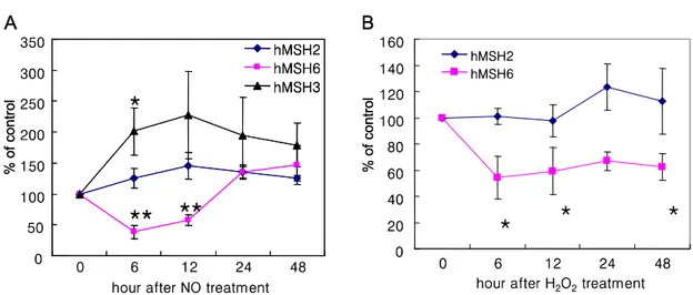
Figure 4. Effect of reactive nitrogen species (RNS) and reactive oxygen species (ROS) on MutS MMR proteins. S-nitroso- N-acetylpenicillamine (SNAP) was used as nitric oxide donor. Hydrogen peroxide was used as ROS stress. Cells were incubated with SNAP (1 mM, 3 hr) or hydrogen peroxide (0.1 mM, 1 hr), and then allowed to recover up to 48 hr. (A) SNAP treatment significantly decreased hMSH6 expression at 6-12 hr, while hMSH3 was increased 6 hr after NO stress. (B) Hydrogen peroxide also selectively decreased hMSH6 at 6 hr, 24 hr and 48 hr, while hMSH2 was not significantly changed. (*P<0.05, **P<0.01)
0 20 40 60 80 100 120 140
0 6 12 24
hour after NO treatment
% change of control
*
0.01 mM 0.1 mM 1.0 mM
0 20 40 60 80 100 120 140
0 6 12 24
hour after NO treatment
% change of control
*
0.01 mM 0.1 mM 1.0 mM 0.01 mM 0.1 mM 1.0 mM
Figure 5. Regulation of hMSH6 by NO in FLS. RA synoviocytes were treated with 0.01, 0.1, 1.0 mM of SNAP for 3 hr and then allowed to recover for various time intervals as described in the Material and Methods. hMSH6 suppression correlated with the concentration of SNAP exposure.
investigated. As shown in Fig. 3, the levels of multi- ple MMR proteins including hMSH2, hMSH3 and hMSH6 as well as a hMLH1 and hPMS2 were similar in RA, OA and normal cultured FLS. Therefore, the differences between OA and RA synovium is likely due to local environmental influences or variations in cellular composition rather than generalized suppres- sion of MMR expression.
Effect of RNS and ROS on MutS MMR proteins in FLS.
Because no differences were observed in basal MMR expression in OA and RA cultured FLS, we studied the role of various mediators on the components of the MMR enzyme complexes in vitro. No changes in hMSH2, -3, or -6 were observed in FLS that had been stimulated with IL-1 or TNF-alpha (data not shown). We then used an in vitro model of oxidative stress to determine if MMR expression in cultured RA FLS is regulated by RNS or ROS. FLS were cultured in the presence of a nitric oxide donor SNAP and the expression of MMR enzymes was determined by western blot analysis (n=5). As shown in Fig. 4, pre-treatment of FLS with SNAP for 3 hr significantly decreased hMSH6 expression 6 to 12 hr after exposure (38% of medium control at 6 hr, P=0.002). The hMSH3 levels increased during the same period of time (200.9% of medium control at 6 hr, P=0.047), indicating that the effect on hMSH6 was not related to non-specific toxicity (Fig. 4A). The hMSH2 expression was not significantly changed although there was a trend towards a modest increase after RNS stress. Alterations in MMR protein expres- sion was transient, and expression returned to base- line within 24~48 hr. No differences were observed
between RA and OA FLS with regard to their re- sponse to SNAP (data not shown). Hydrogen per- oxide treatment also modestly decreased hMSH6 and the effect was more prolonged (Fig. 4B, P<0.05).
hMSH2 was not significantly changed by ROS ex- posure. None of the conditions described caused significant cell death as determined by cell staining for viable cells. These data indicate that the MMR pattern observed in RA synovial tissue, i.e., low hMSH6 and high hMSH3, was reproduced in cul- tured FLS after oxidative stress.
Effect of dose and duration of NO exposure on hMSH6 expression in FLS. Kinetics and dose response experi-
Control 0.5hr 1hr 3hr NO incubation time
hMSH6 Actin 0.5hr 1hr 3hr 0.5hr 1hr 3hr
6 hr 12 hr 24 hr
Control 0.5hr 1hr 3hr NO incubation time
hMSH6 Actin 0.5hr 1hr 3hr 0.5hr 1hr 3hr
6 hr 12 hr 24 hr
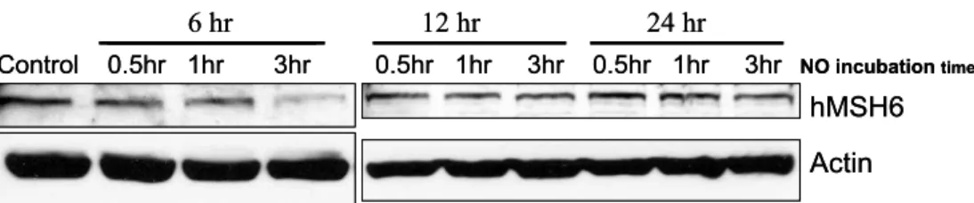
Figure 6. Kinetics of hMSH6 regulation by NO. RA FLS were treated with 1 mM SNAP for 0.5~3 hr and then analyzed hMSH6 over 24 hr. hMSH6 expression was determined by western blot analysis.
ments were then performed to characterize the ef- fects of RNS on MMR expression. To evaluate the dose of NO required to suppress hMSH6, RA FLS cell lines (n=3) were exposed to various concen- trations of SNAP (0.01, 0.1, 1.0 mM) for 3 hr. Mini- mal or no effects were observed for the lower con- centrations, while 1 mM clearly suppressed hMSH6 (P<0.05) (Fig. 5). We then treated RA FLS cell lines with 1 mM of SNAP for 0.5 to 3 hr, and analyzed hMSH6 protein levels. As illustrated in Fig. 6, suppression of hMSH6 was correlated with the dura- tion of exposure to SNAP (P<0.05).
Discussion
Genotoxic adducts, especially ROS and RNS, are released as a result of cell activation and can damage genomic DNA. One such product, nitric oxide (NO), is especially prevalent in chronic inflammatory dis- eases like ulcerative colitis (29) and rheumatoid arthritis (30-32). In the former, local NO production leads to the accumulation of somatic mutations that can ultimately lead to neoplastic transformation (33).
DNA damage and mutation have also been docu- mented in the synovial intimal lining in rheumatoid synovium (34). To determine the mechanisms of DNA damage in inflamed tissues, we evaluated the rheumatoid synovium for broader evidence of muta- genesis. Based on an extensive literature in colon can- cer and inflammatory bowel disease, we examined the RA synovial tissue genome for evidence of micro- satellite instability (MSI). Microsatellites are tandem repeated sequences in the DNA that are primarily (but not exclusively) located in non-coding regions.
MSI is defined as an microsatellite insertion or dele- tion mutation in a target tissue or tumor compared with an autologous control (usually peripheral blood) (18). Many studies demonstrate an association between MSI, DNA damage, and development of neoplasia (19,35,36).
Criteria for high and low MSI phenotypes (MSI-H and MSI-L, respectively) and microsatellite stable phenotype (MSS) have been developed for colon tumor loci (18). Five specific microsatellites can be evaluated for evidence of mutations and the number
of mutations quantified. The phenotypes are: 1) MSI-H, with mutations in ≥40% of loci; 2) MSI-L with mutations in ≤20% of loci; and 3) MSS, with no microsatellite mutations. Our initial studies were performed on RA and OA synovial tissues and used the same loci validated for colon inflammation since the mechanisms of mutagenesis are similar. These experiments demonstrated clear evidence of the MSI- H genotype in RA. In contrast, most OA samples were either MSI-L or MSS. The presence of micro- satellite mutations in RA is consistent with previous studies demonstrating somatic mutations and in- creased DNA strandbreaks (5,34). One previous study suggested that microsatellites were stable in cultured RA synoviocytes, although many of these samples were derived from patients with less severe disease at the time of synovectomy (37). Our study focused on end stage arthritis requiring total joint re- placement.
The mechanisms of MSI have been evaluated in other settings, especially in hereditary non-polyposis colon cancer (HNPCC). These patients have many mutations in the colon and frequently develop colon cancer in early adulthood. The etiology of the mutagenic propensity in HNPCC is defective DNA mismatch repair (MMR) enzymes (22,25,35). This complex system involves many repair enzymes, in- cluding two key heterodimeric complexes known as MutSα and MutSβ. hMSH2 is a common subunit for both of dimers and is the most common defec- tive gene in HNPCC (22). It can pair with hMSH6 to form MutSα while hMSH3 and hMSH2 form MutSβ. MutSα mainly repairs of single base mis- pairs, while MutSβ repairs larger insertion/deletion mispairs (38). Since MSH2 is the common com- ponent, balance between repair of single base misma- tches or longer insertions/deletions depends on the ratio of MSH6 and MSH3. In cells that over-express MSH3, the available MSH2 protein is sequestered into MutS. This leads to degradation of the part- nerless MSH6 and defective repair of single base mismatches (39). Therefore, MMR deficiency can arise not only through mutation or transcriptional silencing of MMR genes, but also as a result of an
imbalance in the relative amounts of the MSH3 and MSH6 proteins.
Based our observation that RA synovium is MSI- H, we assessed the expression of MMR proteins in this tissue. hMSH2 and another MMR enzyme, hMLH1, are reportedly expressed by intimal lining cells in RA as determined by immunohistochemistry (37). However, there is no information on the other key enzymes or their relative amounts compared with OA. Surprisingly, decreased hMSH6 and, to a lesser extent, MSH2 protein was observed in the rheumat- oid synovium compared with OA. However, hMSH3 expression was significantly higher in RA. This pat- tern of MMR enzyme expression provides a potential explanation for the type of mutation observed in RA, where single base pair substitutions account for the vast majority of abnormalities (5).
The MMR differences between OA and RA syno- vium led us to explore the expression and regulation of these enzymes in FLS. Similar levels of MMR pro- teins were expressed in RA and OA FLS, indicating that the abnormalities observed in the tissue were not simply due to a generalized suppression of hMSH6 or other enzymes in RA as occurs in HNPCC. The pattern of MMR expression changed when FLS were stressed by RNS, with induction of hMSH3 and sup- pression of hMSH6. Suppression of hMSH6 was dose dependent and varied directly with the duration of NO exposure. The fact that other MMR proteins or housekeeping genes like actin were unaffected indicates that hMSH6 suppression was not due to non-specific toxicity. The effects on MMR expression were transient in vitro since hMSH3 and hMSH6 expression returned to normal levels after 24 hrs.
While the mechanism of MMR regulation is currently unknown, the protein levels in other cells correlate with mRNA expression and suggests that trans- criptional regulation is likely to participate (40).
The cultured FLS studies showed that the pattern of MMR expression in RA synovium (high hMSH3/
low hMSH6) was reproduced in synoviocytes that were exposed to genotoxic stimuli. This tissue culture model obviously does not completely mimic the situation in chronically inflamed synovium, and many other cell types can contribute to MSI or MMR protein expression in RA. Nevertheless, the levels of RNS and ROS exposure that alter MSH expression are relevant since similar concentrations can be achieved in stressed cells (32,41). Suppression of hMSH6 appears to be paradoxical at first. However, it might represent a mechanism that protects the cell from major DNA damage while permitting single base pair changes. This could provide a short-term survival benefits to the organism by shifting the balance from MutSα to MutSβ. It also might serve
as a host mechanism for establishing a "mutator"
phenotype, which can have additional survival be- nefits (42).
Hyperoxide can also decrease expression of MMR repair enzymes like hMSH6 in certain human tumor cell lines (26). Suppression of MutSα in erythroleu- kemia cells by reactive oxygen suppresses DNA repair activity as determined by functional assays.
This effect can be reversed by the addition of recombinant MMR enzymes to the cell extracts. An analogous situation occurs in non-neoplastic tissues like rheumatoid synovium and primary cells derived from the tissue after exposure to RNS. These data suggest that suppression of hMSH6 in vivo can lead to the accumulation of mutations due to functional impairment of the MMR system.
Exposure to reactive species, especially RNS, can be directly mutagenic and increase the likelihood of single base mismatches in RA through oxidative dea- mination (43). In addition, suppression of MMR me- chanisms could contribute to a "hypermutable" state and increase the likelihood for the accumulation of somatic mutations (17). This situation is probably relevant to chronic inflammation in many sites. In RA, for instance, ROS production is increased by the hypoxic environment mediated by activated leuko- cytes, ischemia-reperfusion injury, an increased meta- bolic rate, and reduced capillary density (44,45).
Expression of iNOS is markedly increased in RA, especially in the synovial intimal linings (31). Local NO production results in nitrosylation of synovial proteins and can produce mutagenic nucleotide ad- ducts (43). The combination of these influences over many years can, therefore, lead to somatic mutations in chronic inflammatory diseases.
Relaxation of MMR in stressed synovium has parallels in other diseases. For instance, MSI has been described in ulcerative colitis which also may be associated with functional MMR suppression (46). RA is the first chronic inflammatory disease associated with both MSI, somatic mutations of key genes, and suppression of DNA repair mechanisms but does not lead to neoplastic disease. Instead, this process might alter the natural history of RA and cause local inv- asion and an aggressive FLS phenotype. It also pro- vides potential therapeutic targets to modify the long-term destructive potential of RA synovium. By limiting local RNS or ROS production, mutagenesis can potentially be minimized. Alternatively, replacing or augmenting MMR expression could mitigate the genotoxicity in the synovium or in other chronically inflamed sites.
References
1. Cifone MA, Fidler IJ: Correlation of patterns of anchorage-
independent growth with in vivo behavior of cells from a murine fibrosarcoma. Proc Natl Acad Sci USA 77;1039- 1043, 1980
2. Lafyatis R, Remmers EF, Roberts AB, Yocum DE, Sporn MB, Wilder RL: Anchorage-independent growth of syno- viocytes from arthritic and normal joints. Stimulation by exogenous platelet-derived growth factor and inhibition by transforming growth factor-beta and retinoids. J Clin Invest 83;1267-1276, 1989
3. Muller-Ladner U, Kriegsmann J, Gay RE, Gay S: Onco- genes in rheumatoid arthritis. Rheum Dis Clin North Am 21;675-690, 1995
4. Muller-Ladner U, Kriegsmann J, Franklin BN, Matsumoto S, Geiler T, Gay RE, Gay S: Synovial fibroblasts of patients with rheumatoid arthritis attach to and invade normal human cartilage when engrafted into SCID mice.
Am J Pathol 149;1607-1615, 1996
5. Firestein GS, Echeverri F, Yeo M, Zvaifler NJ, Green DR:
Somatic mutations in the p53 tumor suppressor gene in rheumatoid arthritis synovium. Proc Natl Acad Sci U S A 94;10895-10900, 1997
6. Inazuka M, Tahira T, Horiuchi T, Harashima S, Sawabe T, Kondo M, Miyahara H, Hayashi K: Analysis of p53 tumour suppressor gene somatic mutations in rheumatoid arthritis synovium. Rheumatology (Oxford) 39;262-266, 2000
7. Kullmann F, Judex M, Neudecker I, Lechner S, Justen HP, Green DR, Wessinghage D, Firestein GS, Gay S, Scholmerich J, Muller-Ladner U: Analysis of the p53 tumor suppressor gene in rheumatoid arthritis synovial fibroblasts. Arthritis Rheum 42;1594-1600, 1999
8. Reme T, Travaglio A, Gueydon E, Adla L, Jorgensen C, Sany J: Mutations of the p53 tumour suppressor gene in erosive rheumatoid synovial tissue. Clin Exp Immunol 111;353-358, 1998
9. Cannons JL, Karsh J, Birnboim HC, Goldstein R: HPRT- mutant T cells in the peripheral blood and synovial tissue of patients with rheumatoid arthritis. Arthritis Rheum 41;1772-1782, 1998
10. Roivainen A, Jalava J, Pirila L, Yli-Jama T, Tiusanen H, Toivanen P: H-ras oncogene point mutations in arthritic synovium. Arthritis Rheum 40;1636-1643, 1997
11. Suzuki H, Harpaz N, Tarmin L, Yin J, Jiang HY, Bell JD, Hontanosas M, Groisman GM, Abraham JM, Meltzer SJ: Microsatellite instability in ulcerative colitis-associated colorectal dysplasias and cancers. Cancer Res 54;4841- 4844, 1994
12. Kern SE, Redston M, Seymour AB, Caldas C, Powell SM, Kornacki S, Kinzler KW: Molecular genetic profiles of colitis-associated neoplasms. Gastroenterology 107;420- 428, 1994
13. Brentnall TA, Crispin DA, Bronner MP, Cherian SP, Hueffed M, Rabinovitch PS, Rubin CE, Haggitt RC, Boland CR: Microsatellite instability in nonneoplastic mu- cosa from patients with chronic ulcerative colitis. Cancer Res 56;1237-1240, 1996
14. Boland CR: Roles of the DNA mismatch repair genes in colorectal tumorigenesis. Int J Cancer 69;47-49, 1996 15. Boland CR, Ricciardiello L: How many mutations does it
take to make a tumor? Proc Natl Acad Sci USA 96;14675- 14677, 1999
16. Loeb LA, Springgate CF, Battula N: Errors in DNA replication as a basis of malignant changes. Cancer Res
34;2311-2321, 1974
17. Loeb LA: Mutator phenotype may be required for multi- stage carcinogenesis. Cancer Res 51;3075-3079, 1991 18. Boland CR, Thibodeau SN, Hamilton SR, Sidransky D,
Eshleman JR, Burt RW, Meltzer SJ, Rodriguez-Bigas MA, Fodde R, Ranzani GN, Srivastava S: A National Cancer Institute Workshop on Microsatellite Instability for cancer detection and familial predisposition: development of in- ternational criteria for the determination of microsatellite instability in colorectal cancer. Cancer Res 58;5248-5257, 1998
19. Boland CR, Sato J, Saito K, Carethers JM, Marra G, Laghi L, Chauhan DP: Genetic instability and chromosomal aberrations in colorectal cancer: a review of the current models. Cancer Detect Prev 22;377-382, 1998
20. Thibodeau SN, Bren G, Schaid D: Microsatellite instability in cancer of the proximal colon. Science 260;816-819, 1993
21. Ionov Y, Peinado MA, Malkhosyan S, Shibata D, Perucho M: Ubiquitous somatic mutations in simple repeated se- quences reveal a new mechanism for colonic carcino- genesis. Nature 363;558-561, 1993
22. Fishel R, Lescoe MK, Rao MR, Copeland NG, Jenkins NA, Garber J, Kane M, Kolodner R: The human mutator gene homolog MSH2 and its association with hereditary nonpolyposis colon cancer. Cell 75;1027-1038, 1993 23. Leach FS, Nicolaides NC, Papadopoulos N, Liu B, Jen
J, Parsons R, Peltomaki P, Sistonen P, Aaltonen LA, Nystrom-Lahti M: Mutations of a mutS homolog in hereditary nonpolyposis colorectal cancer. Cell 75;1215- 1225, 1993
24. Papadopoulos N, Nicolaides NC, Wei YF, Ruben SM, Carter KC, Rosen CA, Haseltine WA, Fleischmann RD, Fraser CM, Adams MD, Venter JC, Hamilton SR, Peter- sen GM, Watson P, Lynch HT, Peltomaki P, Mecklin J-P, Chapelle ADL, Kinzler KW, Vogelstein B: Mutation of a mutL homolog in hereditary colon cancer. Science 263;
1625-1629, 1994
25. Nicolaides NC, Papadopoulos N, Liu B, Wei YF, Carter KC, Ruben SM, Rosen CA, Haseltine WA, Fleischmann RD, Fraser CM, Adams MD, Venter JC, Dunlop MG, Hamilton SR, Petersent GM, Chapelle ADL, Vogelstein B, Kinzler KW: Mutations of two PMS homologues in hereditary nonpolyposis colon cancer. Nature 371;75-80, 1994
26. Chang CL, Marra G, Chauhan DP, Ha HT, Chang DK, Ricciardiello L, Randolph A, Carethers JM, Boland CR:
Oxidative stress inactivates the human DNA mismatch repair system. Am J Physiol Cell Physiol 283;C148-154, 2002
27. Arnett FC, Edworthy SM, Bloch DA, McShane DJ, Fries JF, Cooper NS, Healey LA, Kaplan SR, Liang MH, Luthra HS, Medsger TA, Mitchelle DM, Neustadt DH, Pinals RS, Schaller JG, Sharp JT, Wilder RL, Hunder GG.: The American Rheumatism Association 1987 revised criteria for the classification of rheumatoid arthritis. Arthritis Rheum 31;315-324, 1988
28. Alvaro-Gracia JM, Zvaifler NJ, Firestein GS: Cytokines in chronic inflammatory arthritis. V. Mutual antagonism between interferon-gamma and tumor necrosis factor- alpha on HLA-DR expression, proliferation, collagenase production, and granulocyte macrophage colony-stimulat- ing factor production by rheumatoid arthritis synoviocytes.
J Clin Invest 86;1790-1798, 1990
29. Singer, II, Kawka DW, Scott S, Weidner JR, Mumford RA, Riehl TE, Stenson WF: Expression of inducible nitric oxide synthase and nitrotyrosine in colonic epithelium in inflammatory bowel disease. Gastroenterology 111;871- 885, 1996
30. Sakurai H, Kohsaka H, Liu MF, Higashiyama H, Hirata Y, Kanno K, Saito I, Miyasaka N: Nitric oxide production and inducible nitric oxide synthase expression in inflam- matory arthritides. J Clin Invest 96;2357-2363, 1995 31. Grabowski PS, Wright PK, Van't Hof RJ, Helfrich MH,
Ohshima H, Ralston SH: Immunolocalization of inducible nitric oxide synthase in synovium and cartilage in rheuma- toid arthritis and osteoarthritis. Br J Rheumatol 36;651- 655, 1997
32. Hilliquin P, Borderie D, Hernvann A, Menkes CJ, Ekin- djian OG: Nitric oxide as S-nitrosoproteins in rheumatoid arthritis. Arthritis Rheum 40;1512-1517, 1997
33. Hussain SP, Amstad P, Raja K, Ambs S, Nagashima M, Bennett WP, Shields PG, Ham AJ, Swenberg JA, Marrogi AJ, Harris CC: Increased p53 mutation load in noncan- cerous colon tissue from ulcerative colitis: a cancer-prone chronic inflammatory disease. Cancer Res 60;3333-3337, 2000
34. Firestein GS, Yeo M, Zvaifler NJ: Apoptosis in rheu- matoid arthritis synovium. J Clin Invest 96;1631-1638, 1995
35. Dietmaier W, Wallinger S, Bocker T, Kullmann F, Fishel R, Ruschoff J: Diagnostic microsatellite instability: defini- tion and correlation with mismatch repair protein expres- sion. Cancer Res 57;4749-4756, 1997
36. Wong NA, Harrison DJ: Colorectal neoplasia in ulcerative colitis-recent advances. Histopathology 39;221-234, 2001 37. Kullmann F, Widmann T, Kirner A, Justen HP, Wessing-
hage D, Dietmaier W, Ruschoff J, Gay S, Scholmerich J, Muller-Ladner U: Microsatellite analysis in rheumatoid arthritis synovial fibroblasts. Ann Rheum Dis 59;386-389, 2000
38. Kolodner RD, Marsischky GT: Eukaryotic DNA mis- match repair. Curr Opin Genet Dev 9;89-96, 1999 39. Marra G, Iaccarino I, Lettieri T, Roscilli G, Delmastro P,
Jiricny J: Mismatch repair deficiency associated with overexpression of the MSH3 gene. Proc Natl Acad Sci USA 95;8568-8573, 1998
40. Chang DK, Ricciardiello L, Goel A, Chang CL, Boland CR: Steady-state regulation of the human DNA mismatch repair system. J Biol Chem 275;18424-18431, 2000 41. Borderie D, Le Marechal H, Ekindjian OG, Hernvann A:
Nitric oxide modifies glycolytic pathways in cultured human synoviocytes. Cell Biol Int 24;285-289, 2000 42. Chang DK, Metzgar D, Wills C, Boland CR: Micro-
satellites in the eukaryotic DNA mismatch repair genes as modulators of evolutionary mutation rate. Genome Res 11;1145-1146, 2001
43. Wiseman H, Halliwell B: Damage to DNA by reactive oxygen and nitrogen species: role in inflammatory disease and progression to cancer. Biochem J 313(Pt 1);17-29, 1996
44. Mapp PI, Grootveld MC, Blake DR: Hypoxia, oxidative stress and rheumatoid arthritis. Br Med Bull 51;419-436, 1995
45. Tak PP, Zvaifler NJ, Green DR, Firestein GS: Rheuma- toid arthritis and p53: how oxidative stress might alter the course of inflammatory diseases. Immunol Today 21;78- 82, 2000
46. Jackson AL, Chen R, Loeb LA: Induction of microsatellite instability by oxidative DNA damage. Proc Natl Acad Sci USA 95;12468-12473, 1998