• 검색 결과가 없습니다.

Schwannomas, arising from schwann cells of a nerve sheath, are the most common benign tumors of the peripheral nerves. They are also known as neurile momas, and malignant transformation is rare.

N/A
N/A
Protected

Academic year: 2021

Share "Schwannomas, arising from schwann cells of a nerve sheath, are the most common benign tumors of the peripheral nerves. They are also known as neurile momas, and malignant transformation is rare."

Copied!
5
0
0

로드 중.... (전체 텍스트 보기)

전체 글

(1)

Introduction

Schwannomas, arising from schwann cells of a nerve sheath, are the most common benign tumors of the peripheral nerves. They are also known as neurile momas, and malignant transformation is rare.

1-3

Benign solitary

schwannomas involving upper limb account for 73%, and were common in brachial plexus (40%), ulnar nerve (20%), and median nerve (9%).

4

Schwannoma is slow growing mass, and it does not cause pain or neurologic sign, unless the tumor is large enough. We reported an unusual case that schwannoma of median nerve at upper arm mimicked symptoms of ulnar neuropathy, which was diagnosed by Electromyography (EMG) and Magnetic resonance imaging (MRI).

Case Report

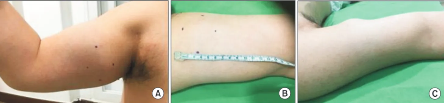
A 42-year-old man complained of sudden pain on his

근위부 정중신경에 발생한 신경초종에 의한 척골신경 압박에 대한 증례보고

한미향

1

, 오자영

1

, 윤정윤

1

, 이상욱

2

, 김재민

1

가톨릭대학교 의과대학 인천성모병원 1재활의학과, 2정형외과

Schwannoma Involving the Proximal Median Nerve with Compression of the Ulnar Nerve

Mi-Hyang Han

1

, Ja-Young Oh

1

, Jung-Yoon Yoon

1

, Sang-Uk Lee

2

, Jae Min Kim

1

Departments of

1

Rehabilitation Medicine and

2

Orthopedic Surgery, Incheon St. Mary’s Hospital, College of Medicine, The Catholic University of Korea, Incheon, Korea

Received May 31, 2016

Revised (1st) July 14, 2016, (2nd) August 12, 2016 Accepted August 12, 2016

Corresponding Author: Jae Min Kim

Department of Rehabilitation Medicine, Incheon St. Mary’s Hospital, College of Medicine, The Catholic University of Korea, 56 Dongsu-ro, Bupyeong-gu, Incheon 21431, Korea

Tel: 82-32-280-5207, Fax: 82-32-280-5040, E-mail: jaeminmd@gmail.com

Schwannomas, arising from Schwann cells of a nerve sheath, are the most common benign tumors of the peripheral nerves. Schwannoma cause symptoms including paresthesia and motor weakness depending on involved nerves or pain after growing enough to compress surrounding soft tissue. Here in this case, schwannoma was originated from the median nerve at the mid humerus, but the tumor did not cause median neuropathic symptom but ulnar neuropathic symptom by compressing the ulnar nerve.

Key Words: schwannoma, median nerve, inching study

Copyright © by Korean Association of EMG Electrodiagnostic Medicine

This is an Open Ac cess article distributed under the terms of the Creative Commons Attribution Non-Commercial License (http://creativecommons.org/licenses/by-nc/4.0) which permits unrestricted non-commercial use, distribution, and reproduction in any medium, provided the original work is properly cited.

ISSN 1229-6066 https://doi.org/10.18214/jkaem.2016.18.2.66 J Korean Assoc EMG Electrodiagn Med 18(2):66-70, 2016

J Korean Assoc

Electrodiagn Med EMG

(2)

right wrist and tingling sensation on his right 4th and 5th fingers. He also presented with palpable mass on his right upper arm. In his history, when he exercised at the gym 10 days ago, suddenly he felt pain in his right upper arm, wrist and was aware of palpable mass on his medial upper arm. On physical examination, there was sensory disturbance in volar fifth and medial fourth digits of right hand, but motor weakness was not shown. There was painless oval shaped palpable mass about 4 × 3 cm at the medial side of middle humerus (Fig. 1). Tinel sign and Phalen’s signs at the wrist and elbow were negative but, percussion over the mass elicited tingling sensation on medial hand.

Considering history of sudden pain and palpable

mass after exercise, we suspected that the muscle tear like biceps muscle injury at first, and torn muscle could cause the symptoms of tingling sensation of 4

th

and 5

th

fingers by compressing the nearby ulnar nerve.

To confirm the injury of ulnar nerve, compatible with symptoms, nerve conduction study (NCS) and needle EMG were performed. In NCS findings, the motor and sensory NCSs of ulnar nerve were normal, but the sensory nerve action potential (SNAP) of median nerve showed decreased amplitude up to 40% compared with the left side and normal latency of SNAP (Table 1). To determine the location of mass and involved nerve area, inching study on median nerve of upper arm segment was performed. The decreased amplitude

A B C

Fig. 1. The oval shaped mass about 4 × 3 cm at the medial side of middle humerus, right. (A) Medial view with arm flexion posture, (B) Medial view with arm extension posture, (C) Superior oblique view.

Table 1. Result of the Nerve Conduction Studies

Nerve Stimulation

site

Recording site

Latency Amplitude CV (m/s)

Rt Lt Rt Lt Rt Lt

Median (S) Wrist Digit III 2.90 2.90 27.3 44.4 - -

Ulnar (S) Wrist Digit V 3.10 3.30 20.2 16.0 - -

Ulnar (M) Wrist ADM 2.20 2.55 9.2 9.9 - -

B. Elbow 6.15 6.45 8.7 7.5 55.7 61.5

A. Elbow 7.50 8.7 74.1

Axilla 9.45 8.1 74.4

Median (M) Wrist APB 2.6 2.75 9.8 9.6 - -

Elbow 7.0 6.75 9.6 9.5 56.8 58.7

Above elbow 2 cm 7.35 9.6 57.1

Above elbow 4 cm 1.75 9.0 50.0

Above elbow 6 cm 8.1 8.8 57.1

Above elbow 8 cm 8.5 8.5 50.0

Above elbow 10 cm* 8.85 8.4 57.1

Above elbow 12 cm

9.7 4.7 23.5

Axilla 11.1 8.4 64.3

All motor latencies are onset latencies and all sensory latencies are peak latencies Amplitudes are measured in millivolt (mV, motor) and in microvolt (μV, sensory)

M, motor study; S, sensory study; CV, conduction velocity; Rt, right; Lt, left; ADM, abductor digiti minimi; APB, abductor pollicis brevis

*Before the mass site.

Along the mass site

(3)

and conduction velocity of compound motor action

potential (CMAP) was founded between 10 cm and 12 cm proximal to the elbow, where the mass was palpable (Table 1, Fig. 2). In needle EMG, there was normal configuration without abnormal spontaneous activity in muscles that innervated by median, ulnar nerves.

Based on the results of EMG, MRI was performed to identify the mass and the acute injury of median nerve.

The MRI exhibited that 4 × 3.3 × 4.9 cm well circum- scribed round mass was placed at the mid shaft level of right humerus, and it was observed along the median nerve. The rim of round lesion was enhanced and the center of the lesion was low signal in T1 weighted sequence after contrast medium administration (Fig.

3), suggesting the schwannoma. After excluding other causes of symptoms and definite nerve injury,

A B

Fig. 3. The MRI images (A) T1 enhance Axial view (B) T1 enhance Coronal view of schwannoma of the median nerve. About 4 × 3.3 × 4.9 cm cystic schwannoma with hemorrhagic change is shown along median nerve between biceps and brachialis muscle at right mid humerus level. MRI findings demonstrated that schwannoma (black arrows) com pressed ulnar nerve (white arrow).

A B C

Fig. 4. (A) Well capsulated oval shaped mass was attached to the median nerve (black arrow). (B) Median nerve was preserved after excision of the mass (white arrow). (C) The size was 4 × 3.3 × 4.9 cm.

Fig. 2. Compound motor action potentials of medina nerve through inching study, 1

st

line; wirst, 2

nd

line; elbow, 3

rd

line; Above elbow 2 cm, 4

th

line; Above elbow 4 cm, 5

th

line; Above elbow 6 cm, 6

th

line;

Above elbow 8 cm, 7

th

line; Above elbow 10 cm (before the mass), 8

th

line; Above elbow 12 cm (along the mass), 9

th

line; Axilla.

(4)

the patient underwent surgery to remove the mass. A curvilineal skin incision was outlined over the palpable mass surface. The mass was founded between biceps brachii and brachialis muscles, and encapsulated with median fascicles. The ulnar nerve was compressed by the mass, neurolysis was performed. The nerve fascicles surrounding mass and the median nerve were dissected, and the mass was totally extracted (Fig. 4). Histopathologic examination confirmed the schwannoma with microcalcifications of the median nerve. After operation, the patient’s symptoms, that mimic ulnar neuropathy, resolved.

Discussion

Schwannomas are encapsulated benign nerve sheath tumor, present as a solitary lesion of peripheral nerves.

The prevalence of schwannoma was the highest in adults between 20 and 50 years of age, regardless of gender.

3

The incidence of upper limb schwannomas accounts for 12 to 19% and is higher in the flexor surface of the upper limb, because of the concentration of nerve.

5

The common origins of nerves are reported as ulnar, median and radial nerves in order.

6

Clinically, most of the schwannomas could be diagnosed on physical examination and symptoms.

The typical symptoms of schwannoma are the pain, paresthesia and motor weakness with movable mass in the longitudinal direction along the involved nerve. If the affected nerve is motor nerve, the weakness can be prominent. The paresthesia of involved nerve region after percussion of the tumor is a high predictive value (87.5% sensitivity) for schwannoma.

5

However, in clinical grounds, diagnosis of schwannoma can be difficult. Many physicians are likely to misdiagnose focal neurologic deficits like paresthesia and motor weakness as entrapment syndrome of peripheral nerve.

In addition, the slow growth pattern of schwannoma makes nervous adaptation to the increased volume.

Thus, schwannoma presents as asymptomatic swelling and can be detected delayed before other symptoms

appear.

3

To complement the lack of specific clinical sign, EMG, MRI, and USG (ultrasonography) are needed to diagnose. For the confirmation, histopathologic examination of the neoplasm is essential. EMG may reveal delayed latency and diminished or absent amplitudes in nerve conduction studies.

3

This is because the schwannoma, consisted of nerve fascicle, can cause delay of nerve conduction and reduction of axonal potentials. MRI gives information about tumor morphology, size, extent, anatomical location and relationship of peripheral nerve and surrounding soft tissue. In MRI, schwannomas show low-intense signals on T1-seight images and hyperintense signals on T2- weighted images. And the findings called as target sign, the central low signal intensity of fibrous component and peripheral high signal intensity of myxomatous elements on T2-weighted images, is the typical MRI image of schwannoma.

5

In USG, schwannoma is eccentrically located to the nerve fibers and appears as hypoechoic lesion with regular margin. The USG offers images depend on dynamic position of upper extremity during active and passive flexion and extension that can be useful to detect lesion which presents different symptoms according to posture.

1

Our case is unusual, in that the neurological symp- toms are not coincidence with corresponding nerve.

The patient presented pain and tingling sensation of

volar fifth and medial fourth digits, which is the sensory

distribution of ulnar nerve. But, in NCS findings, there

was decreased amplitude of SNAP in right median

nerve stimulating at wrist. In inching study, there was

decreased amplitude and conduction velocity of CMAP

in right median nerve stimulating at between 10 cm and

12 cm proximal to the elbow, which is because the mass

effect of schwannoma restricts supramaximal stimulation

of median nerve. The MRI showed the exact location, size

and the relationship with median nerve of mass. Through

surgery, we founded the mass encapsulated with median

fascicles and compressing the ulnar nerve. His symptoms

were due to compression of ulnar nerve because of the

(5)

large size of the tumor. But, there was no abnormality

in ulnar nerve in NCS and EMG studies, which may be because the mass effect of schwannoma was not sufficient to make neuropraxia of ulnar nerve.

Median nerve schwannomas are rare and often noticed incidentally as asymptomatic palpable mass with or without symptoms of median nerve neuropathy.

1,7,8

According to location of schwannoma in median nerve, the symptoms are various and easily to be confused with other lesions. A schwannoma at the wrist can be easily misdiagnosed as carpal tunnel syndrome. There are some cases that lesion at the elbow caused pronator syndrome by compressing surrounding soft tissues.

9

A proximal median schwannoma can cause symptoms of ulnar neuropathy because of near position. There is one case that show symptom like ulnar neuropathy caused by proximal median schwannoma, which is similar with our case. This case showed a single proximal median nerve schwannoma made median neuropathy, including ulnar neuropathy by compressing the nerve and this was confirmed by electrodiagnostic findings.

7

The need of operative treatment depends on expected improvement of pain and neurologic symptoms caused by space-occupying mass or cosmetic reasons. The en bloc resection that spares the adjacent nerve is the treatment of choice of schwannoma and enucleation of the involving nerve is essential to save its function.

10

In our case, the neurolysis and dissection were performed to save the function of ulnar and median nerve. After this surgery, patient’s symptom such as tingling sensation and pain of his hand was resolved.

In this case, the patient complained about palpable mass on upper arm and tingling sensation on his right hand of the ulnar nerve branch domain. However, EMG and MRI showed that the cause is not the ulnar

nerve but the median nerve. During the surgery, it was identified schwannoma of the median nerve, and the mass compressed the ulnar nerve, which might cause the symptom. Our findings suggest that EMG and MRI are helpful to find the exact cause of symptoms and to determine the treatment plan.

References

1. Mariottini A, Carangelo B, Peri G, Tacchini D, Mormouras V, Muya M, et al.: Schwannoma of median nerve at the elbow.

Case report and short review of the literature. G Chir 2011:

32: 55-58

2. Malizos K, Ioannou M, Kontogeorgakos V: Ancient schwan- noma involving the median nerve: a case report and review of the literature. Strategies Trauma Limb Reconstr 2013: 8:

63-66

3. Boufettal M, Azouz M, Rhanim A, Abouzahir M, Mahfoud M, Bardouni AE, et al.: Schwannoma of the median nerve:

diagnosis sometimes delayed. Clin Med Insights Case Rep 2014: 7: 71-73

4. Knight DM, Birch R, Pringle J: Benign solitary schwannomas:

a review of 234 cases. J Bone Joint Surg Br 2007: 89: 382-387 5. Tang CY, Fung B, Fok M, Zhu J: Schwannoma in the upper

limbs. Biomed Res Int 2013: 2013: 167196

6. Gosk J, Gutkowska O, Urban M, Wnukiewicz W, Reichert P, Ziółkowski P: Results of surgical treatment of schwannomas arising from extremities. Biomed Res Int 2015: 2015: 547926 7. DiTrapani R, Rubin DI: An unusual presentation of a

proximal median nerve schwannoma. Muscle Nerve 2012:

46: 983-984

8. Kang HJ, Shin SJ, Kang ES: Schwannomas of the upper extremity. J Hand Surg Br 2000: 25: 604-607

9. Afshar A: Pronator Syndrome Due to Schwannoma. J Hand Microsurg 2015: 7: 119-122

10. Hubert J, Landes G, Tardif M: Schwannoma of the median

nerve. J Plast Surg Hand Surg 2013: 47: 75-77

수치

Fig. 1.	The	oval	shaped	mass	about	4	×	3	cm	at	the	medial	side	of	middle	humerus,	right.	(A)	Medial	view	with	arm	flexion	posture,	(B)	Medial	 view	with	arm	extension	posture,	(C)	Superior	oblique	view.

참조

관련 문서

Sometimes the big reason, people lack energy is because they are confused about their purpose in life and they are not doing the work they feel they should be doing..

(1.1 points) Which of the following is the best example of the great damage in (a) The damage they do is particularly great when they are relied upon as guide in the process

1 John Owen, Justification by Faith Alone, in The Works of John Owen, ed. John Bolt, trans. Scott Clark, "Do This and Live: Christ's Active Obedience as the

The stepmother cannot break common notion that they are the evildoer since the children was killed from the trick of stepmother in the novel and there are

The purpose of this study is to analyze the situations of violating the right to learn and violence in student-athletes from an ethnographic view and

Purpose: Calcaneal fracture is a rare fracture, which accounts for about 2% of all fractures, but is one of the most common fractures in the ankle bone.. There is

There are various legal grounds about finance bills and the budgetary system. For example, there are some nations of which the authority of the legislature is simply

Purpose: Giant cell tumor of the tendon sheath are the most common tumors after ganglionic cysts in benign soft tissue tumors which could be recurred after surgical