Vein resection in patients with adenocarcinoma of the head of pancreas adherent to the portomesenteric venous axis is
beneficial despite a high rate of R1 resection
Ramkiran Cherukuru1, Sanjay Govil1, Mukul Vij1, and Mohamed Rela1,2
1Gleneagles Global Hospital and Health City, Chennai, India,
2Institute of Liver Studies, King’s College Hospital, London, UK
Backgrounds/Aims: En-bloc vein resection (VR) for pancreatic ductal adenocarcinoma (PDAC) of the head of pancreas adherent to the portomesenteric axis benefits patients when the vein wall is not infiltrated by tumour and an R0 re- section is achieved, albeit at the expense of greater morbidity and mortality. Methods: A retrospective review of pan- creaticoduodenectomy for PDAC over 6 years was conducted. Patients were divided into a standard resection group (Group SR) and simultaneous vein resection group (Group VR) and compared for outcome. Results: The study group consisted of 41 patients (Group SR 15, Group VR 26). VR was performed by end-to-end reconstruction in 12 patients and with interposition grafts in 13 cases (autologous vein in 10, PTFE in 3). R1 resections occurred in 49% patients, with the superior mesenteric artery margin most commonly involved. Patients with Ishikawa grade III and IV vein in- volvement were more likely to carry a positive SMA margin (p=0.04). Involvement of the splenoportal junction was associated with a significantly greater risk of pancreatic transection margin involvement. No difference in morbidity was seen between the groups. Median survival in the entire group of patients was 17 months and did not vary sig- nificantly between the groups. The only significant predictor of survival was lymph node status. Conclusions: Venous involvement by proximal PDAC is indicative of tumor location rather than tumor biology. VR improves outcomes in patients with tumor adhesion to the portomesenteric venous axis despite a high incidence of R1 resections and greater operative mortality. (Ann Hepatobiliary Pancreat Surg 2018;22:261-268)
Key Words: Pancreatic cancer; Vein involvement; Borderline resectable; Vein resection; Survival
Received: November 28, 2017; Revised: April 24, 2018; Accepted: April 26, 2018 Corresponding author: Ramkiran Cherukuru
Institute of Liver Disease and Transplantation, Gleneagles Global Hospital and Health City, 439, Cheran Nagar, Perumbakkam, Chennai - 600 100, India
Tel: +91-96180-28290, Fax: +91-44-4477-7000, E-mail: cramkay@gmail.com
Copyright Ⓒ 2018 by The Korean Association of Hepato-Biliary-Pancreatic Surgery
This is an Open Access article distributed under the terms of the Creative Commons Attribution Non-Commercial License (http://creativecommons.org/
licenses/by-nc/4.0) which permits unrestricted non-commercial use, distribution, and reproduction in any medium, provided the original work is properly cited.
Annals of Hepato-Biliary-Pancreatic Surgery ∙ pISSN: 2508-5778ㆍeISSN: 2508-5859
INTRODUCTION
Fewer than 20% of patients diagnosed with pancreatic ductal adenocarcinoma (PDAC) involving the head of the pancreas present with resectable disease.1 An additional 5-10% of patients present with borderline resectable pan- creatic cancer (BRPC) and are likely to benefit from re- section after neoadjuvant therapy (NAT).1,2 Pancreatic re- section with simultaneous resection of the portomesenteric venous axis improves resectability and consequently sur- vival in patients with PDAC with operative morbidity and mortality comparable to standard resections.3-6 The benefit is clear and consistent when compared to survival after surgical bypass procedures.3,7 It yields maximal benefit in
patients with short segment venous involvement8 and those without histologically demonstrable vein wall infiltration.6,9 However, a few reviews have raised con- cerns that venous involvement by PDAC indicates ad- vanced disease stage4,5 and questioned the rationale for this procedure unless the vein wall was free of tumor and R0 resection was achieved.9
Conventional preoperative imaging is unreliable in pre- dicting vein wall infiltration and determining the need for vein resection (VR).9,10 Differentiation between tumor ad- hesion and infiltration of the vein is unreliable even dur- ing surgery. Only about 40% (range 17 to 78%) of pa- tients who undergo VR during pancreatic resection mani- fest true vein wall infiltration.6 Nonetheless, Delpero et
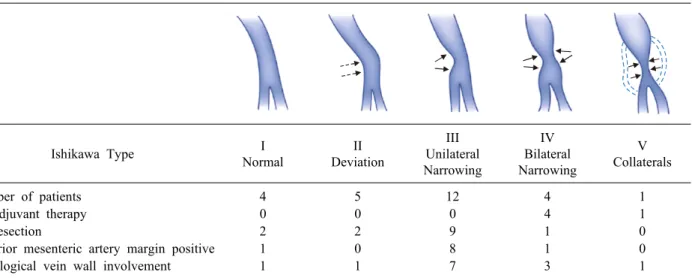
Table 1. Characteristics by Ishikawa type
Ishikawa Type I
Normal
II Deviation
III Unilateral Narrowing
IV Bilateral Narrowing
V Collaterals
Number of patients 4 5 12 4 1
Neoadjuvant therapy 0 0 0 4 1
R1 resection 2 2 9 1 0
Superior mesenteric artery margin positive 1 0 8 1 0
Histological vein wall involvement 1 1 7 3 1
al.5 while reporting the findings of a survey from the Association Francaise de Chirurgie recommended NAT for all patients scheduled for VR during pancreatic re- section, including those with venous involvement in the form of impingement, abutment, narrowing, thrombosis or occlusion observed in cross-sectional imaging studies.9
This study presents the experience of the authors with VR in patients with PDAC of the head of pancreas.
MATERIALS AND METHODS
A retrospective analysis of patients undergoing re- section for proximal pancreatic cancer between July 2010 and July 2016 was performed. The study group consisted of all patients with histologically confirmed PDAC fol- lowing resection. The cohort was divided into two sub- groups based on whether or not they underwent VR.
Patients who underwent standard pancreatic resection con- stituted Group SR. Those who underwent an additional VR were included in Group VR. The preoperative patient characteristics, radiological imaging, operative findings, hospital stay, morbidity and 90-day mortality, tumor path- ology including TNM stage, margin status and histo- logical vascular invasion, were recorded and compared between the groups.
All patients underwent MDCT with pancreatic protocol.
Patients with BRPC as per the MD Anderson Cancer Center definition (‘short segment occlusion of the portal vein (PV), superior mesenteric vein (SMV) or spleno- portal junction with reconstructable, healthy vein proximal
and distal to the area of tumor involvement’)1,2,9 were ad- vised NAT. For purposes of this study, computed tomog- raphy (CT) scans were reviewed and venous involvement was further assessed using the Ishikawa et al.11 classi- fication (Table 1). Patients diagnosed with BRPC under- went endobiliary stenting and endoscopic ultrasonography (EUS)-guided biopsy of the tumor, followed by NAT as advised by a medical oncologist. Preoperative biopsies and endobiliary stenting were performed selectively in pa- tients who did not undergo NAT. Laparoscopy was per- formed on all patients prior to laparotomy.
All patients were treated with classical Whipple’s pan- creaticoduodenectomy and standard lymphadenectomy. A combined posterior and uncinate dissection along the su- perior mesenteric artery (SMA) was used to completely mobilise the head of pancreas prior to transection of the pancreatic neck.12 An isolated Roux loop was used to cre- ate an infracolic gastrojejunostomy to the left of the mid- dle colic vessels. The pancreatic and biliary reconstruction was performed on a single jejunal loop. The preferred pancreatic anastomosis was a duct-to-mucosa pancreatico- jejunostomy. VR was performed when a trial dissection demonstrated venous involvement. Reconstruction was ei- ther by direct end-to-end anastomosis of the vein or by interposition with autologous vein graft (left internal jug- ular vein) or 10 mm polytetrafluoroethylene (PTFE). The splenic vein (SV) was reimplanted when possible. Portal flow was documented after reconstruction with intra- operative Doppler ultrasound. Anticoagulation with low molecular weight heparin for 2 weeks was advised only
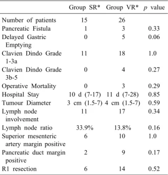
Table 2. Group SR vs Group VR* - comparison of outcomes Group SR* Group VR* p value
Number of patients 15 26
Pancreatic Fistula 1 3 0.33
Delayed Gastric Emptying
0 5 0.06
Clavien Dindo Grade 1-3a
11 18 1.0
Clavien Dindo Grade 3b-5
0 4 0.27
Operative Mortality 0 3 0.29
Hospital Stay 10 d (7-17) 11 d (7-28) 0.85 Tumour Diameter 3 cm (1.5-7) 4 cm (1.5-7) 0.59 Lymph node
involvement
11 17 0.34
Lymph node ratio 33.9% 13.8% 0.16
Superior mesenteric artery margin positive
6 10 1.0
Pancreatic duct margin positive
2 9 0.17
R1 resection 6 14 0.52
*Group SR, Standard pancreatic resection (no vascular re- section); *Group VR, Additional vascular resection for patients with PTFE interposition grafts.
Drain fluid amylase was monitored routinely on day 3, prior to removal of drains, and as needed, based on clinical suspicion of a pancreatic leak. International Study Group definitions were used to diagnose pancreatic leaks,13 haemor- rhage14 and delayed gastric emptying (DGE).15 Chylous leaks were diagnosed when drain fluid amylase was normal and drain fluid triglycerides were elevated on a normal diet.
All patients were recommended adjuvant chemotherapy.
Adjuvant radiotherapy was not prescribed for any patient.
Pancreaticoduodenectomy specimens were evaluated as per the Leeds Protocol16 using serial axial specimen slic- ing after inking the different margins. A positive margin was defined as one in which tumor cells were within 1mm of the inked surface of the specimen.
IBM SPSS Statistic v.20 for Windows (IBM, Armonk NY) was used for data entry and analysis. Continuous da- ta were evaluated using medians, ranges and Mann- Whitney U test. Categorical data were evaluated by Fisher’s exact test. Multivariate analysis was performed using multiple regression to determine the factors predict- ing 90-day survival. A p-value below 0.05 was considered statistically significant for all tests of comparison. Survival was estimated using Kaplan-Meier method and compared using the Log rank test.
RESULTS
We performed 94 pancreaticoduodenectomies between July 2010 and July 2016, 41 of which were indicated for PDAC and constituted the study group. An additional 7 patients with PDAC did not proceed to resection due to metastatic disease diagnosed at laparoscopy or laparotomy.
The median age of the patients in the study group was 59 years (range: 42-79 years) and included 17 women.
Of these, 5 patients were diagnosed with BRPC and re- ceived NAT. Four patients received a gemcitabine-capeci- tabine combination, and one received FOLFIRINOX (5-fluorouracil, leucovorin, irinotecan, and oxaliplatin) regimen. Three patients also underwent preoperative radi- ation therapy. None experienced down-sizing of the tumor with NAT.
Operative details of vascular resection
The extent of vein involvement by Ishikawa Classification
is described in Table 1.
Segmental resection was performed in all but one pa- tient who underwent sleeve resection. Reconstruction was carried out via primary anastomosis in 12 patients, inter- position autologous left internal jugular vein graft in 10, and interposition PTFE graft in 3 patients. Of the 11 pa- tients with tumor involvement of the splenoportal junc- tion, the SV was ligated in 6, reconstructed to the neo portal vein in 3, and 2 patients underwent total pan- createctomy with splenectomy.
Arterial reconstruction was performed in a patient with limited involvement of the common hepatic artery ad- jacent to the gastroduodenal artery.
Morbidity, mortality and hospital stay
Of the 4 pancreatic fistulae, 3 were Grade A, and 1 was Grade B requiring percutaneous drainage of an intra-ab- dominal collection. Of the 5 patients with DGE, all from group VR, 3 were Grade A, and 1 each Grade B and Grade C. One patient underwent re-exploration for bleed- ing from the mesentery on day 1 after resection in the VR group. Details are provided in Table 2.
All three deaths occurred in the VR group. One death was attributed to hepatic ischemia in a patient with in-
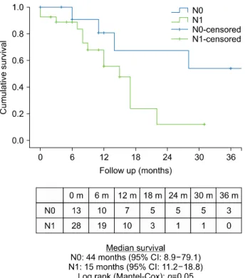
Fig. 2. Comparison of patient survival according to lymph node involvement.
Fig. 1. Comparison of patient survival between the standard resection (SR) group and vein resection (VR) group.
adequate portal flow following prolonged PTFE re- construction with compromised hepatic arterial flow.
Another patient died from pulmonary sepsis following re-suturing of her abdominal wound for wound dehiscence.
She also had Grade C DGE. The third patient also died from sepsis, the source of which is unclear. However, drain amylase was normal and CT scans did not reveal any intra-abdominal collections. Of the three deaths, two occurred in patients who underwent PTFE reconstruction.
Histopathology
The median maximum tumor diameter of the entire group was 3.6 cm (range 1.5-7cm). The median number of lymph nodes excised was 23 (range 1-77). Twenty-eight patients (68.3%) had lymph node metastases. The lymph node ratio (LNR) for the entire cohort was 19.25%.
Positive microscopic resection margins were noted in 20 (48.8%) patients. The superior mesenteric artery mar- gin (39%) was the most frequently involved followed by the pancreatic transection margin (27%). Forty per cent of patients with positive margins showed multiple margin involvement. Patients with Ishikawa grade III and IV vein involvement were more likely to have a positive SMA margin than those with lesser degrees of involvement
(p=0.04). Tumour infiltration of the splenoportal junction was associated with a significantly greater risk of pancre- atic transection margin involvement (p=0.03). Four of 5 patients who received NAT had R0 resections, despite Ishikawa grade IV and V venous involvement. Details are presented in Table 1 and 2.
Histological venous invasion was demonstrated in 13 patients (50%), and was significantly more common in pa- tients with Ishikawa III and IV grade venous involvement (p=0.04); it was associated with increasing Ishikawa type (Table 2).
Survival
The median survival for the entire group of patients was 17 months (95% CI 13.3-20.7) and was not different between Groups SR (14 months, 95%CI 8.7-19.3) and VR (17months, 95%CI 7.1-26.9), (Log Rank p=0.91) (Fig. 1).
Median survival was significantly better in patients with- out lymph node metastases (44 months vs. 15 months, Log Rank p=0.05) (Fig. 2) and worse in patients with his- tologically demonstrable vein wall infiltration by tumor (22 months vs. 11 months, Breslow Wilcoxon p=0.04, Log Rank 0.14) (Fig. 3). No statistically significant differ- ence was found in survival between patients who under-
Fig. 4. Comparison of patient survival following R0 vs. R1 resection.
Fig. 3. Comparison of patient survival according to histo-
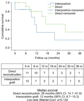
logical vein wall involvement. Fig. 5. Comparison of patient survival according to type of vascular reconstruction.
went R0 or R1 resections (22 months vs. 12 months, p=0.28) (Fig. 4). Survival was not significantly better in those who underwent direct venous reconstruction, in-
dicative of a shorter resection, compared to those who re- quired an interposition graft (28 months vs 12 months, p=0.12) (Fig. 5).
Multivariate analysis revealed that the 90-day survival was not significantly affected by age, tumor size, lymph node ratio, completeness of resection (R0 or R1), compli- cations (DGE, pancreatic leak) or concomitant vascular resection. However, this finding may be attributed to the small sample size.
DISCUSSION
Early reports extolling the benefits of VR along with pancreaticoduodenectomy found that vein involvement was a function of tumor location rather than an indication of aggressive tumor.17 This observation was supported by the latest relevant meta-analysis of 9005 patients from 27 selected trials.6 However two large, recently published series from the Association Francaise de Chirurgie5 and the Japanese Multicentre Study group for Hepatobiliary Surgery4 state that tumors requiring VR were more likely to be stage T4,4,5 poorly differentiated,5 and have a greater incidence of lymph node metastases compared with those
undergoing standard resections.4,5 The present series sup- ports the meta-analysis data. Tumor size and lymph node involvement did not correlate with venous involvement.
Small tumors measuring 1.5 cm in maximal diameter re- quired VR whereas others as large as 7 cm in maximal diameter did not. Lymph node metastases were more com- mon and the LNR higher in patients undergoing standard resections (SR). Patients with lymph node metastases showed significantly poorer survival after resection in the present series. Improved survival in the VR group there- fore likely reflects the lower LNR rather than any benefit from VR itself.
In a meta-analysis, Giovinazzo et al.6 reported sig- nificantly greater overall morbidity (OR 1.34), reopera- tions (OR 1.4) and postoperative bleeding (OR 1.61) in pa- tients undergoing VR compared with standard resections.
The incidence of pancreatic fistula and DGE was not in- creased after VR. The mean operative mortality in their analysis was 3.9% in patients undergoing VR and 3% in those undergoing standard resections (p=0.02). Although the overall morbidity in the present series was greater fol- lowing VR, all the complications in the SR group, and all but one in the VR group were minor. The difference in morbidity was wholly accounted for by the 3 post- operative deaths in the VR group, 2 of which occurred in the 3 patients who underwent PTFE reconstruction.
Since autologous vein graft was the preferred mode of re- construction by the authors, the use of PTFE may reflect either the need for an unplanned or uncontrolled re- construction that did not provide enough time for harvest- ing an autologous vein, or a very long reconstruction.
These situations are known to be associated with in- creased operative mortality.8,18
Factors associated with poor survival post VR include the need for reconstruction longer than 3 cm,8 true vein wall invasion on histology,6,9 and R1 resection.9 Although shorter venous reconstructions, as indicated by the ability to perform primary reconstructions were associated with improved survival compared with those requiring inter- position grafts, the difference was not significant in the present series. True vein wall invasion was associated with significantly poorer early survival as indicated by the Breslow Wilcoxon test (p=0.04) though not the Log Rank test, and may be predicted reliably in patients with Ishikawa types 3, 4 and 5. VR may thus be beneficial in
those patients with higher grades of radiological portome- senteric venous tumor involvement (Ishikawa type 3 and above). This finding together with the 48% R1 resection rate in the present series suggest that all patients planned for VR are recommended NAT despite the lack of reliable evidence suggesting that R0 resections prolong survival compared with R1 resections either in the present series or in other studies.19
The risk of isolated local recurrence in patients with R1 resections is less than 10%.20 Evolving consensus suggest that margin involvement might be more indicative of the quality of pathological examination of the resected speci- men21 and tumor biology.22 A review of specimen histology using the axial slicing technique described by Verbecke et al.16 revised the R1 resection rate from 14% to 76%,23 and 53% to 85%16 respectively, in 2 large independent series even in patients without VR. The importance of tumor bi- ology in the present series is supported by the high rate of R1 resection in the SR group, the fact that 40% of pa- tients with R1 resections had more than one margin in- volved by tumor, and that survival advantage from R0 re- section was not apparent even with tumor-free margins of 1 mm.
SMA margin is the most frequently (15 to 45%) in- volved margin after VR:,6 and was involved in 39% of patients in the present series. SMA-first dissection12 and periadventitial dissection of the SMA24 are recommended in order to improve the likelihood of R0 resections along this margin, without clear evidence to support.25 The other commonly involved margin is the pancreatic transection margin (27% in the present series) especially in patients with tumor overlying the splenopancreatic junction.26 Pancreatic transection to the left of the SMV26 (at the splenic artery origin) in such patients, as well as addi- tional resections based on frozen section biopsy of the margin are desirable to ensure clear resection margins.
Although the ‘Whipple At The Splenic Artery’ or ‘WATSA’,26 advocated by Strasberg, appears sensible and also permits better control of the splenic vein in such patients, the role of additional resection dependent on frozen section biopsy is disputed. Kooby et al.,27 and others,19 determined that involvement of this margin reflected adverse pathological factors such as tumor size, lymph node involvement and perineural infiltration, with an overwhelming effect on survival despite additional pancreatic resection with clear
margins.
This study suffers from being retrospective in nature.
Therefore, additional details such as the true length of vein resection, the use of SMA first dissection and peri- adventitial resection of the SMA, the proportion of pa- tients completing adjuvant therapy and the nature of re- currence (local or distant) are not available. The numbers are relatively small, although the proportion of patients undergoing VR is high, reflecting a referral bias.
In conclusion, vein involvement by proximal PDAC is indicative of location rather than tumor biology. VR bene- fits patients with tumor adhesion to the portomesenteric venous axis despite the risk of R1 resection and operative mortality. Primary reconstruction or interposition of autol- ogous vein grafts appears to yield better outcomes com- pared with PTFE. Increased use of NAT in patients with high preoperative suspicion for vein resection may be beneficial, although the survival benefit derived from R0 resection is not clearly established in the present series.
REFERENCES
1. Katz MH, Pisters PW, Evans DB, Sun CC, Lee JE, Fleming JB, et al. Borderline resectable pancreatic cancer: the importance of this emerging stage of disease. J Am Coll Surg 2008;206:
833-846.
2. Katz MH, Crane CH, Varadachary G. Management of borderline resectable pancreatic cancer. Semin Radiat Oncol 2014;24:
105-112.
3. Ravikumar R, Sabin C, Abu Hilal M, Bramhall S, White S, Wigmore S, et al. Portal Vein resection in borderline resectable pancreatic cancer: a United Kingdom multicentre study. J Am Coll Surg 2014;218:401-411.
4. Murakami Y, Satoi S, Motoi M, Sho M, Kawai M, Matsumoto I, et al. Portal or superior mesenteric vein resection in pan- creatoduodenectomy for pancreatic head cancer. Br J Surg 2015;
102:837-846.
5. Delpero JR, Boher JM, Sauvanet A, Le Truet YP, Sa-Cunha A, Mabrut JY, et al. Pancreatic adenocarcinoma with vein involve- ment: is up front synchronous portal-superior mesenteric vein re- section still justified? a survey of the association Francaise de Chirurgie. Ann Surg Oncol 2015;22:1874-1883.
6. Giovinazzo F, Turri G, Katz MH, Heaton N, Ahmed I. Meta- analysis of benefits of portal-superior mesenteric vein resection in pancreatic resection for ductal adenocarcinoma. Br J Surg 2016;103:179-191.
7. Tol JA, Eshuis WJ, Besselink MG, van Gulik TM, Busch OR, Gouma DJ. Non-radical resection versus bypass procedure for pancreatic cancer - a consecutive series and systematic review.
Eur J Surg Oncol 2015;41:220-227.
8. Pan G, Xie KL, Wu H. Vascular resection in pancreatic ad- enocarcinoma with portal or superior mesenteric vein invasion.
World J Gastroenterol 2013;19:8740-8744.
9. Barreto SG, Windsor JA. Justifying vein resection with pan- creatoduodenectomy. Lancet Oncol 2016. doi: 10.1016/S1470-
2045(15)00463-5. [in press]
10. Al-Hawary MM, Francis IR, Chari ST, Fishman EK, Hough DM, Lu DS, et al. Pancreatic ductal adenocarcinoma radiology report- ing template: consensus statement of the society of abdominal radiology and the american pancreatic association. Radiology 2014;270:248-260.
11. Ishikawa O, Ohigashi H, Imaoka S, Furukawa H, Sasaki Y, Fujita M, et al. Preoperative indications for extended pan- createctomy for locally advanced pancreas cancer involving the portal vein. Ann Surg 1992;215:231-236.
12. Sanjay P, Takaori K, Govil S, Shrikhande SV, Windsor JA.
Artery-first approaches to pancreatoduodenectomy. Br J Surg 2012;99:1027-1035.
13. Bassi C, Dervenis C, Butturini G, Fingerhut A, Yeo C, Izbicki J, et al. Postoperative pancreatic fistula: an international study group (ISGPF) definition. Surgery 2005;138:8-13.
14. Wente MN, Veit JA, Bassi C, Dervenis C, Fingerhut A, Gouma DJ, et al. Postpancreatectomy hemorrhage (PPH): an interna- tional study group of pancreatic surgery (ISGPS) definition.
Surgery 2007;142:20-25.
15. Wente MN, Bassi C, Dervenis C, Fingerhut A, Gouma DJ, Izbicki JR, et al. Delayed gastric emptying (DGE) after pancre- atic surgery: a suggested definition by the international study group of pancreatic surgery (ISGPS). Surgery 2007;142:761-768.
16. Verbecke CS, Leitch D, Menon KV, McMohan MJ, Guillou PJ, Anthoney A. Redefining the r1 resection in pancreatic cancer.
Br J Surg 2006;93:1232-1237.
17. Fuhrman GM, Leach SD, Staley CA, Cusack JC, Charnsangavej C, Cleary KR, et al. Rationale for en bloc vein resection in the treatment of pancreatic adenocarcinoma adherent to the superior mesenteric-portal vein confluence. pancreatic tumour study group. Ann Surg 1996;223:154-162.
18. Kim PT, Wei AC, Atenafu EG, Cavallucci D, Cleary SP, Moulton CA, et al. Planned versus unplanned portal vein re- sections during pancreaticoduodenectomy for adenocarcinoma.
Br J Surg 2013;100:1349-1356.
19. Ethun CG, Kooby DA. The importance of surgical margins in pancreatic cancer. J Surg Oncol 2016;113:283-288.
20. Van den Broeck A, Sergeant G, Ectors N, Van Steenbergen W, Aerts R, Topal B. Patterns of recurrence after curative resection of pancreatic ductal adenocarcinoma. Eur J Surg Oncol 2009;35:600-604.
21. Verbecke CS. Resection margins in pancreatic cancer. Surg Clin North Am 2013;93:647-662.
22. Kimbrough CW, St Hill CR, Martin RC, Mcmasters KM, Scoggins CR. Tumor-positive resection margins reflect an ag- gressive tumor biology in pancreatic cancer. J Surg Oncol 2013;107:602-607.
23. Esposito I, Kleeff J, Bergmann F, Reiser C, Herpel E, Friess H, et al. Most pancreatic cancer resections are r1 resections. Ann Surg Oncol 2008;15:1651-1660.
24. Mizuno S, Isaji S, Tanemura A, Kishiwada M, Murata Y, Azumi Y, et al. Anterior approach to the superior mesenteric artery by using nerve plexus hanging maneuver for borderline resectable pancreatic head carcinoma. J Gastrointest Surg 2014;18:1209- 1215.
25. Butler JR, Ahmad SA, Katz MH, Cioffi JL, Zyromski NJ. A sys- tematic review of the role of periadventitial dissection of the su- perior mesenteric artery in affecting margin status after pan- creatoduodenectomy for pancreatic adenocarcinoma. HPB (Oxford) 2016;18:305-311.
26. Strasberg SM, Sanchez LA, Hawkins WG, Fields RC, Linehan DC. Resection of tumors of the neck of the pancreas with venous invasion: the “whipple at the splenic artery (WATSA)” procedure.
J Gastrointest Surg 2012;16:1048-1054.
27. Kooby DA, Lad NL, Squires MH, Maithel SK, Sarmiento JM, Staley CA, et al. Value of intraoperative neck margin analysis
during whipple for pancreatic adenocarcinoma: a multicenter analysis of 1399 patients. Ann Surg 2014;260:494-501; dis- cussion 501-503.