Backgrounds/Aims: Hepatic portal venous gas (HPVG) is a rare condition, with poor prognosis and a mortality rate of up to 75%. Indications for surgical and non-surgical management of HPVG including associated complications and mortality remain to be clarified. Methods: From January 2008 to December 2014, 18 patients with HPVG diagnosed through abdominal computed tomography (CT) imaging were retrospectively identified. Clinical symptoms, laboratory data, underlying diseases, treatment, and mortality rate were analyzed. Patients were classified into 2 groups: surgical management recommended (SR, n=10) and conservative management (CM, n=8). The SR group was further sub- divided into patients who underwent surgical management (SM-SR, n=5) and those who were managed conservatively (NS-SR, n=5). Results: Conditions underlying HPVG included mesenteric ischemia (38.9%), intestinal obstruction (22.2%), enteritis (22.2%), duodenal ulcer perforation (5.6%), necrotizing pancreatitis (5.6%), and diverticulitis (5.6%).
In terms of mortality, 2 patients (40%) died in the SM-SR group, 1 (12.5%) in the CM group, and 100% in the NS-SR group. Higher scores from Acute Physiology and Chronic Health Evaluation (APACHE) II predicted the mortality rates of the NS-SR and CM groups. Conclusions: Identification of HPVG requires careful consideration for surgical management. If surgical management is indicated, prompt laparotomy should be performed. However, even in the non-surgical management condition, aggressive laparotomy can improve survival rates for patients with high APACHE II scores. (Korean J Hepatobiliary Pancreat Surg 2015;19:181-187)
Key Words: Portal venous gas; Pneumatosis intestinalis; Mesenteric ischemia; Computed tomography; APACHE II
Received: October 27, 2015; Revised: October 30, 2015; Accepted: November 1, 2015 Corresponding author: Sang Hwy Kwon
Department of Surgery, Daegu Fatima Hospital, Ayang Street 99, Dong-gu, Daegu 41199, Korea Tel: +82-53-940-7234, Fax: +82-53-940-7239, E-mail: lovehwik@daum.net
Copyright Ⓒ 2015 by The Korean Association of Hepato-Biliary-Pancreatic Surgery
This is an Open Access article distributed under the terms of the Creative Commons Attribution Non-Commercial License (http://creativecommons.org/
licenses/by-nc/4.0) which permits unrestricted non-commercial use, distribution, and reproduction in any medium, provided the original work is properly cited.
Korean Journal of Hepato-Biliary-Pancreatic Surgery ∙ pISSN: 1738-6349ㆍeISSN: 2288-9213
INTRODUCTION
Hepatic portal venous gas (HPVG) is a rare radiological finding, which is associated with a poor prognosis and a mortality rate as high as 75%.1,2 However, with recent de- velopments in imaging modalities, even very small amounts of gas can now be detected, which allows conservative treatment to be considered as a sufficient intervention that can be implemented early.
A surgical study of 22 patients with HPVG identified a higher mortality rate to be associated with higher Acute Physiology and Chronic Health Evaluation (APACHE) II scores and longer length of bowel resection.3 Another study documented physiological shock and pneumatosis intestinalis (PI) to be significant predictive factors for mortality; however, this study did not differentiate out- comes based on treatment method, namely surgical versus
non-surgical management.4 To date, the risk factors affect- ing the rate of mortality in patients with HPVG who are managed non-surgically have not been clearly evaluated.
Therefore, the aim of our study was to identify these fac- tors in non-surgical patients with HPVG.
MATERIALS AND METHODS
A search was conducted in the radiological databases of Daegu Fatima Hospital in South Korea of the computed tomography (CT) reports of patients over the age of 18 years who had undergone abdominal CT scan. The fol- lowing key words were used to identify cases of HPVG:
hepatic portal venous gas, mesenteric venous gas, air por- togram, PI, portal vein air. Of a total of 41941 patients who underwent abdominal CT from January 2008 to December 2014, 37 patients with the above key words
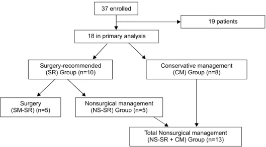
Fig. 1. Allocation of the 18 patients for the primary analysis based on need for surgery and treatment method. SR, Surgical recom- mended; CM, Conservative man- agement; SM-SR, Surgical man- agement – Surgical recommended;
NS-SR, Non-surgical management – Surgical recommended.
were identified. Of these, 19 cases were excluded as CT scans showed only a PI without HPVG. A retrospective review of the medical records of the 18 remaining cases was performed, including a review of their CT scans by two radiology specialists to screen for the associated main portal venous gas (PVG), superior mesenteric vein (SMV) gas, PI, and other intra-abdominal pathology.
As part of the retrospective review of medical records, patients’ early symptoms and underlying diseases and co- morbidities were identified. Clinical condition was de- termined from the physical signs obtained during each pa- tient’s visit to the emergency department including the fol- lowing: blood pressure, pulse rate, respiration rate, body temperature, level of consciousness, and degree of motor response to pain. The physical status for each patient was determined based on the classification system of the American Society of Anesthesiologists (ASA). The se- verity of each patient’s status and expected risk of mortal- ity were investigated by calculating the Acute Physiology and Chronic Health Evaluation (APACHE) II score.
For patients who underwent surgery, the worst labo- ratory test results obtained on the day of admission were selected for analysis. Similarly, for patients who did not undergo surgery, the worst laboratory test results obtained within 24 h after admission were used. Laboratory data included complete blood count (CBC) with differential, as- partate transaminase (AST), alanine transaminase (ALT), blood urea nitrogen (BUN), serum creatinine (SCr), elec- trolytes (sodium, potassium), prothrombin time (PT), parti- al thromboplastin time (PTT), and arterial blood gas analy-
sis (ABGA).
Based on the information obtained from the medical re- cords, patients were divided into 2 groups based on clin- ical management status. These groups were the surgical management recommended (SR, n=10) and conservative management (CM, n=8) groups. The SR group was further subdivided into patients who underwent surgical manage- ment (SM-SR, n=5) and those who could not undergo sur- gery (NS-SR, n=5). Group allocation is summarized in Fig. 1.
For analysis, shock was defined as a systolic blood pressure (BP) of less than 90 mmHg despite proper hydration. Mortality was defined as death within 48 h of hospital admission. In all patients, the ASA classification, Glasgow Coma Scale (GCS) score, and APACHE II score were analyzed as prognostic factors for mortality.
Statistical analyses were performed using SPSS soft- ware (SPSS Inc., Chicago. IL, USA). Between-group dif- ferences for categorical variables were evaluated by chi-square or Fisher’s exact tests, whereas between-group differences for continuous variables were evaluated by Student’s t- or Welch’s t-tests. For multivariate analysis, a logistic regression test was performed.
RESULTS
The study was conducted over a 7-year period and in- cluded 18 patients. The mean age of our study group was 67.3±11.9 years, and the male to female ratio was 1:1.
The distribution of gas in our study group included: 13
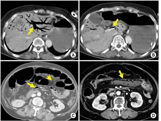
Fig. 2. Portomesenteric venous gas at different anatomical loca- tions in various patients.
Computed tomography reveals massive hepatic portal venous gas (A: arrow) and main portal venous gas (B: arrow), superior mesenteric venous gas (C: ar- row), mesenteric venous branch gas (C: arrow head), and pneu- matosis intestinalis (D: arrow).
patients (72.2%) with PI, 10 (55.6%) with main PVG, and 8 (44.4%) with SMV gas (Fig. 2). The ileum was the most frequent site of PI (9 cases, 45%), followed by the colon (4 cases, 20%), jejunum (3 cases, 15%), cecum (3 cases, 15%), and rectum (1 case, 5%).
Based on ASA physical status classification, 14 (77.8%) patients were identified as having a poor general condition (corresponding to grades III and IV on the clas- sification scale), and the mean APACHE II score for the group was 19.9±10.3.
Disease causes and symptoms
Mesenteric ischemia accounted for 7 cases (38.9%), making it the most frequent cause of HPVG in our study group. Of these cases, 3 were caused by superior mesen- teric artery (SMA) occlusion, 3 by non-occlusive mesen- teric ischemia, and 1 by SMA stenosis. For the remaining patients, intestinal obstruction was identified in 4 cases (22.2%); enteritis in 4 cases (22.2%); and 1 case each of duodenal ulcer perforation (5.6%), necrotizing pancreatitis (5.6%), and diverticulitis (5.6%). The most frequently re- ported symptoms of HPVG were abdominal pain (83.3%), followed by nausea (61.1%), vomiting (61.1%), abdomi- nal distension (38.9%), and diarrhea (16.7%).
Descriptive outcomes of the SM-SR group In our study group, 10 patients were identified for whom surgical treatment was recommended based on the presence of intraperitoneal free air or bowel necrosis as confirmed by CT scan, or signs of peritoneal irritation such as rebound tenderness discovered by physical examination. However, surgery was performed in only 5 of these patients, with the other 5 unable to undergo sur- gery because of serious hemodynamic instability upon ad- mission or other issues such as their caregiver’s refusal to consent to surgery (Fig. 3).
Emergency laparotomy was performed in the 5 patients in the SM-SR group. For 3 of these patients, small bowel resection (248 cm, 220 cm, and 39 cm, respectively) and end-to-end anastomosis were performed. One patient needed ileostomy and resection of the small bowel and colon (small bowel: 120 cm, colon: 38cm). The remaining patient had a duodenal perforation that required duodenal exclusion and gastrojejunostomy. Among the 5 patients who underwent surgery, 2 (40%) died on post-operative day 1. The first of these patients had alcoholic liver cir- rhosis and died of disseminated intravascular coagulation (DIC), while the second patient died of acute renal failure (ARF) and adult respiratory distress syndrome (ARDS).
Complications in the 3 surviving patients included:
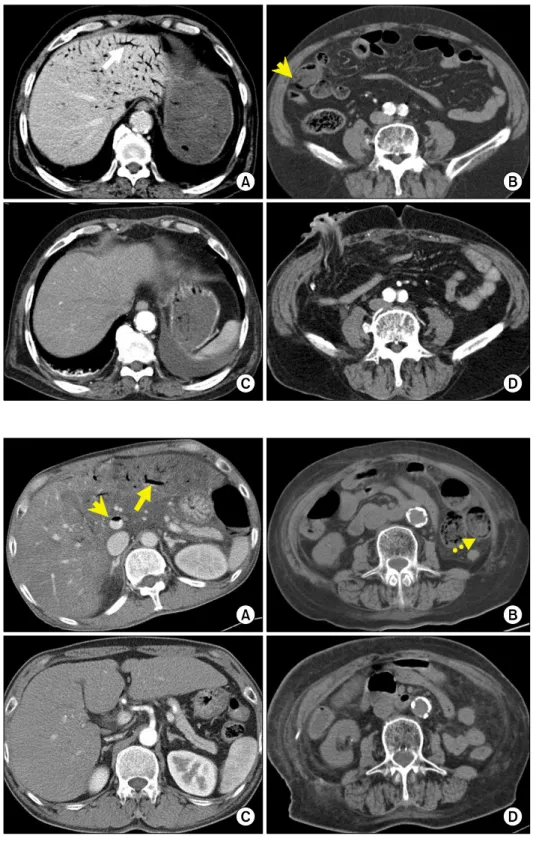
Fig. 3. A 77-year-old man with superior mesenteric artery occlu- sion underwent ascending colon and small bowel resection and loop ileostomy. Before oper- ation, computed tomography demonstrates hepatic portal ve- nous gas (A: arrow), pneuma- tosis intestinalis (B: arrow head).
Computed tomography per- formed at postoperative day 12 shows absence of hepatic portal venous gas (C and D).
Fig. 4. An 81-year-old woman with small bowel enteritis under- went medical management.
Before treatment, computed to- mography shows hepatic portal venous gas (A: arrow), main por- tal venous gas (A: arrow head), and pneumatosis intestinalis (B:
dashed arrow). Computed to- mography performed at day 3 of medical treatment shows radio- logic improvement (C and D).
post-operative atelectasis and wound sepsis in one patient;
another patient suffered from short bowel syndrome with severe diarrhea and malnutrition; and an intra-abdominal abscess formed in the last patient.
Descriptive outcomes of the non-surgically managed patients (NS-SR and CM groups) In our study group, 13 patients were managed non-sur- gically (Fig. 4). Of these patients, 5 had been recom- mended for surgery but were ultimately not suitable can- didates for surgical laparotomy due to non-consent or he-
SMV gas
Shock at admission pH at admission ASA classification III & IV APACHE II score GCS score Surgery Mortality rate
5/10 (50%) 8/10 (80%) 7.25±0.2 9/10 (90%)
24.7±9.4 9.5±3.7 5/10 (50%) 7/10 (70%)
3/8 (37.5%) 2/8 (25.0%)
7.27±0.2 5/8 (62.5%)
14.0±8.5 14.1±1.0
0 1/8 (12.5%)
0.664 0.054 0.848 0.275 0.024 0.003 0.036 0.025 PVG, Portal Venous Gas; SMV, Superior Mesenteric Vein;
ASA, American Society of Anesthesiologists; APACHE, Acute Physiology and Chronic Health Evaluation; GCS, Glasgow Coma Scale
Liver cirrhosis Ischemic heart disease Chronic Renal Failure Laboratory data WBC (/l) AST (U/L) ALT (U/L) Creatinine (mg/dl) pH
Arterial oxygen
1/10 (10%) 2/10 (20%)
0 15,618±11,368
195±273 115±232 2.12±0.93 7.25±0.22 86.56±42.41
2/8 (25%) 1/8 (13%) 3/8 (38%) 13,630±5,440
86±129 20±11 3.08±2.66 7.27±0.22 101.38±32.41
0.56 1.00 0.07 0.657 0.314 0.269 0.358 0.848 0.413 WBC, White Blood Cell; AST, Aspartate Transaminase; ALT, Alanine Transaminase
modynamic instability. For the other 8 patients, a CM ap- proach had been recommended (i.e., the CM group).
Non-surgical management included intravenous fluid ther- apy, antibiotics, and L-tube decompression.
Of this combined group of 13 patients managed non-surgically, 6 (46.2%) died. Of these, only 1 patient was from the CM group (mortality rate 12.5%), with the cause of death of this patient attributable to alcoholic ketoacidosis. In contrast, all patients in the NS-SR group died within 24h of diagnosis resulting in a mortality rate of 100%.
Comparison of the SR and CM groups
Both groups were comparable with regard to the male-to-female ratio; mean age; presence of PI, PVG, or SMV gas; rate and nature of comorbidities; and laboratory data. The frequency of shock and the degree of acid- ification at admission were higher in the SR group as compared with the CM group; however, these be- tween-group differences were not significant. Significant between-group differences were identified for GCS score, APACHE II score, and mortality rate (Tables 1, 2).
Comparison of factors affecting mortality in non- surgically managed (NS-SR and CM) groups For the 13 patients who had HPVG and underwent non-surgical management only, the overall mortality rate was 46.2% (6/13 cases), which was relatively high. From
analysis of prognostic factors affecting mortality, no dif- ference was found between patients in the groups who survived and those who died, based on sex, age, or pres- ence of PI, main PVG, or SMV gas. A higher but non-sig- nificant mortality rate was associated with more severe acidification at admission or with an ASA classification of III or IV. According to univariate analysis, APACHE II scores (p=0.000), GCS (p=0.008), shock at admission (p=0.005), and sign of peritoneal irritation at physical ex- amination (p=0.005) were significant predictive variables of mortality (Tables 3, 4). Only APACHE II scoring re- mained a significant predictive variable in the multivariate analysis.
DISCUSSION
Although HPVG is an ominous radiological finding, it is a non-specific sign, being associated with a range of conditions. HPVG was first reported by Wolfe and Evans in 1955, who described gas in the portal vein system of six infants who had enterocolitis.2 The clinical symptoms of HPVG include non-specific symptoms and signs such as pain, nausea, diarrhea, vomiting, abdominal distension, peritoneal irritation, and acidosis.5 The general nature of these symptoms can delay diagnosis and result in fatality when HPVG is not promptly managed.
Although the mechanism of HPVG has not been estab- lished, HPVG occurs when intestinal gas enters the vein,
Prognostic Factor No. of Patients Mortality p-value Sex
Male Female Peritoneal sign (–)
(+) PI (–) (+) Main PVG (–) (+) SMV gas (–) (+)
ASA classification I, II
III, IV pH <7.30 ≥7.30
Shock at admission (–)
(+)
5 8 8 5 4 9 4 9 7 6 3 10 7 6 6 7
3 (60%) 3 (37.5%) 1 (12.5%) 5 (100%) 1 (25.0%) 5 (55.6%) 1 (25.0%) 5 (55.6%) 2 (28.6%) 4 (66.7%) 0 6 (60%) 5 (71.4%) 1 (16.7%) 0 6 (85.7%)
0.592
0.005
0.559
0.559
0.286
0.192
0.103
0.005
PI, Pneumatosis Intestinalis; PVG, Portal Venous Gas; SMV, Superior Mesenteric Vein; ASA, American Society of Anesthesiologists
Prognostic Factor Survival group Mortality group p-value Age
APACHE score GCS
66.0±14.82 11.86±6.44 14.29±0.95
66.67±12.29 30.17±4.36
7.83±3.76
0.932 0.000 0.008 APACHE, Acute Physiology and Chronic Health Evaluation;
GCS, Glasgow Coma Scale
passing through the mucosal layer (via a damaged muco- sa), due to increased pressure within the intestine resulting from increased intra-abdominal pressure.1 Another possi- ble mechanism involves the proliferation of anaerobic bacteria within the intestine, producing a large amount of gas that enters the venous circulation.6
To identify the cause of HPVG, Kinoshita et al.7 con- ducted a study of 64 patients, reporting the following un- derlying causes of HPVG: mesenteric ischemia (43%); di- gestive tract dilation (12%); intraperitoneal abscess (11%);
ulcerative colitis (4%); gastric ulcer (4%); complications from endoscopic procedures (4%); intraperitoneal tumor (3%); and other causes (15%). In our hospital, mesenteric ischemia was the cause in 39% of HPVG cases, a finding comparable to that of Kinoshita et al.7
HPVG can be diagnosed by radiographic examination such as through plain abdominal radiography, ultra- sonography (USG), color Doppler flow imaging, or CT.8 Among these, CT is the best diagnostic method because of its high sensitivity to HPVG and its capability in con-
current investigation of underlying diseases or abdominal pathology.9-11 Moreover, in critically ill patients with poor vital signs, CT can be performed quickly, with little influ- ence of practitioner or patient factors on results. In our retro- spective case series, CT was used to diagnose all the patients.
There is an ongoing debate regarding the correlation be- tween the associated SMV, mesenteric vein gas or PI in HPVG patients and mortality rate. Heye et al.12 reported a higher mortality rate in a group of 47 patients with HPVG when gas was present in the SMV. Morris et al.13 advocated that the combination of HPVG and PI increases mortality. Another study reported that HPVG accompanied by PI increased the risk for fulminant bowel infarction.14 In contrast, Faberman and Mayo-Smith9 found no associa- tion between mesenteric vein gas and mortality. Another study reported that PI and HPVG do not reflect the se- verity of bowel ischemia.15 In our study, presence of PI, main PVG, and SMV gas did not affect the mortality rate, leading us to conclude that the distribution of gas in the venous system was not associated with mortality.
Until 1978, when HPVG was first detected, the mortal- ity rate approached 75%.1 As diagnosis of HPVG was based on the conventional abdominal films available at that time, HPVG was diagnosed only when there was a large amount of gas, and therefore bowel necrosis was al- ready present in many cases. Recently, the mortality rate associated with HPVG has decreased to between 29% and 39% as a result of improved diagnosis using CT or USG, which allows detection of even a small amount of HPVG, and improvements in treatment.7,11,12 For all HPVG pa- tients in our study, the mortality rate was 44.4% (8/18), a rate somewhat higher than the rate reported in previous studies. This difference in the mortality rate can be ex- plained by the relatively large number of patients for whom surgical management was recommended but, for various reasons, underwent conservative treatment instead.
would not have tolerated a surgical procedure or who re- fused surgery.
A number of studies have evaluated the risk factors af- fecting mortality rate in patients with HPVG. One study reported that a high APACHE II score and longer re- section of the small bowel were significantly associated with higher mortality.3 Another study reported that shock at admission and PI were significant risk factors for HPVG-related mortality.4 In our study, a high APACHE II score was identified as a predictive factor of mortality among patients receiving only medical treatment.
If HPVG can be detected early in its course by imaging modality, then what is the best course of treatment? Based on our outcomes, we postulate that the decision to pro- ceed with surgical intervention over conservative treat- ment should be carefully considered, taking into account clinical symptoms, laboratory findings, and radiographic findings. Nelson et al. published an “ABC” algorithm, which stresses that operative treatment (Aggressive treat- ment), close monitoring (Be careful), and medical treat- ment (Conservative treatment) should be carried out, de- pending on the patient’s condition.6
In conclusion, when HPVG is detected via imaging, all patients should not necessarily undergo surgery. However, in our study, among patients of the SR group who were managed non-surgically (NS-SR), all (5/5) died within 24h of diagnosis. In patients (13/18) receiving only medical treatment, a higher APACHE II score was identified as a predictive factor of mortality. Thus we propose that, if surgical indication is present, emergent laparotomy is es- sential, and in medically-managed patients with a higher APACHE II score, indications for surgical management should be judiciously evaluated considering that ag- gressive laparotomy can increase survival.
In this study, the prognostic factors affecting the mor- tality rate of patients with HPVG were investigated.
However, our study was limited by its retrospective de- sign and a small sample size not representative of the gen- eral characteristics of all patients with HPVG. Prospective
Radiology) and Hee-Jin Kim (Department of Radiology) who reviewed the CT scans of HPVG patients.
REFERENCES
1. Liebman PR, Patten MT, Manny J, Benfield JR, Hechtman HB.
Hepatic-portal venous gas in adults: etiology, pathophysiology and clinical significance. Ann Surg 1978;187:281-287.
2. Wolfe JN, Evans WA. Gas in the portal veins of the liver in infants; a roentgenographic demonstration with postmortem ana- tomical correlation. Am J Roentgenol Radium Ther Nucl Med 1955;74:486-488.
3. Wu JM, Tsai MS, Lin MT, Tien YW, Lin TH. High APACHE II score and long length of bowel resection impair the outcomes in patients with necrotic bowel induced hepatic portal venous gas. BMC Gastroenterol 2011;11:18.
4. Seak CJ, Hsu KH, Wong YC, Ng CJ, Yen DH, Seak JC, et al.
The prognostic factors of adult patients with hepatic portal ve- nous gas in the ED. Am J Emerg Med 2014;32:972-975.
5. Iannitti DA, Gregg SC, Mayo-Smith WW, Tomolonis RJ, Cioffi WG, Pricolo VE. Portal venous gas detected by computed to- mography: is surgery imperative? Dig Surg 2003;20:306-315.
6. Nelson AL, Millington TM, Sahani D, Chung RT, Bauer C, Hertl M, et al. Hepatic portal venous gas: the ABCs of management. Arch Surg 2009;144:575-581.
7. Kinoshita H, Shinozaki M, Tanimura H, Umemoto Y, Sakaguchi S, Takifuji K, et al. Clinical features and management of hepatic portal venous gas: four case reports and cumulative review of the literature. Arch Surg 2001;136:1410-1414.
8. Abboud B, El Hachem J, Yazbeck T, Doumit C. Hepatic portal venous gas: physiopathology, etiology, prognosis and treatment.
World J Gastroenterol 2009;15:3585-3590.
9. Faberman RS, Mayo-Smith WW. Outcome of 17 patients with portal venous gas detected by CT. AJR Am J Roentgenol 1997;
169:1535-1538.
10. Monneuse O, Pilleul F, Barth X, Gruner L, Allaouchiche B, Valette PJ, et al. Portal venous gas detected on computed tomog- raphy in emergency situations: surgery is still necessary. World J Surg 2007;31:1065-1071.
11. Hou SK, Chern CH, How CK, Chen JD, Wang LM, Lee CH.
Hepatic portal venous gas: clinical significance of computed to- mography findings. Am J Emerg Med 2004;22:214-218.
12. Heye T, Bernhard M, Mehrabi A, Kauczor HU, Hosch W.
Portomesenteric venous gas: is gas distribution linked to etiology and outcome? Eur J Radiol 2012;81:3862-3869.
13. Morris MS, Gee AC, Cho SD, Limbaugh K, Underwood S, Ham B, et al. Management and outcome of pneumatosis intestinalis.
Am J Surg 2008;195:679-682.
14. Bodewes HW, Puylaert JB. Ultrasound in detection of portal ve- nous gas in adults. Gastrointest Radiol 1991;16:35-37.
15. Peloponissios N, Halkic N, Pugnale M, Jornod P, Nordback P, Meyer A, et al. Hepatic portal gas in adults: review of the liter- ature and presentation of a consecutive series of 11 cases. Arch Surg 2003;138:1367-1370.